Department of Biomedicine and Molecular Imaging Center (MIC), University of Bergen, Bergen, Norway.
Department of Biosciences, University of Oslo, Oslo, Norway.
Histochem Cell Biol. 2022 Sep;158(3):241-251. doi: 10.1007/s00418-022-02115-y. Epub 2022 May 23.
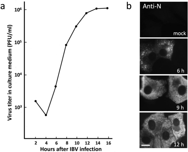
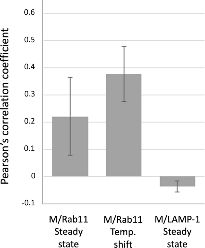
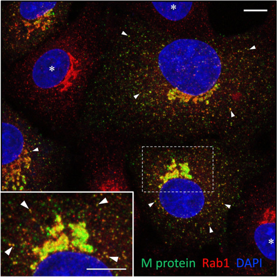
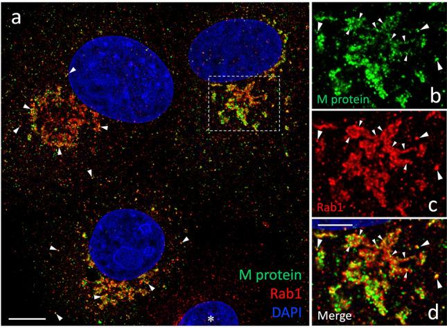
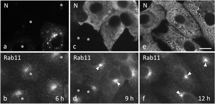
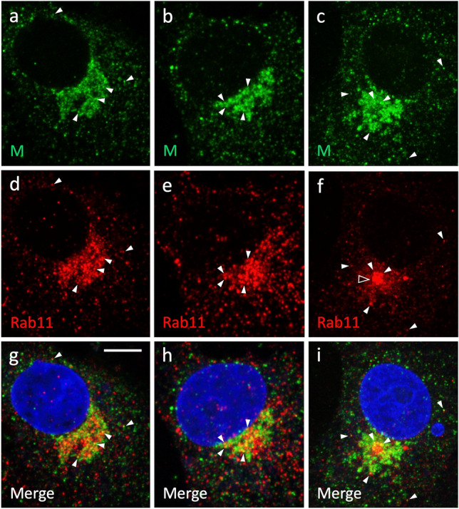
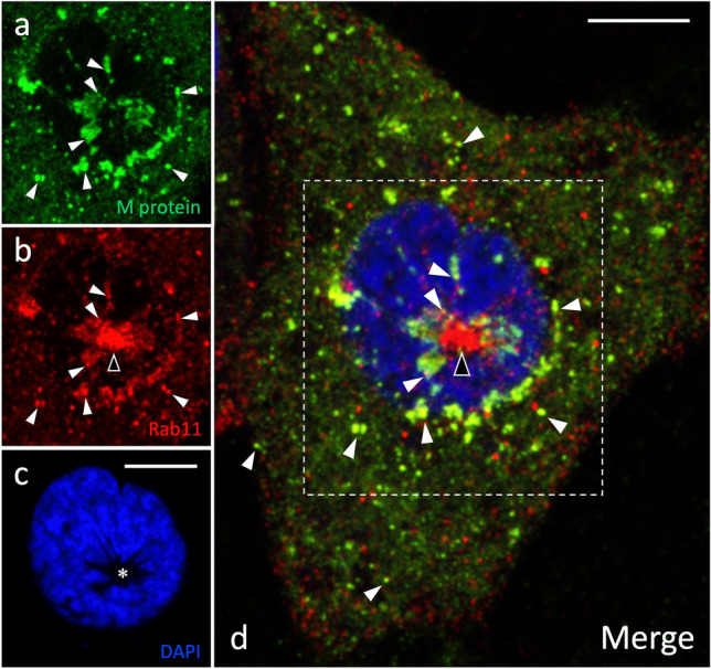
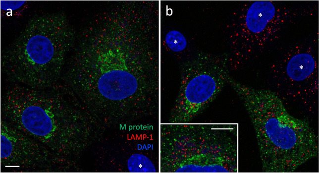
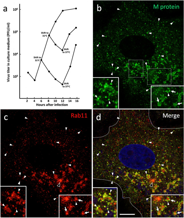
After their assembly by budding into the lumen of the intermediate compartment (IC) at the endoplasmic reticulum (ER)-Golgi interface, coronaviruses (CoVs) are released from their host cells following a pathway that remains poorly understood. The traditional view that CoV exit occurs via the constitutive secretory route has recently been questioned by studies suggesting that this process involves unconventional secretion. Here, using the avian infectious bronchitis virus (IBV) as a well-established model virus, we have applied confocal microscopy to investigate the pathway of CoV egress from epithelial Vero cells. We report a novel effect of IBV infection on cellular endomembranes, namely, the compaction of the pericentrosomal endocytic recycling compartment (ERC) defined by the GTPase Rab11, which coincides with the previously described Golgi fragmentation, as well as virus release. Despite Golgi disassembly, the IC elements containing the major IBV membrane protein (M)-which mostly associates with newly formed virus particles-maintain their close spatial connection with the Rab11-positive endocytic recycling system. Moreover, partial colocalization of the M protein with Rab11 was observed, whereas M displayed negligible overlap with LAMP-1, indicating that IBV egress does not occur via late endosomes or lysosomes. Synchronization of virus release using temperature-shift protocols was accompanied by increased colocalization of M and Rab11 in vesicular and vacuolar structures in the pericentrosomal region and at the cell periphery, most likely representing IBV-containing transport carriers. In conclusion, these results add CoVs to the growing list of viruses exploiting the endocytic recycling apparatus defined by Rab11 for their assembly and/or release.
冠状病毒(CoVs)在组装完成后通过出芽进入内质网(ER)-高尔基体界面的中间隔室(IC)内腔,随后通过一条仍未被充分了解的途径从宿主细胞中释放出来。传统观点认为 CoV 的释放是通过组成型分泌途径发生的,但最近的研究表明,这一过程涉及非常规分泌,这一观点受到了质疑。在这里,我们使用作为成熟模型病毒的禽传染性支气管炎病毒(IBV),应用共聚焦显微镜技术来研究 CoV 从上皮细胞 Vero 中的逸出途径。我们报告了 IBV 感染对细胞内膜系统的一种新的影响,即中心体周围的内体再循环区(ERC)致密化,由 GTPase Rab11 定义,这与之前描述的高尔基体碎片化以及病毒释放同时发生。尽管高尔基体解体,但包含主要 IBV 膜蛋白(M)的 IC 元件——它主要与新形成的病毒颗粒结合——与其密切的空间连接仍保持与 Rab11 阳性的内体再循环系统的连接。此外,观察到 M 蛋白与 Rab11 的部分共定位,而 M 蛋白与 LAMP-1 的重叠程度很小,这表明 IBV 的释放不是通过晚期内体或溶酶体发生的。使用温度骤变方案进行病毒释放同步化时,M 和 Rab11 在中心体周围的囊泡和空泡结构以及细胞边缘中的共定位增加,这很可能代表含有 IBV 的运输载体。总之,这些结果将 CoVs 添加到越来越多的利用 Rab11 定义的内体再循环装置进行组装和/或释放的病毒列表中。