Żurek Michał, Jasak Kamil, Niemczyk Kazimierz, Rzepakowska Anna
Department of Otorhinolaryngology Head and Neck Surgery, Medical University of Warsaw, 1a Banacha Str., 02-097 Warsaw, Poland.
Doctoral School, Medical University of Warsaw, 61 Żwirki i Wigury Str., 02-091 Warsaw, Poland.
J Clin Med. 2022 May 12;11(10):2752. doi: 10.3390/jcm11102752.
Early diagnosis of laryngeal lesions is necessary to begin treatment of patients as soon as possible to preserve optimal organ functions. Imaging examinations are often aided by artificial intelligence (AI) to improve quality and facilitate appropriate diagnosis. The aim of this study is to investigate diagnostic utility of AI in laryngeal endoscopy.
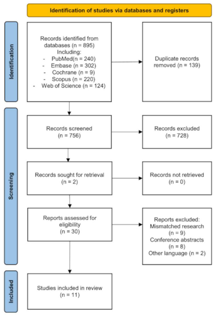
Five databases were searched for studies implementing artificial intelligence (AI) enhanced models assessing images of laryngeal lesions taken during laryngeal endoscopy. Outcomes were analyzed in terms of accuracy, sensitivity, and specificity.
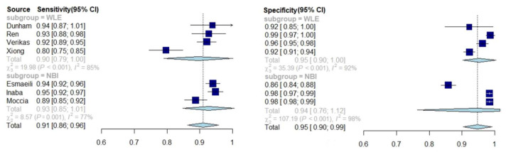
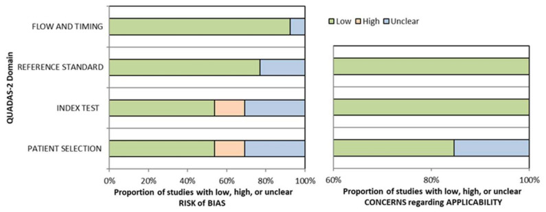
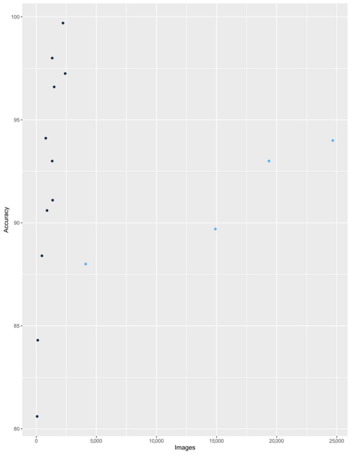
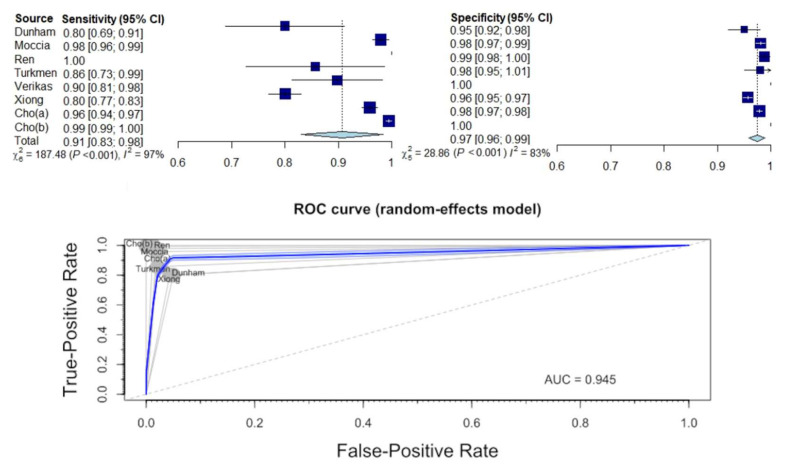
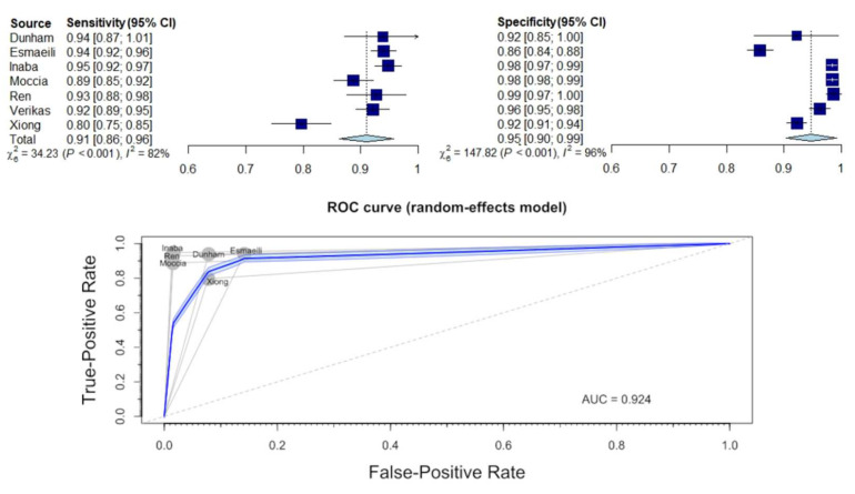
All 11 studies included presented an overall low risk of bias. The overall accuracy of AI models was very high (from 0.806 to 0.997). The accuracy was significantly higher in studies using a larger database. The pooled sensitivity and specificity for identification of healthy laryngeal tissue were 0.91 and 0.97, respectively. The same values for differentiation between benign and malignant lesions were 0.91 and 0.94, respectively. The comparison of the effectiveness of AI models assessing narrow band imaging and white light endoscopy images revealed no statistically significant differences ( = 0.409 and 0.914).
In assessing images of laryngeal lesions, AI demonstrates extraordinarily high accuracy, sensitivity, and specificity.
早期诊断喉部病变对于尽快开始治疗患者以保留最佳器官功能至关重要。影像学检查通常借助人工智能(AI)来提高质量并促进恰当诊断。本研究的目的是探讨AI在喉镜检查中的诊断效用。
检索了五个数据库,以查找实施人工智能(AI)增强模型评估喉镜检查期间拍摄的喉部病变图像的研究。根据准确性、敏感性和特异性对结果进行分析。
纳入的所有11项研究总体偏倚风险较低。AI模型的总体准确性非常高(从0.806到0.997)。在使用更大数据库的研究中,准确性显著更高。识别健康喉部组织的合并敏感性和特异性分别为0.91和0.97。区分良性和恶性病变的相同值分别为0.91和0.94。评估窄带成像和白光内镜图像的AI模型有效性比较显示无统计学显著差异(分别为0.409和0.914)。
在评估喉部病变图像时,AI表现出极高的准确性、敏感性和特异性。