Department of Radiology, Ospedale del Mare-ASL NA1 Centro, Via Enrico Russo 11, 80147 Naples, Italy.
Tomography. 2022 May 23;8(3):1386-1400. doi: 10.3390/tomography8030112.
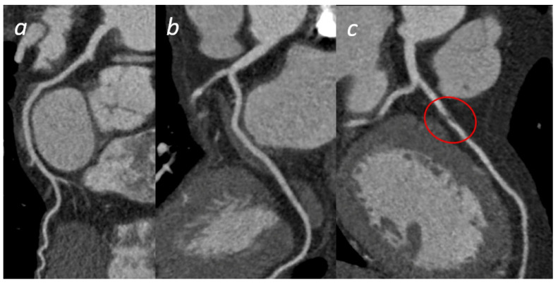
Current strategies for the evaluation of patients with chest pain have significantly changed thanks to the implemented potentiality of CT and MRI. The possible fatal consequences and high malpractice costs of missed acute coronary syndromes lead to unnecessary hospital admissions every year. CT provides consistent diagnostic support, mainly in suspected coronary disease in patients with a low or intermediate pre-test risk. Moreover, it can gain information in the case of cardiac involvement in pulmonary vascular obstructive disease. MRI, on the other hand, has a leading role in the condition of myocardial damage irrespective of the underlying inflammatory or stress related etiology. This article discusses how radiology techniques (CT and MRI) can impact the diagnostic workflow of the most common cardiac and vascular pathologies that are responsible for non-traumatic chest pain admissions to the Emergency Department.
由于 CT 和 MRI 的应用潜力,目前评估胸痛患者的策略发生了重大变化。漏诊急性冠状动脉综合征可能导致致命后果和高额医疗事故成本,每年都会导致不必要的住院治疗。CT 主要在低或中预测试风险的疑似冠心病患者中提供一致的诊断支持。此外,它还可以在肺血管阻塞性疾病累及心脏的情况下获取信息。另一方面,MRI 在心肌损伤的情况下具有主导作用,无论潜在的炎症或应激相关病因如何。本文讨论了放射学技术(CT 和 MRI)如何影响导致非创伤性胸痛急诊就诊的最常见心脏和血管病理学的诊断工作流程。