• 文献检索
  • 文档翻译
  • 深度研究
  • 学术资讯
  • Suppr Zotero 插件Zotero 插件
  • 邀请有礼
  • 套餐&价格
  • 历史记录
应用&插件
Suppr Zotero 插件Zotero 插件浏览器插件Mac 客户端Windows 客户端微信小程序
定价
高级版会员购买积分包购买API积分包
服务
文献检索文档翻译深度研究API 文档MCP 服务
关于我们
关于 Suppr公司介绍联系我们用户协议隐私条款
关注我们

Suppr 超能文献

核心技术专利:CN118964589B侵权必究
粤ICP备2023148730 号-1Suppr @ 2026

文献检索

告别复杂PubMed语法,用中文像聊天一样搜索,搜遍4000万医学文献。AI智能推荐,让科研检索更轻松。

立即免费搜索

文件翻译

保留排版,准确专业,支持PDF/Word/PPT等文件格式,支持 12+语言互译。

免费翻译文档

深度研究

AI帮你快速写综述,25分钟生成高质量综述,智能提取关键信息,辅助科研写作。

立即免费体验

硒蛋白SELENOM和SELENOT在A-172人胶质母细胞瘤细胞系中对细胞凋亡、内质网应激和钙稳态调节中的作用

The Role of Selenoproteins SELENOM and SELENOT in the Regulation of Apoptosis, ER Stress, and Calcium Homeostasis in the A-172 Human Glioblastoma Cell Line.

作者信息

Varlamova Elena G, Goltyaev Michael V, Turovsky Egor A

机构信息

Institute of Cell Biophysics of the Russian Academy of Sciences, Federal Research Center "Pushchino Scientific Center for Biological Research of the Russian Academy of Sciences", 142290 Pushchino, Russia.

出版信息

Biology (Basel). 2022 May 25;11(6):811. doi: 10.3390/biology11060811.

DOI:10.3390/biology11060811
PMID:35741332
原文链接:https://pmc.ncbi.nlm.nih.gov/articles/PMC9220170/
Abstract

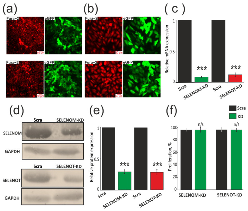
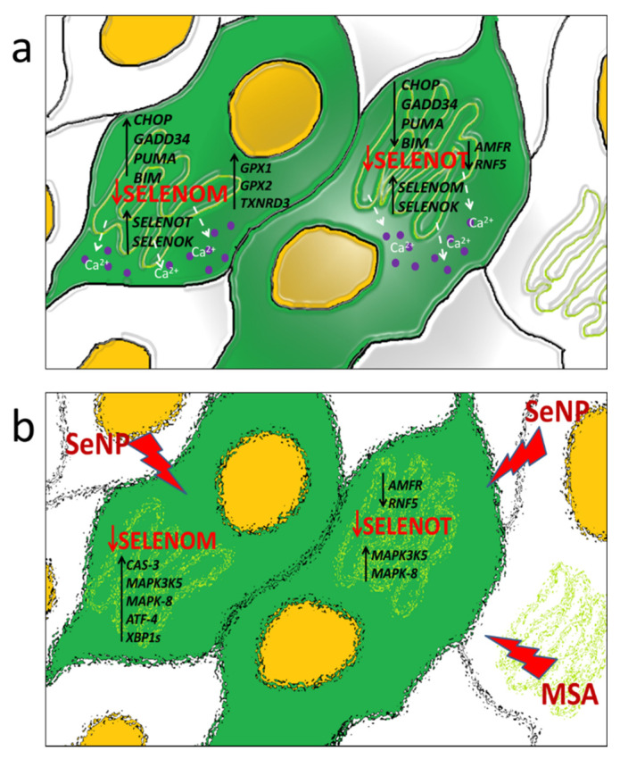
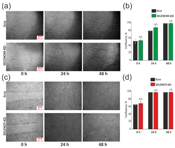
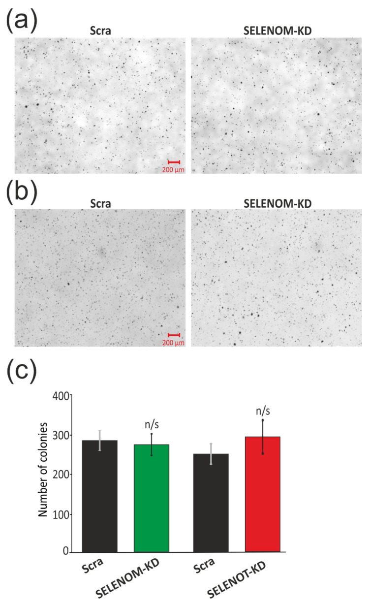
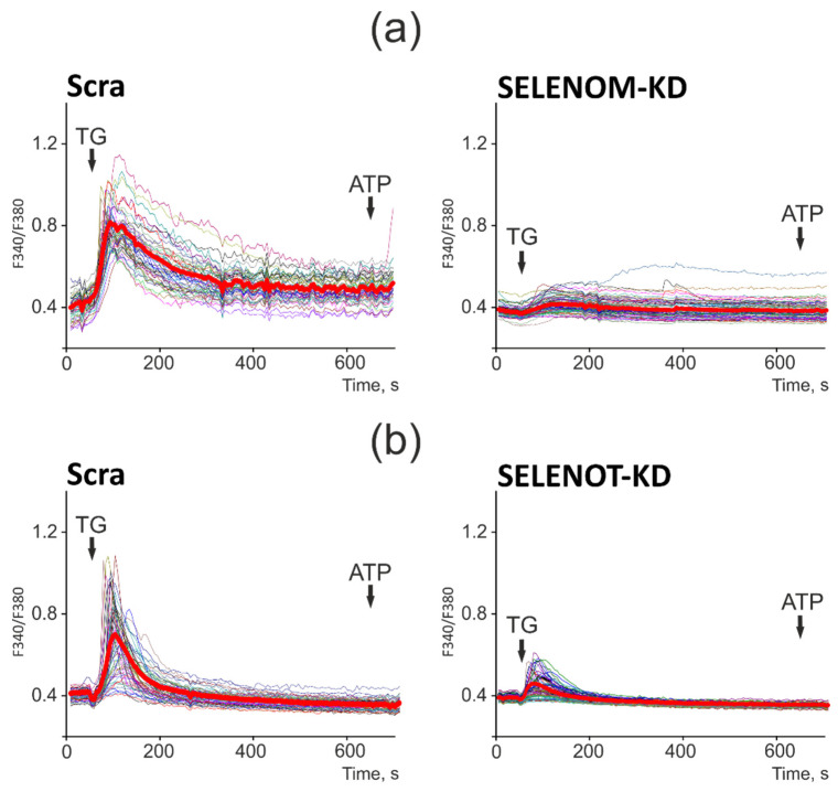
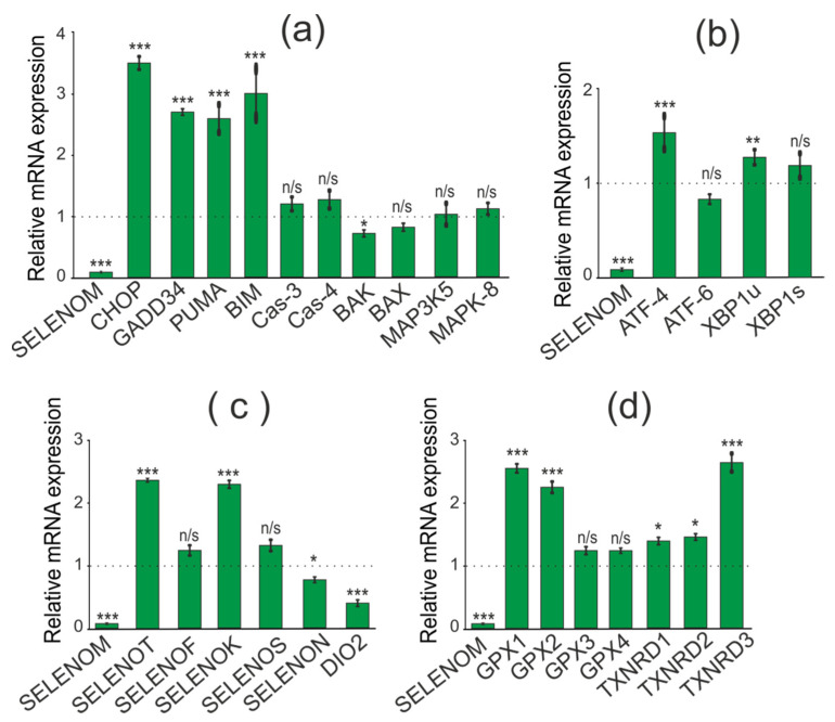
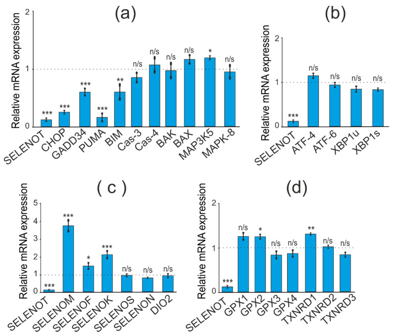
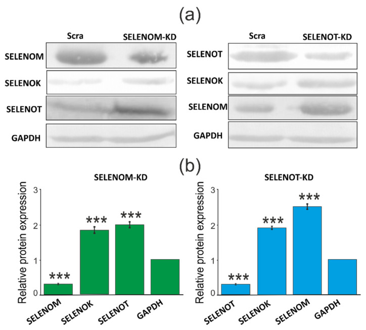
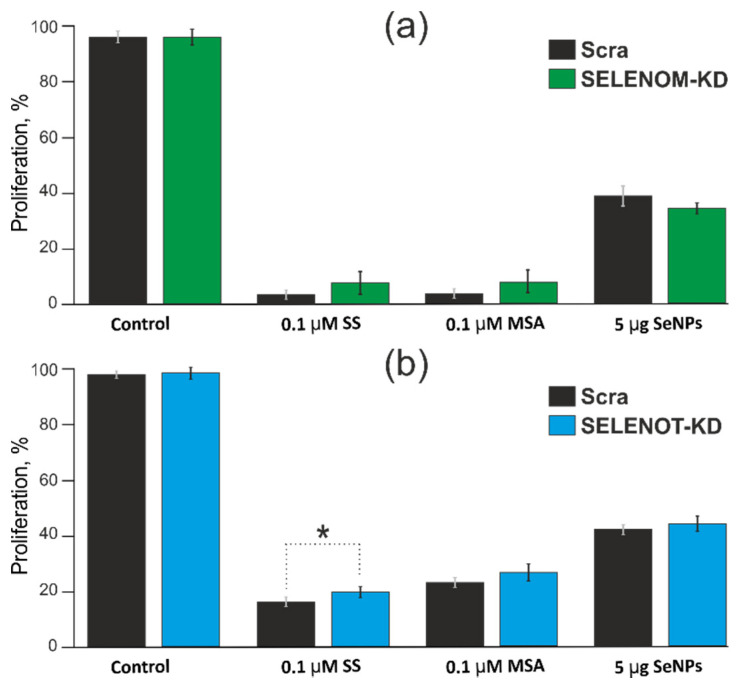
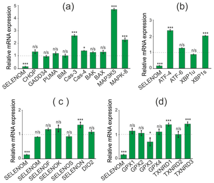
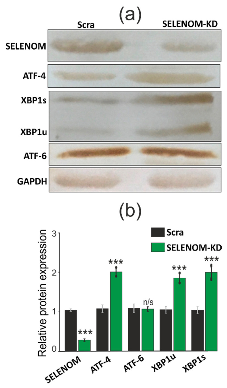
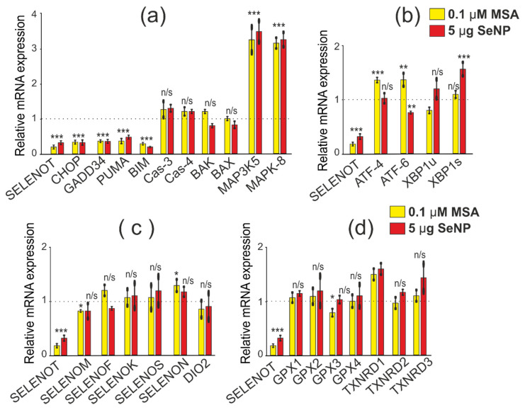
It is known that seven mammalian selenoproteins are localized in the endoplasmic reticulum: SELENOM, SELENOT, SELENOF, SELENOK, SELENOS, SELENON, and DIO2. Among them, SELENOM and SELENOT are the least studied; therefore, the study of their function using the widespread method of suppressing the expression of genes encoding these proteins and the activity of the enzymes themselves by RNA interference is of great interest. We have shown that a decrease in the expression of SELENOM and SELENOT mRNA in the A-172 human glioblastoma cell line by more than 10 times and the quantitative content of enzymes by more than 3 times leads to ER stress, expressed as a decrease in the ER capacity for storing Ca ions. At the level of regulation of apoptotic processes, SELENOM knockdown leads to an increase in the expression of pro-apoptotic CHOP, GADD34, PUMA, and BIM genes, but a compensatory increase in the levels of SELENOT and antioxidant genes from the group of glutathione peroxidases and thioredoxins did not induce cell death. Knockdown of SELENOT had the opposite effect, reducing the expression of pro-apoptotic proteins and regulating the level of a smaller number of genes encoding antioxidant enzymes, which also did not affect the baseline level of apoptosis in the studied cells. At the same time, ER stress induced by MSA or SeNPs induced a more pronounced pro-apoptotic effect in SELENOT knockdown cells through suppression of the expression of selenium-containing antioxidant proteins. Thus, in this work, for the first time, the mechanisms of fine regulation of the processes of apoptosis, cell proliferation, and ER stress by two ER resident proteins, SELENOM and SELENOT, are touched upon, which is not only fundamental but also applied to clinical importance due to the close relationship between the calcium signaling system of cells, folding proteins-regulators of apoptosis and cell survival pathways.

摘要

已知七种哺乳动物硒蛋白定位于内质网

SELENOM、SELENOT、SELENOF、SELENOK、SELENOS、SELENON和DIO2。其中,SELENOM和SELENOT的研究最少;因此,使用广泛的RNA干扰方法抑制编码这些蛋白质的基因表达和酶本身的活性来研究它们的功能具有极大的意义。我们已经表明,在A-172人胶质母细胞瘤细胞系中,SELENOM和SELENOT mRNA的表达降低超过10倍,酶的定量含量降低超过3倍,会导致内质网应激,表现为内质网储存钙离子的能力下降。在凋亡过程的调控水平上,SELENOM基因敲低会导致促凋亡的CHOP、GADD34、PUMA和BIM基因的表达增加,但SELENOT和谷胱甘肽过氧化物酶及硫氧还蛋白组中的抗氧化基因水平的代偿性增加并未诱导细胞死亡。SELENOT基因敲低则产生相反的效果,降低促凋亡蛋白的表达并调节较少数量的编码抗氧化酶的基因水平,这也不影响所研究细胞中的基础凋亡水平。同时,MSA或硒纳米颗粒诱导的内质网应激通过抑制含硒抗氧化蛋白的表达,在SELENOT基因敲低的细胞中诱导出更明显的促凋亡作用。因此,在这项工作中,首次涉及了两种内质网驻留蛋白SELENOM和SELENOT对凋亡、细胞增殖和内质网应激过程的精细调控机制,这不仅具有基础性,而且由于细胞的钙信号系统、折叠蛋白——凋亡调节因子和细胞存活途径之间的密切关系,还具有临床重要性。

https://cdn.ncbi.nlm.nih.gov/pmc/blobs/b2c5/9220170/cec0fa50600e/biology-11-00811-g013.jpg
https://cdn.ncbi.nlm.nih.gov/pmc/blobs/b2c5/9220170/87dfdaa8ded4/biology-11-00811-g001.jpg
https://cdn.ncbi.nlm.nih.gov/pmc/blobs/b2c5/9220170/a33d5a3ea64a/biology-11-00811-g002.jpg
https://cdn.ncbi.nlm.nih.gov/pmc/blobs/b2c5/9220170/30b7c2da9039/biology-11-00811-g003.jpg
https://cdn.ncbi.nlm.nih.gov/pmc/blobs/b2c5/9220170/fda401fc2b18/biology-11-00811-g004.jpg
https://cdn.ncbi.nlm.nih.gov/pmc/blobs/b2c5/9220170/8d32c9d4a070/biology-11-00811-g005.jpg
https://cdn.ncbi.nlm.nih.gov/pmc/blobs/b2c5/9220170/cbc66b4b53a9/biology-11-00811-g006.jpg
https://cdn.ncbi.nlm.nih.gov/pmc/blobs/b2c5/9220170/802aba2862fd/biology-11-00811-g007.jpg
https://cdn.ncbi.nlm.nih.gov/pmc/blobs/b2c5/9220170/9b14bb136368/biology-11-00811-g008.jpg
https://cdn.ncbi.nlm.nih.gov/pmc/blobs/b2c5/9220170/595bda3ee056/biology-11-00811-g009.jpg
https://cdn.ncbi.nlm.nih.gov/pmc/blobs/b2c5/9220170/e6872f0267ee/biology-11-00811-g010.jpg
https://cdn.ncbi.nlm.nih.gov/pmc/blobs/b2c5/9220170/9c0e4cf27bd8/biology-11-00811-g011.jpg
https://cdn.ncbi.nlm.nih.gov/pmc/blobs/b2c5/9220170/5b5159aea1b0/biology-11-00811-g012.jpg
https://cdn.ncbi.nlm.nih.gov/pmc/blobs/b2c5/9220170/cec0fa50600e/biology-11-00811-g013.jpg

相似文献

1
The Role of Selenoproteins SELENOM and SELENOT in the Regulation of Apoptosis, ER Stress, and Calcium Homeostasis in the A-172 Human Glioblastoma Cell Line.硒蛋白SELENOM和SELENOT在A-172人胶质母细胞瘤细胞系中对细胞凋亡、内质网应激和钙稳态调节中的作用
Biology (Basel). 2022 May 25;11(6):811. doi: 10.3390/biology11060811.
2
Endoplasmic reticulum-resident selenoproteins and their roles in glucose and lipid metabolic disorders.内质网定位的硒蛋白及其在糖脂代谢紊乱中的作用。
Biochim Biophys Acta Mol Basis Dis. 2024 Aug;1870(6):167246. doi: 10.1016/j.bbadis.2024.167246. Epub 2024 May 18.
3
Molecular Mechanisms of the Cytotoxic Effect of Recombinant Selenoprotein SELENOM on Human Glioblastoma Cells.重组硒蛋白 SELENOM 对人胶质母细胞瘤细胞细胞毒性作用的分子机制。
Int J Mol Sci. 2023 Mar 30;24(7):6469. doi: 10.3390/ijms24076469.
4
Selenoprotein T: An Essential Oxidoreductase Serving as a Guardian of Endoplasmic Reticulum Homeostasis.硒蛋白 T:一种必需的氧化还原酶,作为内质网稳态的守护者。
Antioxid Redox Signal. 2020 Dec 10;33(17):1257-1275. doi: 10.1089/ars.2019.7931. Epub 2020 Jul 7.
5
Dietary selenium sources differentially regulate selenium concentration, mRNA and protein expression of representative selenoproteins in various tissues of yellow catfish .饮食中的硒源会在不同程度上调节黄颡鱼不同组织中硒浓度、代表性硒蛋白的 mRNA 和蛋白表达。
Br J Nutr. 2022 Feb 28;127(4):490-502. doi: 10.1017/S000711452100194X. Epub 2021 Jun 4.
6
Roles of Selenoproteins in Brain Function and the Potential Mechanism of Selenium in Alzheimer's Disease.硒蛋白在脑功能中的作用及硒在阿尔茨海默病中的潜在机制
Front Neurosci. 2021 Mar 8;15:646518. doi: 10.3389/fnins.2021.646518. eCollection 2021.
7
Dietary Marginal and Excess Selenium Increased Triglycerides Deposition, Induced Endoplasmic Reticulum Stress and Differentially Influenced Selenoproteins Expression in the Anterior and Middle Intestines of Yellow Catfish .膳食中硒缺乏和过量均会增加鲶鱼甘油三酯沉积、诱导内质网应激并对其前肠和中肠中的硒蛋白表达产生不同影响。
Antioxidants (Basel). 2021 Mar 29;10(4):535. doi: 10.3390/antiox10040535.
8
Selenoprotein Gene mRNA Expression Evaluation During Renal Ischemia-Reperfusion Injury in Rats and Ebselen Intervention Effects.硒蛋白基因 mRNA 表达评价在大鼠肾缺血再灌注损伤和依布硒啉干预效果。
Biol Trace Elem Res. 2023 Apr;201(4):1792-1805. doi: 10.1007/s12011-022-03275-7. Epub 2022 May 12.
9
Expression of ER-resident selenoproteins and activation of cancer cells apoptosis mechanisms under ER-stress conditions caused by methylseleninic acid.在甲基硒酸引起的内质网应激条件下,内质网驻留硒蛋白的表达和癌细胞凋亡机制的激活。
Gene. 2020 Sep 10;755:144884. doi: 10.1016/j.gene.2020.144884. Epub 2020 Jun 18.
10
Selenium alleviates endoplasmic reticulum calcium depletion-induced endoplasmic reticulum stress and apoptosis in chicken myocardium after mercuric chloride exposure.硒缓解氯化汞暴露后鸡心肌内质网钙耗竭诱导的内质网应激和细胞凋亡。
Environ Sci Pollut Res Int. 2023 Apr;30(18):51531-51541. doi: 10.1007/s11356-023-25970-1. Epub 2023 Feb 22.

引用本文的文献

1
Exploring the Neuroprotective Role of Selenium: Implications and Perspectives for Central Nervous System Disorders.探索硒的神经保护作用:对中枢神经系统疾病的影响及展望
Exploration (Beijing). 2025 Apr 1;5(4):e20240415. doi: 10.1002/EXP.20240415. eCollection 2025 Aug.
2
Selenium and Selenoproteins: Mechanisms, Health Functions, and Emerging Applications.硒与硒蛋白:作用机制、健康功能及新应用
Molecules. 2025 Jan 21;30(3):437. doi: 10.3390/molecules30030437.
3
Dietary Se-enrich Cardamine violifolia supplementation decreases lipid deposition and improves antioxidant status in the liver of aging laying hens.

本文引用的文献

1
Therapeutic Potential and Main Methods of Obtaining Selenium Nanoparticles.硒纳米粒子的治疗潜力和主要获取方法。
Int J Mol Sci. 2021 Oct 6;22(19):10808. doi: 10.3390/ijms221910808.
2
Mechanism of Ca-Dependent Pro-Apoptotic Action of Selenium Nanoparticles, Mediated by Activation of Cx43 Hemichannels.由Cx43半通道激活介导的硒纳米颗粒钙依赖性促凋亡作用机制
Biology (Basel). 2021 Aug 3;10(8):743. doi: 10.3390/biology10080743.
3
Mechanisms of the Cytotoxic Effect of Selenium Nanoparticles in Different Human Cancer Cell Lines.
日粮添加富硒碎米荠可减少老龄蛋鸡肝脏脂质沉积并改善其抗氧化状态。
Poult Sci. 2025 Jan;104(1):104620. doi: 10.1016/j.psj.2024.104620. Epub 2024 Dec 2.
4
Reovirus infection induces transcriptome-wide unique A-to-I editing changes in the murine fibroblasts.呼肠孤病毒感染诱导鼠成纤维细胞中转录组范围独特的 A 到 I 编辑变化。
Virus Res. 2024 Aug;346:199413. doi: 10.1016/j.virusres.2024.199413. Epub 2024 Jun 13.
5
Selenium Nanoparticles in Protecting the Brain from Stroke: Possible Signaling and Metabolic Mechanisms.纳米硒对脑缺血性卒中的保护作用:潜在的信号转导及代谢机制
Nanomaterials (Basel). 2024 Jan 11;14(2):160. doi: 10.3390/nano14020160.
6
Pilot Study of Cytoprotective Mechanisms of Selenium Nanorods (SeNrs) under Ischemia-like Conditions on Cortical Astrocytes.硒纳米棒(SeNrs)在皮质星形胶质细胞类似缺血条件下的细胞保护机制的初步研究。
Int J Mol Sci. 2023 Jul 30;24(15):12217. doi: 10.3390/ijms241512217.
7
The Role of Selenium Nanoparticles in the Treatment of Liver Pathologies of Various Natures.硒纳米粒子在治疗各种性质的肝脏病变中的作用。
Int J Mol Sci. 2023 Jun 23;24(13):10547. doi: 10.3390/ijms241310547.
8
Selenium and Selenoproteins in Health.硒与硒蛋白在健康中的作用。
Biomolecules. 2023 May 8;13(5):799. doi: 10.3390/biom13050799.
9
Molecular Mechanisms of the Cytotoxic Effect of Recombinant Selenoprotein SELENOM on Human Glioblastoma Cells.重组硒蛋白 SELENOM 对人胶质母细胞瘤细胞细胞毒性作用的分子机制。
Int J Mol Sci. 2023 Mar 30;24(7):6469. doi: 10.3390/ijms24076469.
10
Ca-Dependent Effects of the Selenium-Sorafenib Nanocomplex on Glioblastoma Cells and Astrocytes of the Cerebral Cortex: Anticancer Agent and Cytoprotector.硒索拉非尼纳米复合物对脑胶质瘤细胞和大脑皮质星形胶质细胞的 Ca 依赖性影响:抗癌剂和细胞保护剂。
Int J Mol Sci. 2023 Jan 26;24(3):2411. doi: 10.3390/ijms24032411.
硒纳米粒子对不同人癌细胞系细胞毒性作用的机制。
Int J Mol Sci. 2021 Jul 21;22(15):7798. doi: 10.3390/ijms22157798.
4
THE MAIN CYTOTOXIC EFFECTS OF METHYLSELENINIC ACID ON VARIOUS CANCER CELLS.**标题**:亚硒酸甲酯对各种癌细胞的主要细胞毒性作用。
Int J Mol Sci. 2021 Jun 21;22(12):6614. doi: 10.3390/ijms22126614.
5
Roles of Selenoproteins in Brain Function and the Potential Mechanism of Selenium in Alzheimer's Disease.硒蛋白在脑功能中的作用及硒在阿尔茨海默病中的潜在机制
Front Neurosci. 2021 Mar 8;15:646518. doi: 10.3389/fnins.2021.646518. eCollection 2021.
6
Expression of ER-resident selenoproteins and activation of cancer cells apoptosis mechanisms under ER-stress conditions caused by methylseleninic acid.在甲基硒酸引起的内质网应激条件下,内质网驻留硒蛋白的表达和癌细胞凋亡机制的激活。
Gene. 2020 Sep 10;755:144884. doi: 10.1016/j.gene.2020.144884. Epub 2020 Jun 18.
7
Activation of Signal Pathways of Apoptosis under Conditions of Prolonged ER-Stress Caused by Exposure of Mouse Testicular Teratoma Cells to Selenium-Containing Compounds.在暴露于含硒化合物的情况下,小鼠睾丸畸胎瘤细胞经历延长的内质网应激导致细胞凋亡信号通路的激活。
Dokl Biochem Biophys. 2020 Jan;490(1):9-11. doi: 10.1134/S160767292001007X. Epub 2020 Apr 27.
8
Protein-protein interactions of ER-resident selenoproteins with their physiological partners.内质网定位的硒蛋白与其生理伴侣的蛋白-蛋白相互作用。
Biochimie. 2020 Apr-May;171-172:197-204. doi: 10.1016/j.biochi.2020.03.012. Epub 2020 Mar 15.
9
Selenoprotein T Promotes Proliferation and G1-to-S Transition in SK-N-SH Cells: Implications in Parkinson's Disease.硒蛋白 T 促进 SK-N-SH 细胞的增殖和 G1 期到 S 期的转变:在帕金森病中的意义。
J Nutr. 2019 Dec 1;149(12):2110-2119. doi: 10.1093/jn/nxz199.
10
AMPK Activation of PGC-1α/NRF-1-Dependent SELENOT Gene Transcription Promotes PACAP-Induced Neuroendocrine Cell Differentiation Through Tolerance to Oxidative Stress.AMPK 激活 PGC-1α/NRF-1 依赖性 SELENOT 基因转录通过耐受氧化应激促进 PACAP 诱导的神经内分泌细胞分化。
Mol Neurobiol. 2019 Jun;56(6):4086-4101. doi: 10.1007/s12035-018-1352-x. Epub 2018 Sep 28.