• 文献检索
  • 文档翻译
  • 深度研究
  • 学术资讯
  • Suppr Zotero 插件Zotero 插件
  • 邀请有礼
  • 套餐&价格
  • 历史记录
应用&插件
Suppr Zotero 插件Zotero 插件浏览器插件Mac 客户端Windows 客户端微信小程序
定价
高级版会员购买积分包购买API积分包
服务
文献检索文档翻译深度研究API 文档MCP 服务
关于我们
关于 Suppr公司介绍联系我们用户协议隐私条款
关注我们

Suppr 超能文献

核心技术专利:CN118964589B侵权必究
粤ICP备2023148730 号-1Suppr @ 2026

文献检索

告别复杂PubMed语法,用中文像聊天一样搜索,搜遍4000万医学文献。AI智能推荐,让科研检索更轻松。

立即免费搜索

文件翻译

保留排版,准确专业,支持PDF/Word/PPT等文件格式,支持 12+语言互译。

免费翻译文档

深度研究

AI帮你快速写综述,25分钟生成高质量综述,智能提取关键信息,辅助科研写作。

立即免费体验

联合表观扩散系数使用扩散加权成像方法鉴别原发性中枢神经系统淋巴瘤和高级别胶质瘤的诊断准确性:系统评价与Meta分析

Diagnostic Accuracy of the Diffusion-Weighted Imaging Method Used in Association With the Apparent Diffusion Coefficient for Differentiating Between Primary Central Nervous System Lymphoma and High-Grade Glioma: Systematic Review and Meta-Analysis.

作者信息

Du Xiaoli, He Yue, Lin Wei

机构信息

Department of Radiology, Chengdu First People's Hospital, Chengdu, China.

Department of Orthopedics, Chengdu First People's Hospital, Chengdu, China.

出版信息

Front Neurol. 2022 Jun 24;13:882334. doi: 10.3389/fneur.2022.882334. eCollection 2022.

DOI:10.3389/fneur.2022.882334
PMID:35812103
原文链接:https://pmc.ncbi.nlm.nih.gov/articles/PMC9263097/
Abstract

BACKGROUND

It is difficult to differentiate between a few primary central nervous system lymphoma (PCNSL) and high-grade glioma (HGG) using conventional magnetic resonance imaging techniques. The purpose of this study is to explore whether diffusion-weighted imaging (DWI) can be effectively used to differentiate between these two types of tumors by analyzing the apparent diffusion coefficient (ADC).

RESEARCH DESIGN AND METHODS

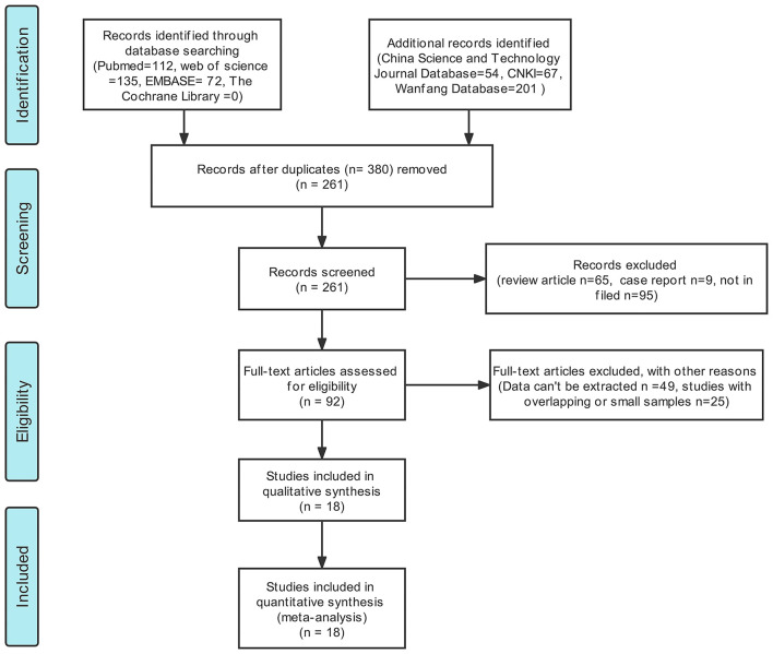
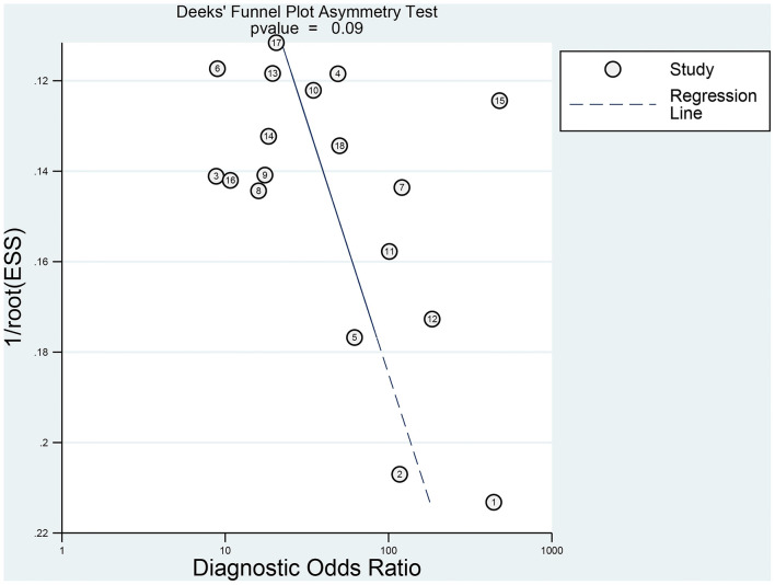
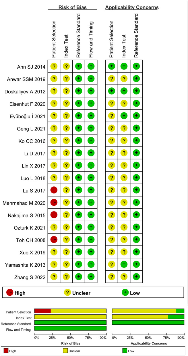
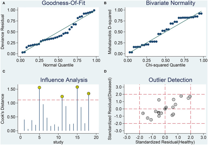
Data presented in Pubmed, Embase, Web of Science, Cochrane Library, China National Knowledge Infrastructure (CNKI), Wanfang Database, and China Science and Technology Journal Database (CQVIP) were analyzed. High-quality literature was included, and the quality was evaluated using the quality assessment of diagnostic accuracy studies-2 (QUADAS-2) tool, and the studies were based on the inclusion and exclusion rules. The pooled sensitivity, pooled specificity, pooled positive likelihood ratio (PLR), pooled negative likelihood ratio (NLR), pooled diagnostic odds ratio (), area under the curve (AUC) of the summary operating characteristic curve (SROC), and corresponding 95% confidence interval () were calculated using the bivariate mixed effect model. Meta-regression analysis and subgroup analysis were used to explore the sources of heterogeneity. The publication bias was evaluated by conducting Deek's test.

RESULTS

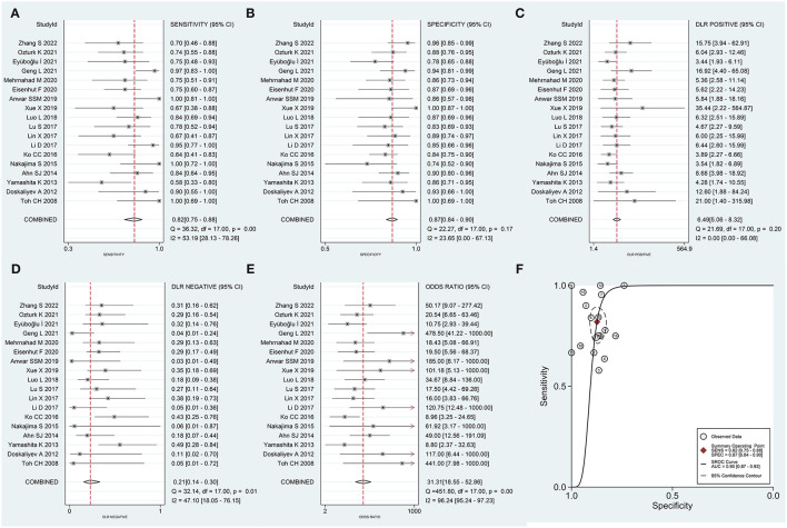
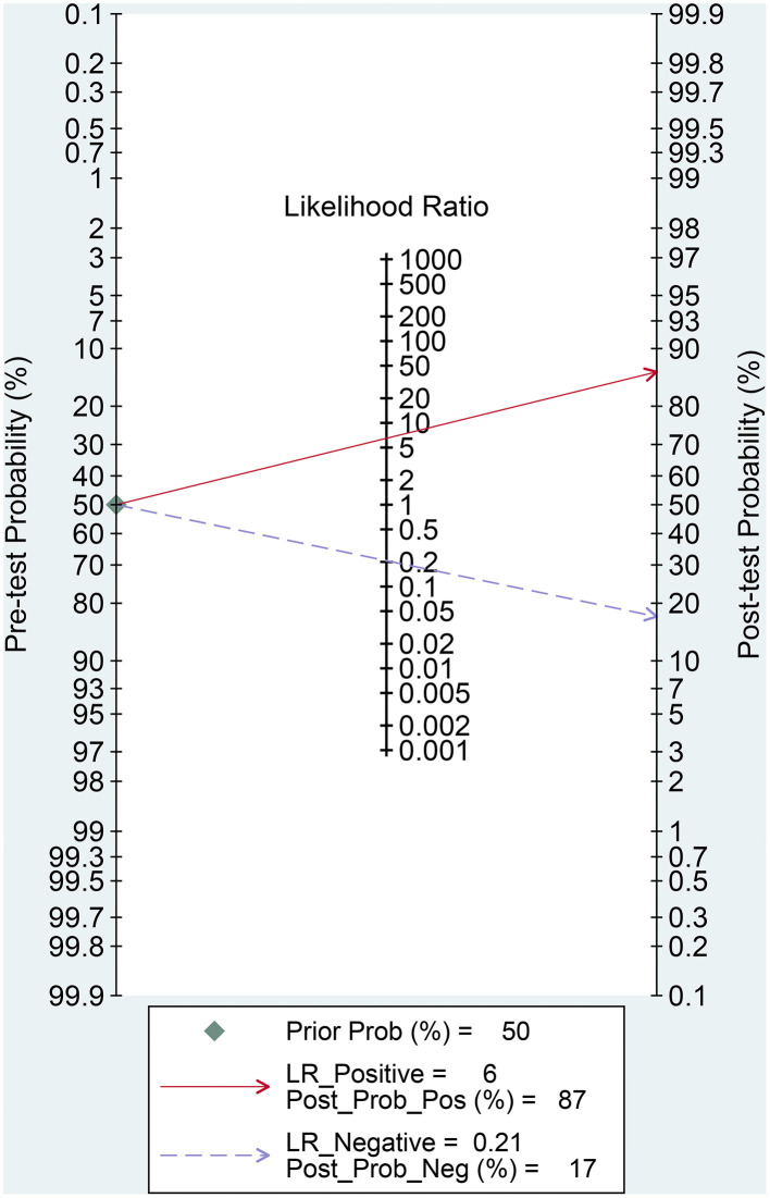
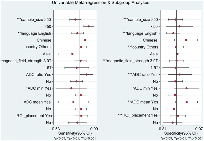
In total, eighteen high-quality studies were included. The pooled sensitivity was 0.82 (95% CI: 0.75-0.88), the pooled specificity was 0.87 (95% CI: 0.84-0.90), the pooled positive likelihood ratio was 6.49 (95% CI: 5.06-8.32), the pooled NLR was 0.21 (95% CI: 0.14-0.30), the pooled was 31.31 (95% CI: 18.55-52.86), and the pooled AUC was 0.90 (95% CI: 0.87-0.92). Sample size, language and country of publication, magnetic field strength, region of interest (ROI), and cut-off values of different types of ADC can potentially be the sources of heterogeneity. There was no publication bias in this meta-analysis.

CONCLUSIONS

The results obtained from the meta-analysis suggest that DWI is characterized by high diagnostic accuracy and thus can be effectively used for differentiating between PCNSL and HGG.

摘要

背景

使用传统磁共振成像技术难以区分原发性中枢神经系统淋巴瘤(PCNSL)和高级别胶质瘤(HGG)。本研究的目的是通过分析表观扩散系数(ADC),探讨扩散加权成像(DWI)是否能有效用于区分这两种类型的肿瘤。

研究设计与方法

分析了PubMed、Embase、Web of Science、Cochrane图书馆、中国知网(CNKI)、万方数据库和中国科技期刊数据库(CQVIP)中的数据。纳入高质量文献,并使用诊断准确性研究质量评估-2(QUADAS-2)工具评估质量,研究基于纳入和排除规则。使用双变量混合效应模型计算合并敏感度、合并特异度、合并阳性似然比(PLR)、合并阴性似然比(NLR)、合并诊断比值比()、汇总操作特征曲线(SROC)的曲线下面积(AUC)及相应的95%置信区间()。采用Meta回归分析和亚组分析探讨异质性来源。通过Deek检验评估发表偏倚。

结果

共纳入18项高质量研究。合并敏感度为0.82(95%CI:0.75-0.88),合并特异度为0.87(95%CI:0.84-0.90),合并阳性似然比为6.49(95%CI:5.06-8.32),合并NLR为0.21(95%CI:0.14-0.30),合并为31.31(95%CI:18.55-52.86),合并AUC为0.90(95%CI:0.87-0.92)。样本量、发表语言和国家、磁场强度、感兴趣区域(ROI)以及不同类型ADC的截断值可能是异质性来源。本Meta分析不存在发表偏倚。

结论

Meta分析结果表明,DWI具有较高的诊断准确性,因此可有效用于区分PCNSL和HGG。

https://cdn.ncbi.nlm.nih.gov/pmc/blobs/9273/9263097/1127c9bd8116/fneur-13-882334-g0007.jpg
https://cdn.ncbi.nlm.nih.gov/pmc/blobs/9273/9263097/186b78bb9c0a/fneur-13-882334-g0001.jpg
https://cdn.ncbi.nlm.nih.gov/pmc/blobs/9273/9263097/c458bef2a352/fneur-13-882334-g0002.jpg
https://cdn.ncbi.nlm.nih.gov/pmc/blobs/9273/9263097/34013ce69f32/fneur-13-882334-g0003.jpg
https://cdn.ncbi.nlm.nih.gov/pmc/blobs/9273/9263097/abcf3bfce615/fneur-13-882334-g0004.jpg
https://cdn.ncbi.nlm.nih.gov/pmc/blobs/9273/9263097/9b5323453a44/fneur-13-882334-g0005.jpg
https://cdn.ncbi.nlm.nih.gov/pmc/blobs/9273/9263097/18a565ce87e1/fneur-13-882334-g0006.jpg
https://cdn.ncbi.nlm.nih.gov/pmc/blobs/9273/9263097/1127c9bd8116/fneur-13-882334-g0007.jpg

相似文献

1
Diagnostic Accuracy of the Diffusion-Weighted Imaging Method Used in Association With the Apparent Diffusion Coefficient for Differentiating Between Primary Central Nervous System Lymphoma and High-Grade Glioma: Systematic Review and Meta-Analysis.联合表观扩散系数使用扩散加权成像方法鉴别原发性中枢神经系统淋巴瘤和高级别胶质瘤的诊断准确性:系统评价与Meta分析
Front Neurol. 2022 Jun 24;13:882334. doi: 10.3389/fneur.2022.882334. eCollection 2022.
2
Use of 18F-FDG-PET/CT in differential diagnosis of primary central nervous system lymphoma and high-grade gliomas: A meta-analysis.18F-FDG-PET/CT在原发性中枢神经系统淋巴瘤和高级别胶质瘤鉴别诊断中的应用:一项荟萃分析。
Front Neurol. 2022 Aug 17;13:935459. doi: 10.3389/fneur.2022.935459. eCollection 2022.
3
Diagnostic performance of DWI for differentiating primary central nervous system lymphoma from glioblastoma: a systematic review and meta-analysis.DWI 对原发性中枢神经系统淋巴瘤与胶质母细胞瘤鉴别诊断的性能:系统评价和荟萃分析。
Neurol Sci. 2019 May;40(5):947-956. doi: 10.1007/s10072-019-03732-7. Epub 2019 Jan 31.
4
Accuracy of ADC derived from DWI for differentiating high-grade from low-grade gliomas: Systematic review and meta-analysis.基于扩散加权成像(DWI)的表观扩散系数(ADC)对高级别与低级别胶质瘤的鉴别诊断准确性:系统评价与Meta分析
Medicine (Baltimore). 2020 Feb;99(8):e19254. doi: 10.1097/MD.0000000000019254.
5
Diagnostic accuracy of arterial spin labeling in differentiating between primary central nervous system lymphoma and high-grade glioma: a systematic review and meta-analysis.动脉自旋标记在鉴别原发性中枢神经系统淋巴瘤和高级别胶质瘤中的诊断准确性:系统评价和荟萃分析。
Expert Rev Anticancer Ther. 2022 Jul;22(7):763-771. doi: 10.1080/14737140.2022.2082948. Epub 2022 May 31.
6
Diagnostic accuracy of diffusion-weighted imaging in differentiating glioma recurrence from posttreatment-related changes: a meta-analysis.弥散加权成像在鉴别胶质瘤复发与治疗后相关改变中的诊断准确性:一项荟萃分析。
Expert Rev Anticancer Ther. 2022 Jan;22(1):123-130. doi: 10.1080/14737140.2022.2000396. Epub 2021 Nov 10.
7
The utility of diffusion MRI with quantitative ADC measurements for differentiating high-grade from low-grade cerebral gliomas: Evidence from a meta-analysis.基于定量表观扩散系数测量的扩散加权磁共振成像在鉴别高级别与低级别脑胶质瘤中的应用:一项荟萃分析的证据
J Neurol Sci. 2017 Feb 15;373:9-15. doi: 10.1016/j.jns.2016.12.008. Epub 2016 Dec 9.
8
A systematic review and meta-analysis of the accuracy of diffusion-weighted MRI in the detection of malignant pulmonary nodules and masses.扩散加权 MRI 检测恶性肺结节和肿块的准确性的系统评价和荟萃分析。
Acad Radiol. 2014 Jan;21(1):21-9. doi: 10.1016/j.acra.2013.09.019.
9
[Diagnostic accuracy of artery peak velocity variation measured by bedside real-time ultrasound for prediction of fluid responsiveness: a Meta-analysis].[床旁实时超声测量动脉峰值速度变化对液体反应性预测的诊断准确性:一项Meta分析]
Zhonghua Wei Zhong Bing Ji Jiu Yi Xue. 2020 Jan;32(1):99-105. doi: 10.3760/cma.j.cn121430-20191226-00018.
10
Diagnostic Performance of Diffusion-Weighted Imaging for Differentiating Malignant From Benign Intraductal Papillary Mucinous Neoplasms of the Pancreas: A Systematic Review and Meta-Analysis.扩散加权成像鉴别胰腺导管内乳头状黏液性肿瘤良恶性的诊断性能:一项系统评价和Meta分析
Front Oncol. 2021 Jul 5;11:637681. doi: 10.3389/fonc.2021.637681. eCollection 2021.

引用本文的文献

1
Intraoperative classification of CNS lymphoma and glioblastoma by AI-based analysis of Stimulated Raman Histology (SRH).通过基于人工智能的受激拉曼组织学(SRH)分析对中枢神经系统淋巴瘤和胶质母细胞瘤进行术中分类。
Brain Spine. 2025 Jan 26;5:104187. doi: 10.1016/j.bas.2025.104187. eCollection 2025.
2
The Prognostic Value of Preoperative Inflammatory Markers for Pathological Grading of Glioma Patients.术前炎症标志物对胶质瘤患者病理分级的预后价值。
Technol Cancer Res Treat. 2024 Jan-Dec;23:15330338241273160. doi: 10.1177/15330338241273160.
3
Imaging Role in Diagnosis, Prognosis, and Treatment Response Prediction Associated with High-grade Glioma.

本文引用的文献

1
Differentiation of high-grade glioma and primary central nervous system lymphoma: Multiparametric imaging of the enhancing tumor and peritumoral regions based on hybrid F-FDG PET/MRI.高级别胶质瘤与原发性中枢神经系统淋巴瘤的鉴别诊断:基于杂交 F-FDG PET/MRI 的增强肿瘤及瘤周区域的多参数成像。
Eur J Radiol. 2022 May;150:110235. doi: 10.1016/j.ejrad.2022.110235. Epub 2022 Mar 7.
2
Primary Central Nervous System Lymphomas.原发性中枢神经系统淋巴瘤。
Hematol Oncol Clin North Am. 2022 Feb;36(1):147-159. doi: 10.1016/j.hoc.2021.09.004.
3
Emerging Landscape of Immunotherapy for Primary Central Nervous System Lymphoma.
影像学在高级别胶质瘤的诊断、预后及治疗反应预测中的作用
J Med Signals Sens. 2024 Mar 27;14:7. doi: 10.4103/jmss.jmss_30_22. eCollection 2024.
4
Radiomics for differentiation of gliomas from primary central nervous system lymphomas: a systematic review and meta-analysis.基于影像组学鉴别胶质瘤与原发性中枢神经系统淋巴瘤的系统评价与Meta分析
Front Oncol. 2024 Feb 14;14:1291861. doi: 10.3389/fonc.2024.1291861. eCollection 2024.
5
Differentiating nasopharyngeal carcinoma from lymphoma in the head and neck region using the apparent diffusion coefficient (ADC) value: a systematic review and meta-analysis.利用表观扩散系数(ADC)值鉴别头颈部区域的鼻咽癌与淋巴瘤:一项系统评价和荟萃分析。
Pol J Radiol. 2023 Oct 17;88:e472-e482. doi: 10.5114/pjr.2023.132172. eCollection 2023.
6
Distinctive magnetic resonance imaging features in primary central nervous system lymphoma: A case report.原发性中枢神经系统淋巴瘤的独特磁共振成像特征:一例报告。
World J Radiol. 2023 Sep 28;15(9):274-280. doi: 10.4329/wjr.v15.i9.274.
7
Use of 18F-FDG-PET/CT in differential diagnosis of primary central nervous system lymphoma and high-grade gliomas: A meta-analysis.18F-FDG-PET/CT在原发性中枢神经系统淋巴瘤和高级别胶质瘤鉴别诊断中的应用:一项荟萃分析。
Front Neurol. 2022 Aug 17;13:935459. doi: 10.3389/fneur.2022.935459. eCollection 2022.
原发性中枢神经系统淋巴瘤免疫治疗的新进展
Cancers (Basel). 2021 Oct 10;13(20):5061. doi: 10.3390/cancers13205061.
4
Differentiation between primary CNS lymphoma and atypical glioblastoma according to major genomic alterations using diffusion and susceptibility-weighted MR imaging.根据弥散和磁化率加权磁共振成像的主要基因组改变区分原发性中枢神经系统淋巴瘤和非典型胶质母细胞瘤。
Eur J Radiol. 2021 Aug;141:109784. doi: 10.1016/j.ejrad.2021.109784. Epub 2021 May 24.
5
Classification of Primary Cerebral Lymphoma and Glioblastoma Featuring Dynamic Susceptibility Contrast and Apparent Diffusion Coefficient.基于动态磁敏感对比和表观扩散系数的原发性脑淋巴瘤和胶质母细胞瘤的分类
Brain Sci. 2020 Nov 20;10(11):886. doi: 10.3390/brainsci10110886.
6
Diagnostic efficacy of apparent diffusion coefficient measurements in differentiation of malignant intra-axial brain tumors.表观扩散系数测量在鉴别恶性脑内肿瘤中的诊断效能。
Turk J Med Sci. 2021 Feb 26;51(1):256-267. doi: 10.3906/sag-2006-1.
7
Differentiating glioblastoma multiforme from cerebral lymphoma: application of advanced texture analysis of quantitative apparent diffusion coefficients.鉴别多形性胶质母细胞瘤与脑淋巴瘤:定量表观扩散系数的高级纹理分析的应用。
Neuroradiol J. 2020 Oct;33(5):428-436. doi: 10.1177/1971400920937382. Epub 2020 Jul 6.
8
Accuracy of ADC derived from DWI for differentiating high-grade from low-grade gliomas: Systematic review and meta-analysis.基于扩散加权成像(DWI)的表观扩散系数(ADC)对高级别与低级别胶质瘤的鉴别诊断准确性:系统评价与Meta分析
Medicine (Baltimore). 2020 Feb;99(8):e19254. doi: 10.1097/MD.0000000000019254.
9
Migraine History: A Predictor of Negative Diffusion-Weighted Imaging in IV-tPA-Treated Stroke Mimics.偏头痛病史:IV-tPA 治疗的卒中样发作模拟物中弥散加权成像呈阴性的预测因素。
J Stroke Cerebrovasc Dis. 2019 Nov;28(11):104282. doi: 10.1016/j.jstrokecerebrovasdis.2019.06.040. Epub 2019 Aug 8.
10
Combined PET/MRI in brain glioma imaging.PET/MRI联合成像在脑胶质瘤中的应用
Br J Hosp Med (Lond). 2019 Jul 2;80(7):380-386. doi: 10.12968/hmed.2019.80.7.380.