Fujian Key Laboratory of Oral Diseases and Fujian Provincial Engineering Research Center of Oral Biomaterial and Stomatological Key Lab of Fujian College and University, School and Hospital of Stomatology, Fujian Medical University, Fuzhou, Fujian, China.
Newland Digital Technology Co., Ltd., Fuzhou, Fujian, China.
Sci Rep. 2022 Jul 27;12(1):12841. doi: 10.1038/s41598-022-16074-w.
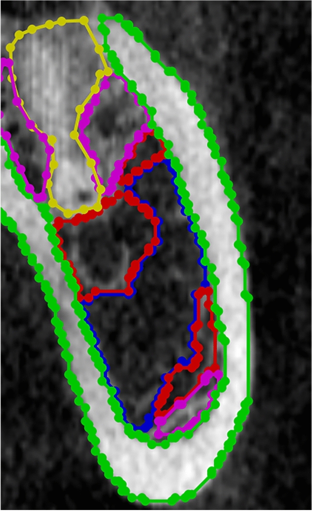
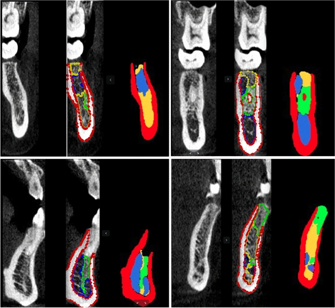
To develop and verify an automatic classification method using artificial intelligence deep learning to determine the bone mineral density level of the implant site in oral implant surgery from radiographic data obtained from cone beam computed tomography (CBCT) images. Seventy patients with mandibular dentition defects were scanned using CBCT. These Digital Imaging and Communications in Medicine data were cut into 605 training sets, and then the data were processed with data standardization, and the Hounsfiled Unit (HU) value level was determined as follows: Type 1, 1000-2000; type 2, 700-1000; type 3, 400-700; type 4, 100-400; and type 5, - 200-100. Four trained dental implant physicians manually identified and classified the area of the jaw bone density level in the image using the software LabelMe. Then, with the assistance of the HU value generated by LabelMe, a physician with 20 years of clinical experience confirmed the labeling level. Finally, the HU mean values of various categories marked by dental implant physicians were compared to the mean values detected by the artificial intelligence model to assess the accuracy of artificial intelligence classification. After the model was trained on 605 training sets, the statistical results of the HU mean values of various categories in the dataset detected by the model were almost the same as the HU grading interval on the data annotation. This new classification provides a more detailed solution to guide surgeons to adjust the drilling rate and tool selection during preoperative decision-making and intraoperative hole preparation for oral implant surgery.
开发并验证一种使用人工智能深度学习的自动分类方法,以从锥形束计算机断层扫描 (CBCT) 图像获得的放射数据确定口腔种植术中种植部位的骨密度水平。对 70 名下颌牙列缺损患者进行 CBCT 扫描。这些数字成像和通信医学数据被切割成 605 个训练集,然后对数据进行标准化处理,并确定亨氏单位 (HU) 值水平如下:1 型,1000-2000;2 型,700-1000;3 型,400-700;4 型,100-400;5 型,-200-100。四名经过培训的牙科种植医生使用 LabelMe 软件手动识别和分类图像中的颌骨密度水平区域。然后,在 LabelMe 生成的 HU 值的协助下,一位具有 20 年临床经验的医生确认了标记级别。最后,将牙科种植医生标记的各个类别的 HU 平均值与人工智能模型检测到的平均值进行比较,以评估人工智能分类的准确性。在对 605 个训练集进行模型训练后,模型检测到的数据集中各个类别的 HU 平均值的统计结果几乎与数据注释中的 HU 分级间隔相同。这种新的分类方法提供了更详细的解决方案,以指导外科医生在术前决策和口腔种植术中的孔准备过程中调整钻孔速度和工具选择。