Department of Orthopaedics, Fujian Institute of Orthopaedics, the First Affiliated Hospital of Fujian Medical University, Fuzhou, 350005, People's Republic of China.
The Centralab, the First Affiliated Hospital of Fujian Medical University, Fuzhou, 350005, People's Republic of China.
Int J Nanomedicine. 2022 Aug 4;17:3483-3495. doi: 10.2147/IJN.S372851. eCollection 2022.
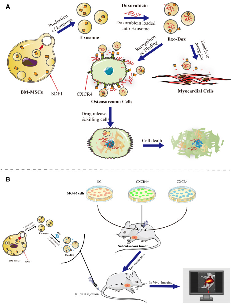
The objective of this study was to investigate the antitumor activity, targeting capability, and mechanism of the developed nanodrug consisting of doxorubicin and exosome (Exo-Dox) derived from mesenchymal stem cells in vitro and in vivo.
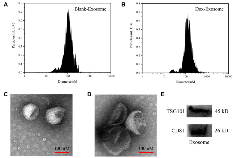
The exosomes were isolated with Exosome Isolation Kit, and the Exo-Dox was prepared by mixing exosome with Dox-HCl, desalinizing with triethylamine and then dialyzing against PBS overnight. The exosome and Exo-Dox were examined by nanoparticle tracking analysis (NTA) and transmission electron microscopy (TEM). The antitumor activity, targeting capability, and mechanism of the developed Exo-Dox were evaluated by cell viability assay, histological and immunofluorescence analysis and in vivo imaging system.
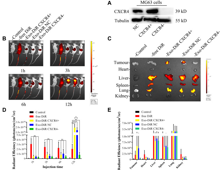
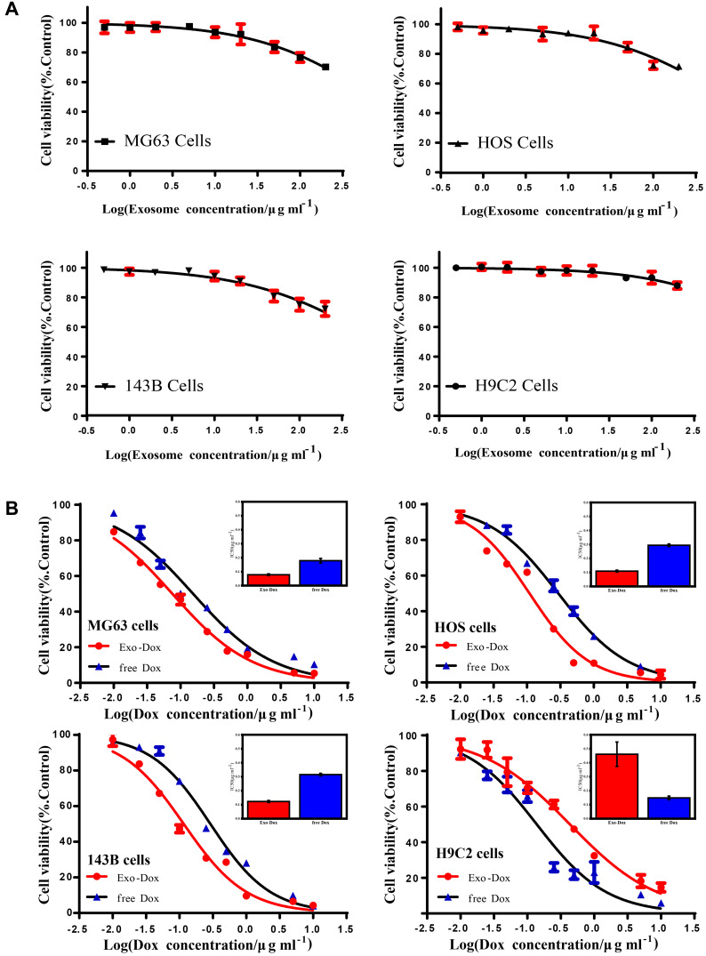
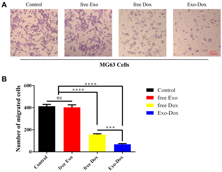
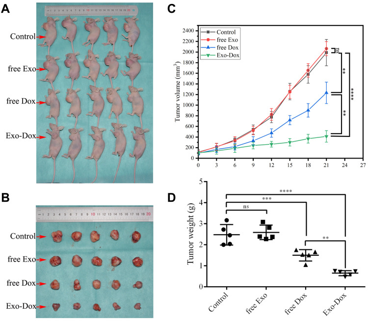
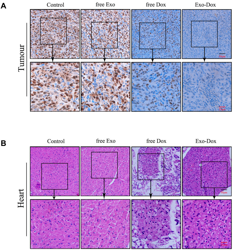
NTA results showed the size of the exosomes had increased from 141.6 nm to 178.1 nm after loading with doxorubicin. Compared with free Dox, the Exo-Dox exhibited higher cytotoxicity against osteosarcoma MG63 cells, HOS cells, and 143B cells than free Dox, the half-maximal inhibitory concentrations (IC50) of Dox, Exo-Dox were calculated to be 0.178 and 0.078 μg mL in MG63 cells, 0.294 and 0.109μg mL in HOS cells, 0.315 and 0.123 μg mL in 143B cells, respectively. The in vivo imaging showed that MSC derived Exo could serve as a highly efficient delivery vehicle for targeted drug delivery. The immunohistochemistry and histology analysis indicated that compared with the free Dox group, the Ki67-positive cells and cardiotoxicity in Exo-Dox group were significantly decreased.
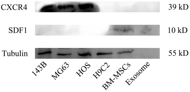
Our results suggested that MSC-derived Exo could be excellent nanocarriers used to deliver chemotherapeutic drug Dox specifically and efficiently in osteosarcoma, resulting in enhanced toxicity against osteosarcoma and less toxicity in heart tissue. We further demonstrated the targeting capability of Exo was due to the chemotaxis of MSC-derived exosomes to osteosarcoma cells via SDF1-CXCR4 axis.
本研究旨在探讨来源于间充质干细胞的阿霉素纳米药物(Exo-Dox)的体内外抗肿瘤活性、靶向能力和作用机制。
采用 Exosome Isolation Kit 分离外泌体,用三乙胺脱盐后用 PBS 透析过夜,将阿霉素盐酸盐(Dox-HCl)与外泌体混合制备 Exo-Dox。采用纳米颗粒跟踪分析(NTA)和透射电子显微镜(TEM)检测外泌体和 Exo-Dox。通过细胞活力测定、组织学和免疫荧光分析以及体内成像系统评价 Exo-Dox 的抗肿瘤活性、靶向能力和作用机制。
NTA 结果显示,载药后外泌体的粒径从 141.6nm 增加到 178.1nm。与游离 Dox 相比,Exo-Dox 对骨肉瘤 MG63 细胞、HOS 细胞和 143B 细胞的细胞毒性更高,MG63 细胞、HOS 细胞和 143B 细胞的 Dox、Exo-Dox 的半数抑制浓度(IC50)分别为 0.178μg/mL 和 0.078μg/mL、0.294μg/mL 和 0.109μg/mL、0.315μg/mL 和 0.123μg/mL。体内成像显示,MSC 来源的外泌体可以作为一种高效的靶向药物递送载体。免疫组化和组织学分析表明,与游离 Dox 组相比,Exo-Dox 组的 Ki67 阳性细胞和心脏毒性明显降低。
我们的研究结果表明,MSC 来源的外泌体可以作为一种高效的载药系统,特异性、有效地递送达药物,从而增强对骨肉瘤的毒性,减少心脏组织的毒性。我们进一步证明了 Exo 的靶向能力是由于 MSC 衍生的外泌体通过 SDF1-CXCR4 轴趋化到骨肉瘤细胞。