School of Computer Science and Engineering, UNSW Sydney, Kensington, NSW, 2052, Australia.
Department of Anatomical Pathology, NSW Health Pathology, St. George Hospital, Kogarah, NSW, 2217, Australia.
Sci Rep. 2022 Aug 25;12(1):14527. doi: 10.1038/s41598-022-18647-1.
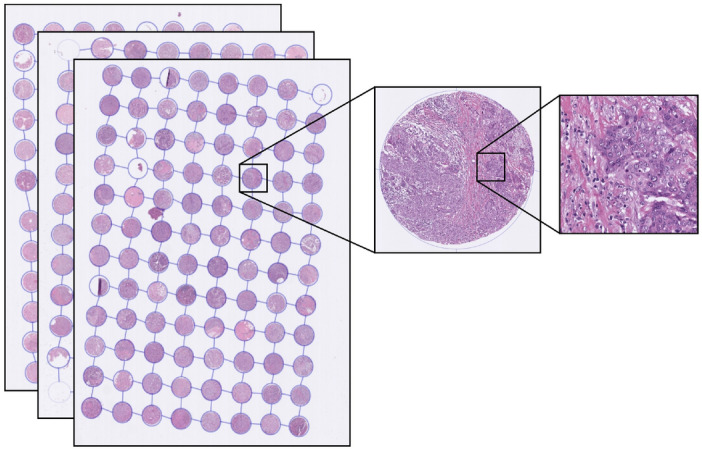
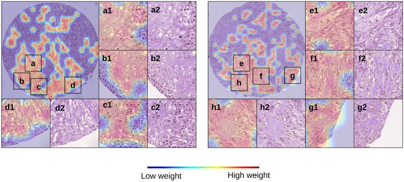
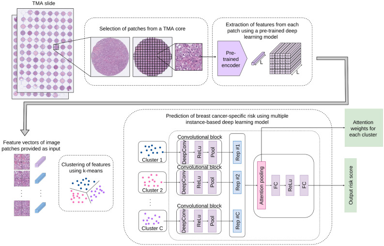
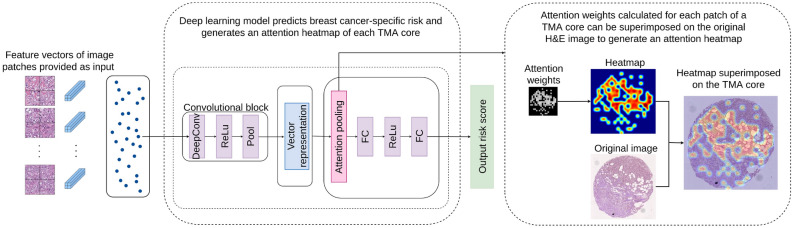
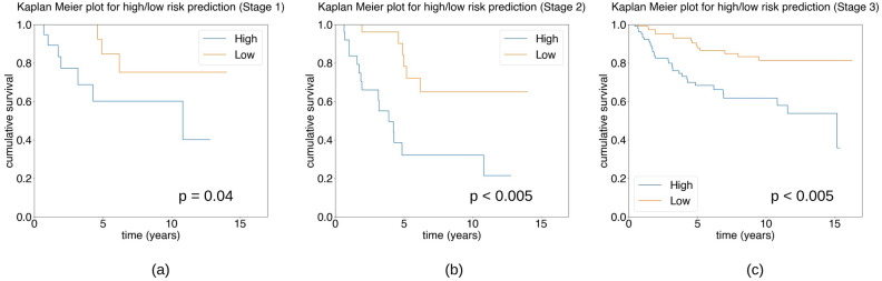
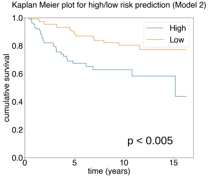
Computational pathology is a rapidly expanding area for research due to the current global transformation of histopathology through the adoption of digital workflows. Survival prediction of breast cancer patients is an important task that currently depends on histopathology assessment of cancer morphological features, immunohistochemical biomarker expression and patient clinical findings. To facilitate the manual process of survival risk prediction, we developed a computational pathology framework for survival prediction using digitally scanned haematoxylin and eosin-stained tissue microarray images of clinically aggressive triple negative breast cancer. Our results show that the model can produce an average concordance index of 0.616. Our model predictions are analysed for independent prognostic significance in univariate analysis (hazard ratio = 3.12, 95% confidence interval [1.69,5.75], p < 0.005) and multivariate analysis using clinicopathological data (hazard ratio = 2.68, 95% confidence interval [1.44,4.99], p < 0.005). Through qualitative analysis of heatmaps generated from our model, an expert pathologist is able to associate tissue features highlighted in the attention heatmaps of high-risk predictions with morphological features associated with more aggressive behaviour such as low levels of tumour infiltrating lymphocytes, stroma rich tissues and high-grade invasive carcinoma, providing explainability of our method for triple negative breast cancer.
计算病理学是一个快速发展的研究领域,因为当前全球范围内通过采用数字工作流程来改变组织病理学。乳腺癌患者的生存预测是一项重要任务,目前依赖于对癌症形态特征、免疫组织化学生物标志物表达和患者临床发现的组织病理学评估。为了促进生存风险预测的手动过程,我们开发了一种使用数字化扫描的苏木精和伊红染色组织微阵列图像进行生存预测的计算病理学框架,这些图像来自临床上侵袭性强的三阴性乳腺癌。我们的结果表明,该模型可以产生平均一致性指数为 0.616。我们的模型预测在单因素分析(危险比=3.12,95%置信区间[1.69,5.75],p<0.005)和使用临床病理数据的多因素分析(危险比=2.68,95%置信区间[1.44,4.99],p<0.005)中进行了独立预后意义的分析。通过对我们模型生成的热图进行定性分析,一位专家病理学家能够将高风险预测的注意力热图中突出的组织特征与形态学特征相关联,这些特征与更具侵袭性的行为相关,如低水平的肿瘤浸润淋巴细胞、富含基质的组织和高级别浸润性癌,为我们的三阴性乳腺癌方法提供了可解释性。