Xu Zhongli, Wang Xinjun, Fan Li, Wang Fujing, Lin Becky, Wang Jiebiao, Trevejo-Nuñez Giraldina, Chen Wei, Chen Kong
Department of Pediatrics, University of Pittsburgh, Pittsburgh, PA, USA.
School of Medicine, Tsinghua University, Beijing, China.
iScience. 2022 Aug 9;25(9):104900. doi: 10.1016/j.isci.2022.104900. eCollection 2022 Sep 16.
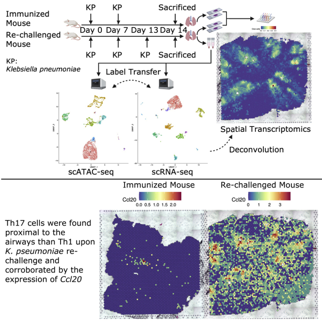
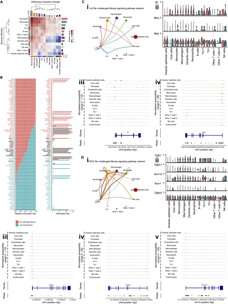
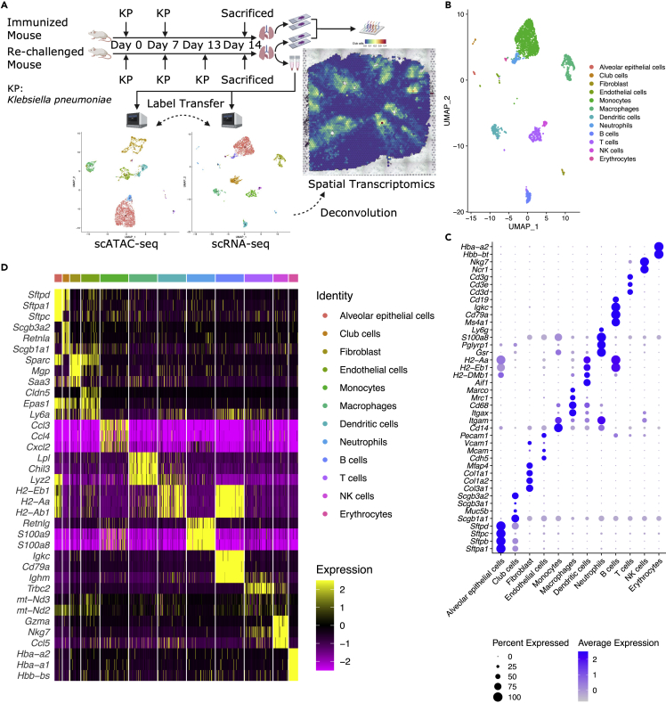
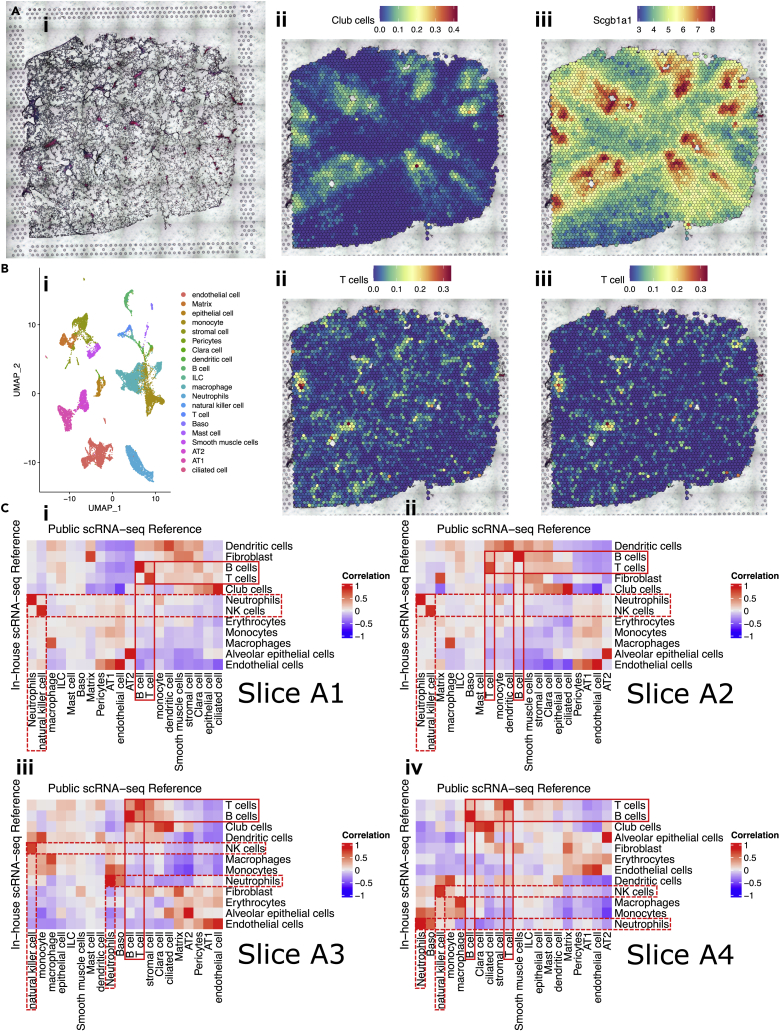
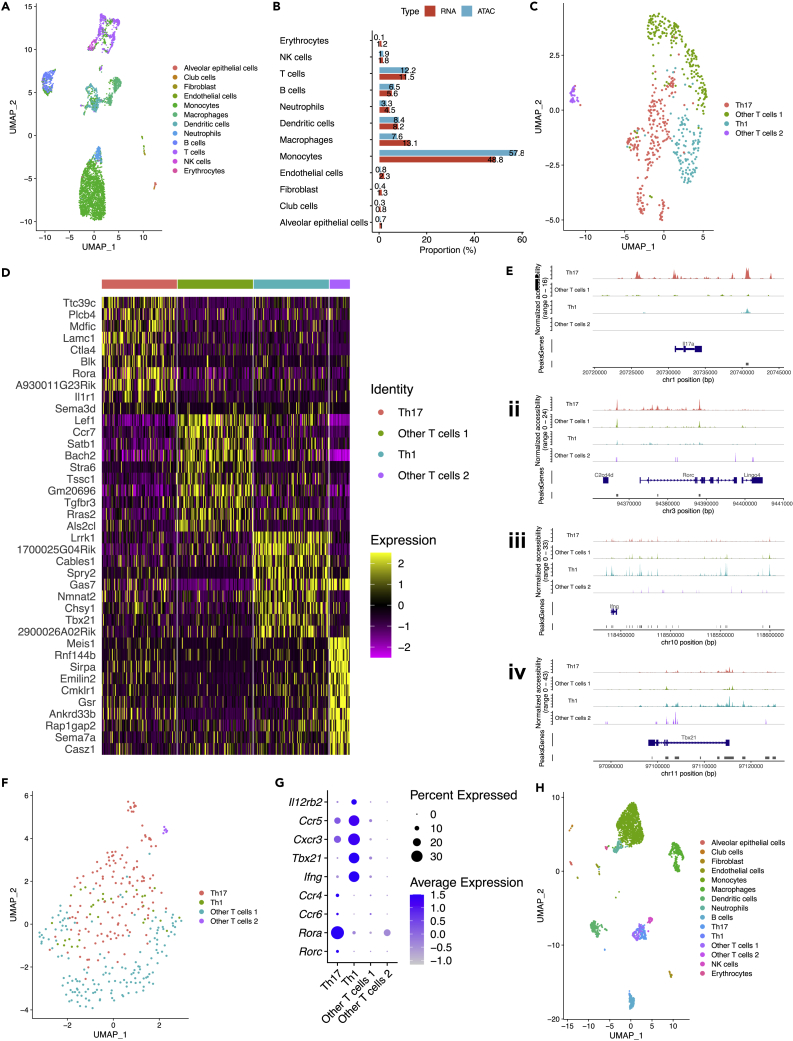
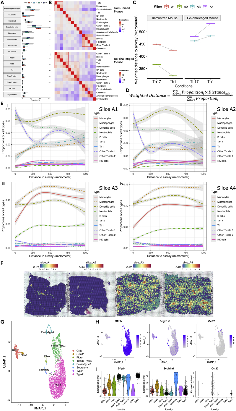
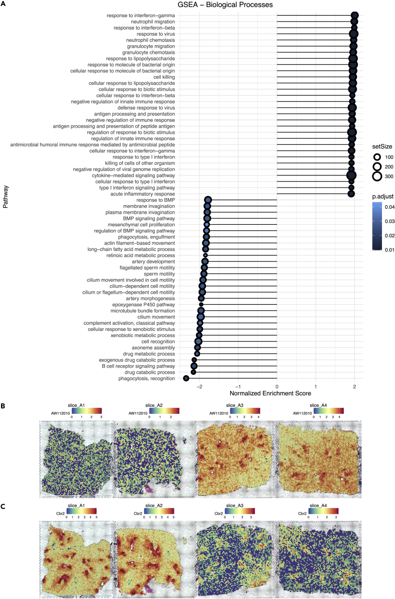
Understanding lung immunity requires an unbiased profiling of tissue-resident T cells at their precise anatomical locations within the lung, but such information has not been characterized in the immunized mouse model. In this pilot study, using 10x Genomics Chromium and Visium platform, we performed an integrative analysis of spatial transcriptome with single-cell RNA-seq and single-cell ATAC-seq on lung cells from mice after immunization using a well-established infection model. We built an optimized deconvolution pipeline to accurately decipher specific cell-type compositions by anatomic location. We discovered that combining scATAC-seq and scRNA-seq data may provide more robust cell-type identification, especially for lineage-specific T helper cells. Combining all three modalities, we observed a dynamic change in the location of T helper cells as well as their corresponding chemokines. In summary, our proof-of-principle study demonstrated the power and potential of single-cell multi-omics analysis to uncover spatial- and cell-type-dependent mechanisms of lung immunity.
要了解肺部免疫,需要在肺部精确的解剖位置对组织驻留T细胞进行无偏差分析,但在免疫小鼠模型中尚未对这类信息进行表征。在这项初步研究中,我们使用10x Genomics Chromium和Visium平台,对使用成熟感染模型免疫后的小鼠肺细胞,进行了空间转录组与单细胞RNA测序和单细胞染色质转座酶可及性测序的综合分析。我们构建了一个优化的反卷积流程,以通过解剖位置准确解读特定细胞类型的组成。我们发现,结合单细胞染色质转座酶可及性测序和单细胞RNA测序数据,可能会提供更可靠的细胞类型识别,特别是对于谱系特异性辅助性T细胞。结合所有这三种方法,我们观察到辅助性T细胞及其相应趋化因子的位置发生了动态变化。总之,我们的原理验证研究证明了单细胞多组学分析在揭示肺部免疫的空间和细胞类型依赖性机制方面的能力和潜力。