• 文献检索
  • 文档翻译
  • 深度研究
  • 学术资讯
  • Suppr Zotero 插件Zotero 插件
  • 邀请有礼
  • 套餐&价格
  • 历史记录
应用&插件
Suppr Zotero 插件Zotero 插件浏览器插件Mac 客户端Windows 客户端微信小程序
定价
高级版会员购买积分包购买API积分包
服务
文献检索文档翻译深度研究API 文档MCP 服务
关于我们
关于 Suppr公司介绍联系我们用户协议隐私条款
关注我们

Suppr 超能文献

核心技术专利:CN118964589B侵权必究
粤ICP备2023148730 号-1Suppr @ 2026

文献检索

告别复杂PubMed语法,用中文像聊天一样搜索,搜遍4000万医学文献。AI智能推荐,让科研检索更轻松。

立即免费搜索

文件翻译

保留排版,准确专业,支持PDF/Word/PPT等文件格式,支持 12+语言互译。

免费翻译文档

深度研究

AI帮你快速写综述,25分钟生成高质量综述,智能提取关键信息,辅助科研写作。

立即免费体验

血小板衍生促凝微泡在视网膜静脉阻塞(RVO)患者中升高。

Platelet-Derived Procoagulant Microvesicles Are Elevated in Patients with Retinal Vein Occlusion (RVO).

作者信息

Marcinkowska Adrianna, Wolska Nina, Luzak Boguslawa, Cisiecki Slawomir, Marcinkowski Karol, Rozalski Marcin

机构信息

Department of Haemostasis and Haemostatic Disorders, Chair of Biomedical Sciences, Medical University of Lodz, Mazowiecka 6/8, 92-215 Lodz, Poland.

Department of Ophthalmology, Karol Jonscher's Municipal Medical Center, 93-113 Lodz, Poland.

出版信息

J Clin Med. 2022 Aug 30;11(17):5099. doi: 10.3390/jcm11175099.

DOI:10.3390/jcm11175099
PMID:36079028
原文链接:https://pmc.ncbi.nlm.nih.gov/articles/PMC9457368/
Abstract

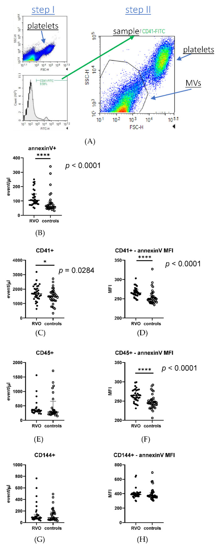
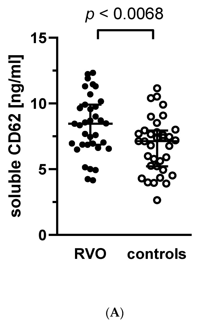
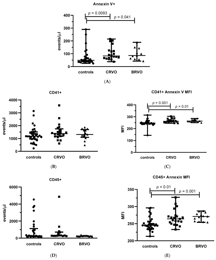
The etiopathogenesis of retinal vein occlusion (RVO) is multifactorial, and the contribution of platelets to RVO development has not been fully elucidated. We aimed to analyze platelet function in RVO patients (n = 35) and controls (n = 35). We found a higher (p < 0.05) level of soluble P-selectin in RVO group vs. controls. Additionally, in RVO patients, the concentration of platelet-derived microvesicles was higher (p < 0.05), and the difference between groups was deeper for the fraction of platelet-derived microvesicles with the procoagulant phenotype (p < 0.0001) and for overall procoagulant microvesicles level (p < 0.0001). The results were similar for the total RVO group and for both RVO types (central- and branched-retinal vein occlusion). We did not find differences in simple platelet parameters (platelet count, mean platelet volume, platelet distribution width, platecrit, reticulated platelets) and inflammatory markers (platelet-lymphocyte ratio, neutrophil-lymphocyte ratio). Similarly, no differences were found for platelet aggregation-stimulated byadenosine diphosphate; collagen; arachidonic acid; and in multiparametric flow cytometry evaluation of P-selectin, PAC-1, and fibrinogen binding for both unstimulated and adenosine diphosphate-, collagen-, and thrombin receptor activating peptide-stimulated platelets. Our results suggest that platelets can contribute to developing RVO by enhancing procoagulant activity through providing a procoagulation surface via platelet-derived microvesicles. The direct role of platelets’ hyperreactivity in developing RVO is less apparent, which is consistent with the complexity and multifactorial background of this disorder.

摘要

视网膜静脉阻塞(RVO)的发病机制是多因素的,血小板在RVO发生发展中的作用尚未完全阐明。我们旨在分析RVO患者(n = 35)和对照组(n = 35)的血小板功能。我们发现RVO组可溶性P-选择素水平高于对照组(p < 0.05)。此外,在RVO患者中,血小板衍生微泡的浓度更高(p < 0.05),并且具有促凝表型的血小板衍生微泡部分(p < 0.0001)和总体促凝微泡水平(p < 0.0001)在两组之间的差异更大。总RVO组以及两种RVO类型(视网膜中央静脉阻塞和视网膜分支静脉阻塞)的结果相似。我们未发现简单血小板参数(血小板计数、平均血小板体积、血小板分布宽度、血小板压积、网织血小板)和炎症标志物(血小板-淋巴细胞比率、中性粒细胞-淋巴细胞比率)存在差异。同样,在由二磷酸腺苷、胶原、花生四烯酸刺激的血小板聚集方面,以及在对未刺激和经二磷酸腺苷、胶原、凝血酶受体激活肽刺激的血小板进行P-选择素、PAC-1和纤维蛋白原结合的多参数流式细胞术评估中,均未发现差异。我们的结果表明,血小板可通过血小板衍生微泡提供促凝表面来增强促凝活性,从而促进RVO的发生发展。血小板高反应性在RVO发生中的直接作用不太明显,这与该疾病的复杂性和多因素背景一致。

https://cdn.ncbi.nlm.nih.gov/pmc/blobs/32d8/9457368/405a5cf67922/jcm-11-05099-g003a.jpg
https://cdn.ncbi.nlm.nih.gov/pmc/blobs/32d8/9457368/662f2412b1a8/jcm-11-05099-g001.jpg
https://cdn.ncbi.nlm.nih.gov/pmc/blobs/32d8/9457368/5bbce5c8e73a/jcm-11-05099-g002a.jpg
https://cdn.ncbi.nlm.nih.gov/pmc/blobs/32d8/9457368/405a5cf67922/jcm-11-05099-g003a.jpg
https://cdn.ncbi.nlm.nih.gov/pmc/blobs/32d8/9457368/662f2412b1a8/jcm-11-05099-g001.jpg
https://cdn.ncbi.nlm.nih.gov/pmc/blobs/32d8/9457368/5bbce5c8e73a/jcm-11-05099-g002a.jpg
https://cdn.ncbi.nlm.nih.gov/pmc/blobs/32d8/9457368/405a5cf67922/jcm-11-05099-g003a.jpg

相似文献

1
Platelet-Derived Procoagulant Microvesicles Are Elevated in Patients with Retinal Vein Occlusion (RVO).血小板衍生促凝微泡在视网膜静脉阻塞(RVO)患者中升高。
J Clin Med. 2022 Aug 30;11(17):5099. doi: 10.3390/jcm11175099.
2
Mean Platelet Volume, Red Cell Distribution Width, and Complete Blood Cell Count Indices in Retinal Vein Occlusions.平均血小板体积、红细胞分布宽度和全血细胞计数指标在视网膜静脉阻塞中的应用。
Ophthalmic Epidemiol. 2021 Feb;28(1):39-47. doi: 10.1080/09286586.2020.1791349. Epub 2020 Jul 10.
3
Platelet activation by collagen is increased in retinal vein occlusion.在视网膜静脉阻塞中,胶原蛋白引起的血小板活化增加。
Thromb Haemost. 2007 Feb;97(2):218-27.
4
Thrombophilic risk factors in different types of retinal vein occlusion in Tunisian patients.突尼斯患者不同类型视网膜静脉阻塞的血栓形成风险因素。
J Stroke Cerebrovasc Dis. 2014 Jul;23(6):1592-8. doi: 10.1016/j.jstrokecerebrovasdis.2013.12.048. Epub 2014 Mar 14.
5
Altered platelet morphological parameters in patients with retinal vein occlusion.视网膜静脉阻塞患者血小板形态学参数的改变
Eur Rev Med Pharmacol Sci. 2016 May;20(10):1934-9.
6
The association of neutrophil-lymphocyte ratio and platelet-lymphocyte ratio with retinal vein occlusion: a systematic review and meta-analysis.中性粒细胞与淋巴细胞比值和血小板与淋巴细胞比值与视网膜静脉阻塞的关联:一项系统评价和荟萃分析。
Acta Ophthalmol. 2022 May;100(3):e635-e647. doi: 10.1111/aos.14955. Epub 2021 Jul 4.
7
ASSOCIATION BETWEEN PLATELET INDICES AND RETINAL VEIN OCCLUSION: A Systematic Review and Meta-Analysis.血小板指数与视网膜静脉阻塞的相关性:系统评价和荟萃分析。
Retina. 2021 Feb 1;41(2):238-248. doi: 10.1097/IAE.0000000000003022.
8
Primary angle closure and primary angle closure glaucoma in retinal vein occlusion.视网膜静脉阻塞中的原发性房角关闭和原发性房角关闭性青光眼。
Acta Ophthalmol. 2019 May;97(3):e364-e372. doi: 10.1111/aos.13879. Epub 2018 Sep 21.
9
Hematologic abnormalities associated with various types of retinal vein occlusion.与各种类型视网膜静脉阻塞相关的血液学异常。
Graefes Arch Clin Exp Ophthalmol. 2002 Mar;240(3):180-96. doi: 10.1007/s00417-001-0421-3. Epub 2002 Feb 9.
10
The Role of Serum Inflammation-Based Factors in Anti-Vascular Endothelial Growth Factor Treatment for Macular Edema Secondary to Retinal Vein Occlusion and Its Subtypes.血清炎症因子在抗血管内皮生长因子治疗视网膜静脉阻塞及其亚型所致黄斑水肿中的作用。
Ophthalmic Res. 2021;64(2):237-245. doi: 10.1159/000509171. Epub 2020 Jun 5.

引用本文的文献

1
"Armed retina"-generating microglial retinal organoids, where are we now?生成“武装视网膜”的小胶质细胞视网膜类器官,我们目前进展如何?
Front Cell Dev Biol. 2025 May 30;13:1574283. doi: 10.3389/fcell.2025.1574283. eCollection 2025.
2
Construction and validation of risk prediction models for different subtypes of retinal vein occlusion.视网膜静脉阻塞不同亚型风险预测模型的构建与验证
Adv Ophthalmol Pract Res. 2025 Mar 17;5(2):107-116. doi: 10.1016/j.aopr.2025.03.003. eCollection 2025 May-Jun.
3
Increased intraocular inflammation in retinal vein occlusion is independent of circulating immune mediators and is involved in retinal oedema.

本文引用的文献

1
Hypertension as a risk factor for retinal vein occlusion in menopausal women: A nationwide Korean population-based study.绝经后女性高血压与视网膜静脉阻塞的相关性:一项基于全国韩国人群的研究。
Medicine (Baltimore). 2021 Oct 29;100(43):e27628. doi: 10.1097/MD.0000000000027628.
2
Platelet and Thrombophilia-Related Risk Factors of Retinal Vein Occlusion.视网膜静脉阻塞的血小板及血栓形成倾向相关危险因素
J Clin Med. 2021 Jul 12;10(14):3080. doi: 10.3390/jcm10143080.
3
ASSOCIATION BETWEEN PLATELET INDICES AND RETINAL VEIN OCCLUSION: A Systematic Review and Meta-Analysis.
视网膜静脉阻塞时眼内炎症增加与循环免疫介质无关,且参与视网膜水肿的形成。
Front Neurosci. 2023 Jul 24;17:1186025. doi: 10.3389/fnins.2023.1186025. eCollection 2023.
血小板指数与视网膜静脉阻塞的相关性:系统评价和荟萃分析。
Retina. 2021 Feb 1;41(2):238-248. doi: 10.1097/IAE.0000000000003022.
4
Assessment of platelet-to-lymphocyte ratio in patients with retinal vein occlusion.视网膜静脉阻塞患者血小板与淋巴细胞比值的评估
Ther Adv Ophthalmol. 2020 Nov 18;12:2515841420971949. doi: 10.1177/2515841420971949. eCollection 2020 Jan-Dec.
5
Mean Platelet Volume, Red Cell Distribution Width, and Complete Blood Cell Count Indices in Retinal Vein Occlusions.平均血小板体积、红细胞分布宽度和全血细胞计数指标在视网膜静脉阻塞中的应用。
Ophthalmic Epidemiol. 2021 Feb;28(1):39-47. doi: 10.1080/09286586.2020.1791349. Epub 2020 Jul 10.
6
Neutrophil/Lymphocyte Ratio and Platelet/Lymphocyte Ratio in Branch Retinal Vein Occlusion.视网膜分支静脉阻塞中的中性粒细胞/淋巴细胞比值和血小板/淋巴细胞比值
J Ophthalmol. 2019 Oct 31;2019:6043612. doi: 10.1155/2019/6043612. eCollection 2019.
7
Neutrophil-to-lymphocyte ratio and platelet-to-lymphocyte ratio in retinal vein occlusion.中性粒细胞与淋巴细胞比值及血小板与淋巴细胞比值与视网膜静脉阻塞的关系。
Clin Exp Optom. 2020 Jul;103(4):490-494. doi: 10.1111/cxo.13008. Epub 2019 Nov 26.
8
Central retinal vein occlusion associated with platelet activation.与血小板活化相关的视网膜中央静脉阻塞
Ther Adv Ophthalmol. 2019 Jul 29;11:2515841419864844. doi: 10.1177/2515841419864844. eCollection 2019 Jan-Dec.
9
Mean platelet volume and central retinal vein occlusion in hypertensive patients.血小板平均体积与高血压患者视网膜中央静脉阻塞。
Can J Ophthalmol. 2019 Apr;54(2):275-279. doi: 10.1016/j.jcjo.2018.04.014. Epub 2018 Jun 22.
10
Assessment of Monocyte/HDL Ratio in Branch Retinal Vein Occlusion.单核细胞/高密度脂蛋白比值评估视网膜分支静脉阻塞。
Ocul Immunol Inflamm. 2020 Apr 2;28(3):463-467. doi: 10.1080/09273948.2019.1569244. Epub 2019 Apr 9.