• 文献检索
  • 文档翻译
  • 深度研究
  • 学术资讯
  • Suppr Zotero 插件Zotero 插件
  • 邀请有礼
  • 套餐&价格
  • 历史记录
应用&插件
Suppr Zotero 插件Zotero 插件浏览器插件Mac 客户端Windows 客户端微信小程序
定价
高级版会员购买积分包购买API积分包
服务
文献检索文档翻译深度研究API 文档MCP 服务
关于我们
关于 Suppr公司介绍联系我们用户协议隐私条款
关注我们

Suppr 超能文献

核心技术专利:CN118964589B侵权必究
粤ICP备2023148730 号-1Suppr @ 2026

文献检索

告别复杂PubMed语法,用中文像聊天一样搜索,搜遍4000万医学文献。AI智能推荐,让科研检索更轻松。

立即免费搜索

文件翻译

保留排版,准确专业,支持PDF/Word/PPT等文件格式,支持 12+语言互译。

免费翻译文档

深度研究

AI帮你快速写综述,25分钟生成高质量综述,智能提取关键信息,辅助科研写作。

立即免费体验

基于乳腺 X 线摄影的放射组学特征在前哨淋巴结阴性三阴性乳腺癌中的应用。

Application of mammography-based radiomics signature for preoperative prediction of triple-negative breast cancer.

机构信息

The First Affiliated Hospital of Soochow University, Soochow, 215006, People's Republic of China.

Suzhou Municipal Hospital, Soochow, 215006, People's Republic of China.

出版信息

BMC Med Imaging. 2022 Sep 14;22(1):166. doi: 10.1186/s12880-022-00875-6.

DOI:10.1186/s12880-022-00875-6
PMID:36104679
原文链接:https://pmc.ncbi.nlm.nih.gov/articles/PMC9472401/
Abstract

OBJECTIVE

This study is aimed to explore the value of mammography-based radiomics signature for preoperative prediction of triple-negative breast cancer (TNBC).

MATERIALS AND METHODS

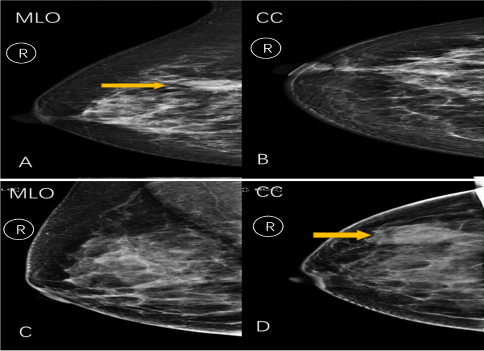
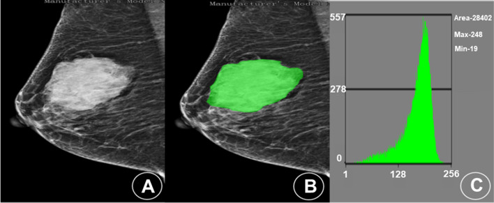
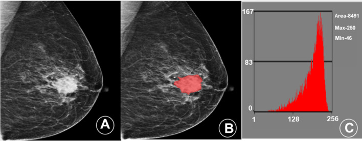
Initially, the clinical and X-ray data of patients (n = 319, age of 54 ± 14) with breast cancer (triple-negative-65, non-triple-negative-254) from the First Affiliated Hospital of Soochow University (n = 211, as a training set) and Suzhou Municipal Hospital (n = 108, as a verification set) from January 2018 to February 2021 are retrospectively analyzed. Comparing the mediolateral oblique (MLO) and cranial cauda (CC) mammography images, the mammography images with larger lesion areas are selected, and the image segmentation and radiomics feature extraction are then performed by the MaZda software. Further, the Fisher coefficients (Fisher), classification error probability combined average correlation coefficients (POE + ACC), and mutual information (MI) are used to select three sets of feature subsets. Moreover, the score of each patient's radiomics signature (Radscore) is calculated. Finally, the receiver operating characteristic curve (ROC) is analyzed to calculate the AUC, accuracy, sensitivity, specificity, positive predictive value, and negative predictive value of TNBC.

RESULTS

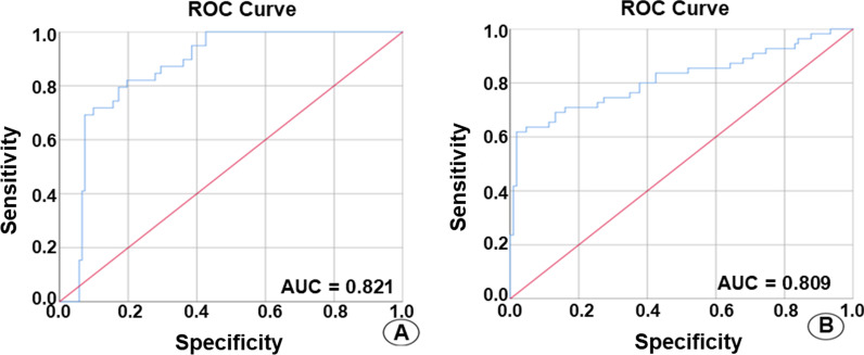
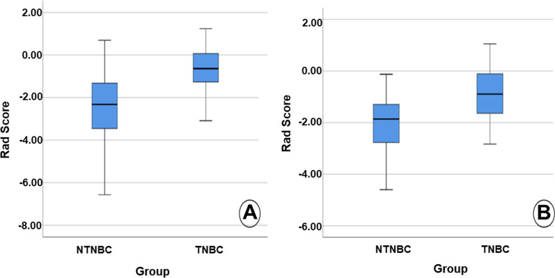
A significant difference in the mammography manifestation between the triple-negative and the non-triple-negative groups (P < 0.001) is observed. The (POE + ACC)-NDA method showed the highest accuracy of 88.39%. The Radscore of triple-negative and non-triple-negative groups in the training set includes - 0.678 (- 1.292, 0.088) and - 2.536 (- 3.496, - 1.324), respectively, with a statistically significant difference (Z = - 6.314, P < 0.001). In contrast, the Radscore in the validation set includes - 0.750 (- 1.332, - 0.054) and - 2.223 (- 2.963, - 1.256), with a statistically significant difference (Z = - 4.669, P < 0.001). In the training set, the AUC, accuracy, sensitivity, specificity, positive predictive value and negative predictive value of TNBC include 0.821 (95% confidence interval 0.752-0.890), 74.4%, 82.5%, 72.5%, 41.2%, and 94.6%, respectively. In the validation set, the AUC, accuracy, sensitivity, specificity, positive predictive value and negative predictive value of TNBC are of 0.809 (95% confidence interval 0.711-0.907), 80.6%, 72.0%, 80.7%, 55.5%, and 93.1%, respectively.

CONCLUSION

In summary, we firmly believe that this mammography-based radiomics signature could be useful in the preoperative prediction of TNBC due to its high value.

摘要

目的

本研究旨在探讨基于乳腺 X 线摄影的放射组学特征在术前预测三阴性乳腺癌(TNBC)中的价值。

材料与方法

回顾性分析 2018 年 1 月至 2021 年 2 月苏州大学附属第一医院(n=211,作为训练集)和苏州市立医院(n=108,作为验证集)的 319 例(年龄 54±14 岁)乳腺癌患者(三阴性 65 例,非三阴性 254 例)的临床和 X 射线数据。比较了内外斜位(MLO)和头尾位(CC)乳腺 X 线摄影图像,选择病变面积较大的乳腺 X 线摄影图像,然后使用 MaZda 软件进行图像分割和放射组学特征提取。进一步采用 Fisher 系数(Fisher)、分类错误概率结合平均相关系数(POE+ACC)和互信息(MI)分别选择三组特征子集。此外,计算每位患者放射组学特征的评分(Radscore)。最后,通过绘制受试者工作特征曲线(ROC),计算 TNBC 的 AUC、准确率、敏感度、特异度、阳性预测值和阴性预测值。

结果

三阴性和非三阴性组之间的乳腺 X 线表现存在显著差异(P<0.001)。(POE+ACC)-NDA 方法的准确率最高,为 88.39%。训练集中三阴性和非三阴性组的 Radscore 分别为-0.678(-1.292,0.088)和-2.536(-3.496,-1.324),差异具有统计学意义(Z=-6.314,P<0.001)。相比之下,验证集中的 Radscore 分别为-0.750(-1.332,-0.054)和-2.223(-2.963,-1.256),差异具有统计学意义(Z=-4.669,P<0.001)。在训练集中,TNBC 的 AUC、准确率、敏感度、特异度、阳性预测值和阴性预测值分别为 0.821(95%置信区间为 0.752-0.890)、74.4%、82.5%、72.5%、41.2%和 94.6%。在验证集中,TNBC 的 AUC、准确率、敏感度、特异度、阳性预测值和阴性预测值分别为 0.809(95%置信区间为 0.711-0.907)、80.6%、72.0%、80.7%、55.5%和 93.1%。

结论

综上所述,我们坚信,由于其高价值,基于乳腺 X 线摄影的放射组学特征可用于术前预测 TNBC。

https://cdn.ncbi.nlm.nih.gov/pmc/blobs/2457/9472401/5dec5b36a974/12880_2022_875_Fig5_HTML.jpg
https://cdn.ncbi.nlm.nih.gov/pmc/blobs/2457/9472401/02a254d04365/12880_2022_875_Fig1_HTML.jpg
https://cdn.ncbi.nlm.nih.gov/pmc/blobs/2457/9472401/700459318373/12880_2022_875_Fig2_HTML.jpg
https://cdn.ncbi.nlm.nih.gov/pmc/blobs/2457/9472401/598cf4ec8303/12880_2022_875_Fig3_HTML.jpg
https://cdn.ncbi.nlm.nih.gov/pmc/blobs/2457/9472401/b4407b527acf/12880_2022_875_Fig4_HTML.jpg
https://cdn.ncbi.nlm.nih.gov/pmc/blobs/2457/9472401/5dec5b36a974/12880_2022_875_Fig5_HTML.jpg

相似文献

1
Application of mammography-based radiomics signature for preoperative prediction of triple-negative breast cancer.基于乳腺 X 线摄影的放射组学特征在前哨淋巴结阴性三阴性乳腺癌中的应用。
BMC Med Imaging. 2022 Sep 14;22(1):166. doi: 10.1186/s12880-022-00875-6.
2
A pilot study of radiomics technology based on X-ray mammography in patients with triple-negative breast cancer.基于 X 射线钼靶的放射组学技术在三阴性乳腺癌患者中的初步研究。
J Xray Sci Technol. 2019;27(3):485-492. doi: 10.3233/XST-180488.
3
Predictive value of triple negative breast cancer based on DCE-MRI multi-phase full-volume ROI clinical radiomics model.基于 DCE-MRI 多期全容积 ROI 临床放射组学模型的三阴性乳腺癌预测价值。
Acta Radiol. 2024 Feb;65(2):173-184. doi: 10.1177/02841851231215145. Epub 2023 Nov 28.
4
Diagnosis of triple negative breast cancer based on radiomics signatures extracted from preoperative contrast-enhanced chest computed tomography.基于术前增强胸部 CT 提取的放射组学特征诊断三阴性乳腺癌。
BMC Cancer. 2020 Jun 22;20(1):579. doi: 10.1186/s12885-020-07053-3.
5
Contrast-Enhanced Mammography Radiomics Analysis for Preoperative Prediction of Breast Cancer Molecular Subtypes.对比增强乳腺 X 线摄影影像组学分析在乳腺癌分子亚型术前预测中的应用。
Acad Radiol. 2024 Jun;31(6):2228-2238. doi: 10.1016/j.acra.2023.12.005. Epub 2023 Dec 23.
6
[The value of CT radiomics in the prediction of EGFR mutation in lung cancer].[CT影像组学在预测肺癌表皮生长因子受体突变中的价值]
Zhonghua Yi Xue Za Zhi. 2020 Mar 10;100(9):690-695. doi: 10.3760/cma.j.issn.0376-2491.2020.09.009.
7
Diagnostic Value of Radiomics Analysis in Contrast-Enhanced Spectral Mammography for Identifying Triple-Negative Breast Cancer.基于对比增强光谱乳腺摄影的影像组学分析在三阴性乳腺癌诊断中的价值
Front Oncol. 2021 Dec 23;11:773196. doi: 10.3389/fonc.2021.773196. eCollection 2021.
8
Utilizing grayscale ultrasound-based radiomics nomogram for preoperative identification of triple negative breast cancer.利用基于灰阶超声的放射组学列线图术前识别三阴性乳腺癌。
Radiol Med. 2024 Jan;129(1):29-37. doi: 10.1007/s11547-023-01739-x. Epub 2023 Nov 2.
9
Mammography-based radiomics for predicting the risk of breast cancer recurrence: a multicenter study.基于乳腺 X 线摄影的放射组学预测乳腺癌复发风险:一项多中心研究。
Br J Radiol. 2021 Nov 1;94(1127):20210348. doi: 10.1259/bjr.20210348. Epub 2021 Sep 14.
10
Radiomics Analysis Based on Automatic Image Segmentation of DCE-MRI for Predicting Triple-Negative and Nontriple-Negative Breast Cancer.基于 DCE-MRI 自动图像分割的放射组学分析预测三阴性和非三阴性乳腺癌。
Comput Math Methods Med. 2021 Aug 10;2021:2140465. doi: 10.1155/2021/2140465. eCollection 2021.

引用本文的文献

1
Radiologic imaging biomarkers in triple-negative breast cancer: a literature review about the role of artificial intelligence and the way forward.三阴性乳腺癌的放射影像学生物标志物:关于人工智能作用及未来发展方向的文献综述
BJR Artif Intell. 2024 Nov 13;1(1):ubae016. doi: 10.1093/bjrai/ubae016. eCollection 2024 Jan.
2
Decoding Breast Cancer: Using Radiomics to Non-Invasively Unveil Molecular Subtypes Directly from Mammographic Images.解读乳腺癌:利用影像组学从乳腺X线图像中直接无创揭示分子亚型
J Imaging. 2024 Sep 4;10(9):218. doi: 10.3390/jimaging10090218.
3
Breast Cancer Molecular Subtype Prediction: A Mammography-Based AI Approach.

本文引用的文献

1
Multidisciplinary considerations in the treatment of triple-negative breast cancer.三阴性乳腺癌治疗中的多学科考量
CA Cancer J Clin. 2020 Nov;70(6):432-442. doi: 10.3322/caac.21643. Epub 2020 Sep 28.
2
Nomogram predicting survival as a selection criterion for postmastectomy radiotherapy in patients with T1 to T2 breast cancer with 1 to 3 positive lymph nodes.列线图预测 T1 至 T2 期伴 1 至 3 个阳性淋巴结乳腺癌患者术后放疗的生存选择标准。
Cancer. 2020 Aug 15;126 Suppl 16:3857-3866. doi: 10.1002/cncr.32963.
3
Texture Analysis with 3.0-T MRI for Association of Response to Neoadjuvant Chemotherapy in Breast Cancer.
乳腺癌分子亚型预测:一种基于乳腺X线摄影的人工智能方法。
Biomedicines. 2024 Jun 20;12(6):1371. doi: 10.3390/biomedicines12061371.
4
Utilizing grayscale ultrasound-based radiomics nomogram for preoperative identification of triple negative breast cancer.利用基于灰阶超声的放射组学列线图术前识别三阴性乳腺癌。
Radiol Med. 2024 Jan;129(1):29-37. doi: 10.1007/s11547-023-01739-x. Epub 2023 Nov 2.
5
A Review of AI-Based Radiomics and Computational Pathology Approaches in Triple-Negative Breast Cancer: Current Applications and Perspectives.基于人工智能的放射组学和计算病理学在三阴性乳腺癌中的研究进展:当前的应用与展望。
Clin Breast Cancer. 2023 Dec;23(8):800-812. doi: 10.1016/j.clbc.2023.06.004. Epub 2023 Jun 21.
6
Artificial Intelligence in the Diagnosis and Treatment of Pancreatic Cystic Lesions and Adenocarcinoma.人工智能在胰腺囊性病变和腺癌诊断与治疗中的应用
Cancers (Basel). 2023 Apr 22;15(9):2410. doi: 10.3390/cancers15092410.
7
An ultrasound-based radiomics model to distinguish between sclerosing adenosis and invasive ductal carcinoma.一种基于超声的影像组学模型,用于鉴别硬化性腺病和浸润性导管癌。
Front Oncol. 2023 Mar 7;13:1090617. doi: 10.3389/fonc.2023.1090617. eCollection 2023.
基于 3.0T MRI 的纹理分析对乳腺癌新辅助化疗反应的相关性研究。
Radiology. 2020 Jan;294(1):31-41. doi: 10.1148/radiol.2019182718. Epub 2019 Nov 26.
4
Overview of radiomics in breast cancer diagnosis and prognostication.乳腺癌诊断和预后中的放射组学概述。
Breast. 2020 Feb;49:74-80. doi: 10.1016/j.breast.2019.10.018. Epub 2019 Nov 6.
5
A pilot study of radiomics technology based on X-ray mammography in patients with triple-negative breast cancer.基于 X 射线钼靶的放射组学技术在三阴性乳腺癌患者中的初步研究。
J Xray Sci Technol. 2019;27(3):485-492. doi: 10.3233/XST-180488.
6
Stand-Alone Artificial Intelligence for Breast Cancer Detection in Mammography: Comparison With 101 Radiologists.孤立人工智能在乳腺钼靶摄影中的乳腺癌检测:与 101 位放射科医生的比较。
J Natl Cancer Inst. 2019 Sep 1;111(9):916-922. doi: 10.1093/jnci/djy222.
7
Radiomics: the facts and the challenges of image analysis.放射组学:图像分析的现状与挑战
Eur Radiol Exp. 2018 Nov 14;2(1):36. doi: 10.1186/s41747-018-0068-z.
8
Targeting the androgen receptor in triple-negative breast cancer.靶向三阴性乳腺癌中的雄激素受体。
Curr Probl Cancer. 2016 Mar-Aug;40(2-4):141-150. doi: 10.1016/j.currproblcancer.2016.09.004. Epub 2016 Sep 20.
9
Breast Cancer Epidemiology and Risk Factors.乳腺癌流行病学与风险因素
Clin Obstet Gynecol. 2016 Dec;59(4):651-672. doi: 10.1097/GRF.0000000000000239.
10
Refinement of Triple-Negative Breast Cancer Molecular Subtypes: Implications for Neoadjuvant Chemotherapy Selection.三阴性乳腺癌分子亚型的细化:对新辅助化疗选择的影响
PLoS One. 2016 Jun 16;11(6):e0157368. doi: 10.1371/journal.pone.0157368. eCollection 2016.