• 文献检索
  • 文档翻译
  • 深度研究
  • 学术资讯
  • Suppr Zotero 插件Zotero 插件
  • 邀请有礼
  • 套餐&价格
  • 历史记录
应用&插件
Suppr Zotero 插件Zotero 插件浏览器插件Mac 客户端Windows 客户端微信小程序
定价
高级版会员购买积分包购买API积分包
服务
文献检索文档翻译深度研究API 文档MCP 服务
关于我们
关于 Suppr公司介绍联系我们用户协议隐私条款
关注我们

Suppr 超能文献

核心技术专利:CN118964589B侵权必究
粤ICP备2023148730 号-1Suppr @ 2026

文献检索

告别复杂PubMed语法,用中文像聊天一样搜索,搜遍4000万医学文献。AI智能推荐,让科研检索更轻松。

立即免费搜索

文件翻译

保留排版,准确专业,支持PDF/Word/PPT等文件格式,支持 12+语言互译。

免费翻译文档

深度研究

AI帮你快速写综述,25分钟生成高质量综述,智能提取关键信息,辅助科研写作。

立即免费体验

利用无监督对抗学习进行肺部 CT 扫描中的病变分割。

Lesion segmentation in lung CT scans using unsupervised adversarial learning.

机构信息

Department of Mathematics and Computer Science, University of Calabria, Rende, Italy.

Department of Electronics, Information and Bioengineering (DEIB), Politecnico di Milano, Milan, Italy.

出版信息

Med Biol Eng Comput. 2022 Nov;60(11):3203-3215. doi: 10.1007/s11517-022-02651-8. Epub 2022 Sep 20.

DOI:10.1007/s11517-022-02651-8
PMID:36125656
原文链接:https://pmc.ncbi.nlm.nih.gov/articles/PMC9486778/
Abstract

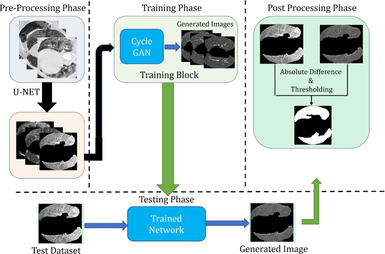
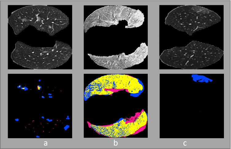
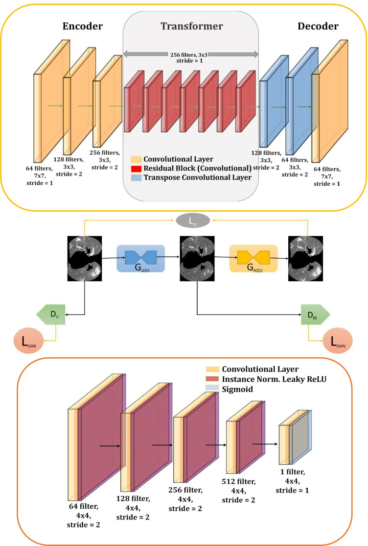
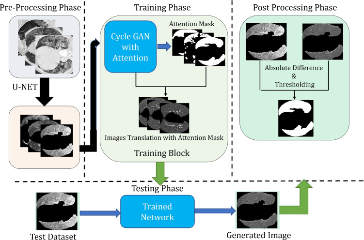
Lesion segmentation in medical images is difficult yet crucial for proper diagnosis and treatment. Identifying lesions in medical images is costly and time-consuming and requires highly specialized knowledge. For this reason, supervised and semi-supervised learning techniques have been developed. Nevertheless, the lack of annotated data, which is common in medical imaging, is an issue; in this context, interesting approaches can use unsupervised learning to accurately distinguish between healthy tissues and lesions, training the network without using the annotations. In this work, an unsupervised learning technique is proposed to automatically segment coronavirus disease 2019 (COVID-19) lesions on 2D axial CT lung slices. The proposed approach uses the technique of image translation to generate healthy lung images based on the infected lung image without the need for lesion annotations. Attention masks are used to improve the quality of the segmentation further. Experiments showed the capability of the proposed approaches to segment the lesions, and it outperforms a range of unsupervised lesion detection approaches. The average reported results for the test dataset based on the metrics: Dice Score, Sensitivity, Specificity, Structure Measure, Enhanced-Alignment Measure, and Mean Absolute Error are 0.695, 0.694, 0.961, 0.791, 0.875, and 0.082 respectively. The achieved results are promising compared with the state-of-the-art and could constitute a valuable tool for future developments.

摘要

医学图像中的病变分割对于正确的诊断和治疗至关重要。在医学图像中识别病变既昂贵又耗时,并且需要高度专业化的知识。出于这个原因,已经开发了监督和半监督学习技术。然而,医学成像中常见的注释数据缺乏是一个问题;在这种情况下,有趣的方法可以使用无监督学习来准确地区分健康组织和病变,而无需使用注释来训练网络。在这项工作中,提出了一种无监督学习技术,用于自动分割二维轴向 CT 肺部切片上的 2019 年冠状病毒病(COVID-19)病变。所提出的方法使用图像翻译技术来生成基于感染肺部图像的健康肺部图像,而无需病变注释。使用注意力掩模进一步提高分割质量。实验表明,所提出的方法能够分割病变,并且在一系列无监督病变检测方法中表现出色。基于指标的测试数据集的平均报告结果:骰子分数、灵敏度、特异性、结构度量、增强对齐度量和平均绝对误差分别为 0.695、0.694、0.961、0.791、0.875 和 0.082。与最先进的方法相比,所取得的结果很有希望,并且可以成为未来发展的有价值的工具。

https://cdn.ncbi.nlm.nih.gov/pmc/blobs/d367/9537132/fd270b1ab434/11517_2022_2651_Fig6_HTML.jpg
https://cdn.ncbi.nlm.nih.gov/pmc/blobs/d367/9537132/17e5adcdb1ba/11517_2022_2651_Fig1_HTML.jpg
https://cdn.ncbi.nlm.nih.gov/pmc/blobs/d367/9537132/c5d03ac5f606/11517_2022_2651_Fig2_HTML.jpg
https://cdn.ncbi.nlm.nih.gov/pmc/blobs/d367/9537132/92eb192adb78/11517_2022_2651_Fig3_HTML.jpg
https://cdn.ncbi.nlm.nih.gov/pmc/blobs/d367/9537132/9bc661647b12/11517_2022_2651_Fig4_HTML.jpg
https://cdn.ncbi.nlm.nih.gov/pmc/blobs/d367/9537132/e72392642f84/11517_2022_2651_Fig5_HTML.jpg
https://cdn.ncbi.nlm.nih.gov/pmc/blobs/d367/9537132/fd270b1ab434/11517_2022_2651_Fig6_HTML.jpg

相似文献

1
Lesion segmentation in lung CT scans using unsupervised adversarial learning.利用无监督对抗学习进行肺部 CT 扫描中的病变分割。
Med Biol Eng Comput. 2022 Nov;60(11):3203-3215. doi: 10.1007/s11517-022-02651-8. Epub 2022 Sep 20.
2
Label-free coronavirus disease 2019 lesion segmentation based on synthetic healthy lung image subtraction.基于合成健康肺图像减影的无标签 2019 年冠状病毒病病变分割。
Med Phys. 2022 Jul;49(7):4632-4641. doi: 10.1002/mp.15661. Epub 2022 Apr 22.
3
Self-supervised region-aware segmentation of COVID-19 CT images using 3D GAN and contrastive learning.基于 3D GAN 和对比学习的 COVID-19 CT 图像自监督区域感知分割。
Comput Biol Med. 2022 Oct;149:106033. doi: 10.1016/j.compbiomed.2022.106033. Epub 2022 Aug 27.
4
Lung Lesion Localization of COVID-19 From Chest CT Image: A Novel Weakly Supervised Learning Method.COVID-19 肺部病变的 CT 图像定位:一种新的弱监督学习方法。
IEEE J Biomed Health Inform. 2021 Jun;25(6):1864-1872. doi: 10.1109/JBHI.2021.3067465. Epub 2021 Jun 3.
5
Unsupervised segmentation and quantification of COVID-19 lesions on computed Tomography scans using CycleGAN.基于 CycleGAN 的 CT 扫描 COVID-19 病变的无监督分割与定量分析。
Methods. 2022 Sep;205:200-209. doi: 10.1016/j.ymeth.2022.07.007. Epub 2022 Jul 8.
6
Unsupervised Deep Learning for Stroke Lesion Segmentation on Follow-up CT Based on Generative Adversarial Networks.基于生成对抗网络的无监督深度学习在基于 CT 随访的脑卒中病灶分割。
AJNR Am J Neuroradiol. 2022 Aug;43(8):1107-1114. doi: 10.3174/ajnr.A7582. Epub 2022 Jul 28.
7
A novel adaptive cubic quasi-Newton optimizer for deep learning based medical image analysis tasks, validated on detection of COVID-19 and segmentation for COVID-19 lung infection, liver tumor, and optic disc/cup.一种用于深度学习的新型自适应三次拟牛顿优化器,在 COVID-19 检测和 COVID-19 肺部感染、肝脏肿瘤以及视盘/杯分割等医学图像分析任务中得到验证。
Med Phys. 2023 Mar;50(3):1528-1538. doi: 10.1002/mp.15969. Epub 2022 Oct 6.
8
HFCF-Net: A hybrid-feature cross fusion network for COVID-19 lesion segmentation from CT volumetric images.HFCF-Net:一种用于从 CT 容积图像中分割 COVID-19 病变的混合特征交叉融合网络。
Med Phys. 2022 Jun;49(6):3797-3815. doi: 10.1002/mp.15600. Epub 2022 Mar 30.
9
Active contour regularized semi-supervised learning for COVID-19 CT infection segmentation with limited annotations.基于主动轮廓正则化半监督学习的有限标注 COVID-19 CT 感染分割。
Phys Med Biol. 2020 Dec 18;65(22):225034. doi: 10.1088/1361-6560/abc04e.
10
Multiscale unsupervised domain adaptation for automatic pancreas segmentation in CT volumes using adversarial learning.基于对抗学习的 CT 容积中多尺度无监督域自适应自动胰腺分割。
Med Phys. 2022 Sep;49(9):5799-5818. doi: 10.1002/mp.15827. Epub 2022 Jul 27.

引用本文的文献

1
Cross-domain lung opacity detection via adversarial learning and box fusion.通过对抗学习和框融合进行跨域肺实质混浊检测。
Sci Rep. 2024 Dec 28;14(1):31353. doi: 10.1038/s41598-024-82719-7.

本文引用的文献

1
Exploiting probability density function of deep convolutional autoencoders' latent space for reliable COVID-19 detection on CT scans.利用深度卷积自动编码器潜在空间的概率密度函数在CT扫描上进行可靠的COVID-19检测。
J Supercomput. 2022;78(9):12024-12045. doi: 10.1007/s11227-022-04349-y. Epub 2022 Feb 24.
2
Unsupervised domain adaptation based COVID-19 CT infection segmentation network.基于无监督域自适应的新冠肺炎CT感染分割网络。
Appl Intell (Dordr). 2022;52(6):6340-6353. doi: 10.1007/s10489-021-02691-x. Epub 2021 Sep 7.
3
COVIDetectioNet: COVID-19 diagnosis system based on X-ray images using features selected from pre-learned deep features ensemble.
COVIDetectioNet:基于X射线图像的COVID-19诊断系统,使用从预学习深度特征集成中选择的特征。
Appl Intell (Dordr). 2021;51(3):1213-1226. doi: 10.1007/s10489-020-01888-w. Epub 2020 Sep 18.
4
Weakly unsupervised conditional generative adversarial network for image-based prognostic prediction for COVID-19 patients based on chest CT.基于胸部 CT 的 COVID-19 患者图像预后预测的弱无监督条件生成对抗网络。
Med Image Anal. 2021 Oct;73:102159. doi: 10.1016/j.media.2021.102159. Epub 2021 Jul 11.
5
Data augmentation using generative adversarial neural networks on brain structural connectivity in multiple sclerosis.基于生成对抗神经网络的多发性硬化脑结构连接数据扩充。
Comput Methods Programs Biomed. 2021 Jul;206:106113. doi: 10.1016/j.cmpb.2021.106113. Epub 2021 Apr 24.
6
Automatic detection of coronavirus disease (COVID-19) using X-ray images and deep convolutional neural networks.使用X射线图像和深度卷积神经网络自动检测冠状病毒病(COVID-19)。
Pattern Anal Appl. 2021;24(3):1207-1220. doi: 10.1007/s10044-021-00984-y. Epub 2021 May 9.
7
Automated Quantification of CT Patterns Associated with COVID-19 from Chest CT.胸部CT中与COVID-19相关的CT模式的自动定量分析
Radiol Artif Intell. 2020 Jul 29;2(4):e200048. doi: 10.1148/ryai.2020200048. eCollection 2020 Jul.
8
Imaging Profile of the COVID-19 Infection: Radiologic Findings and Literature Review.新型冠状病毒肺炎感染的影像学表现:放射学发现与文献综述
Radiol Cardiothorac Imaging. 2020 Feb 13;2(1):e200034. doi: 10.1148/ryct.2020200034. eCollection 2020 Feb.
9
Label-Free Segmentation of COVID-19 Lesions in Lung CT.肺 CT 中 COVID-19 病变的无标记分割。
IEEE Trans Med Imaging. 2021 Oct;40(10):2808-2819. doi: 10.1109/TMI.2021.3066161. Epub 2021 Sep 30.
10
An Open-Source COVID-19 CT Dataset with Automatic Lung Tissue Classification for Radiomics.一个用于放射组学的具有自动肺组织分类功能的开源COVID-19 CT数据集。
Bioengineering (Basel). 2021 Feb 16;8(2):26. doi: 10.3390/bioengineering8020026.