Department of Radiology, Kawasaki Medical School, 577 Matsushima, Kurashiki, Okayama, 701-0192, Japan.
Philips Japan, Tokyo, Japan.
Sci Rep. 2022 Sep 27;12(1):16070. doi: 10.1038/s41598-022-20518-8.
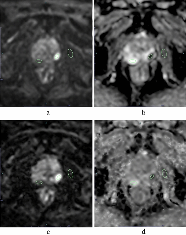
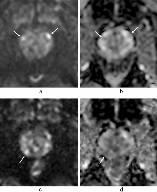
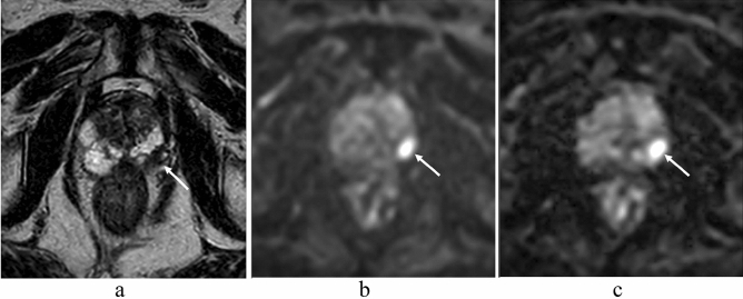
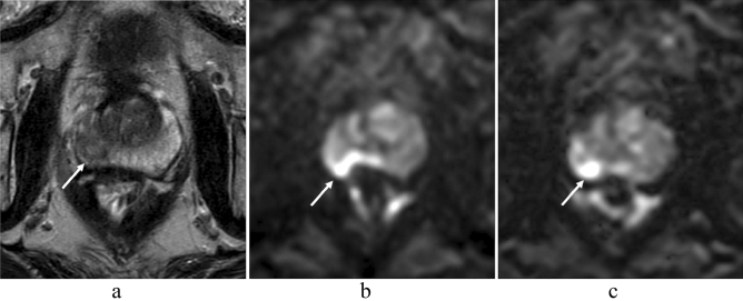
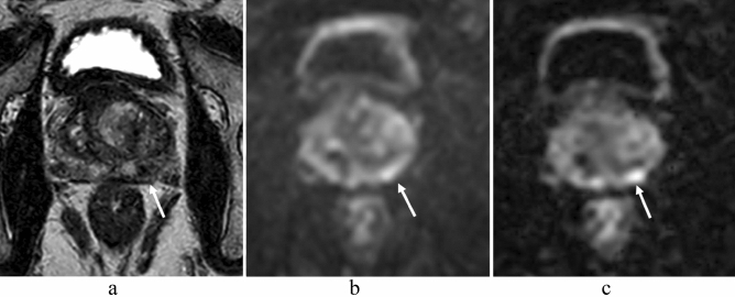
In prostate MRI, single-shot EPI (ssEPI) DWI still suffers from distortion and blurring. Multi-shot EPI (msEPI) overcomes the drawbacks of ssEPI DWI. The aim of this article was to compare the image quality and diagnostic performance for clinically significant prostate cancer (csPC) between ssEPI DWI and msEPI DWI. This retrospective study included 134 patients with suspected PC who underwent 3.0 T MRI and subsequent MRI-guided biopsy. Three radiologists independently assessed anatomical distortion, prostate edge clarity, and lesion conspicuity score for pathologically confirmed csPC. Lesion apparent diffusion coefficient (ADC) and benign ADC were also calculated. In 17 PC patients who underwent prostatectomy, three radiologists independently assessed eight prostate regions by DWI score in PI-RADS v 2.1. Anatomical distortion and prostate edge clarity were significantly higher in msEPI DWI than in ssEPI DWI in the three readers. Lesion conspicuity score was significantly higher in msEPI DWI than in ssEPI DWI in reader 1 and reader 3. Regarding discrimination ability between PC with GS ≤ 3 + 4 and PC with GS ≥ 4 + 3 using lesion ADC, AUC was comparable between ssEPI DWI and msEPI DWI. For diagnostic performance of csPC using DWI score, AUC was comparable between msEPI DWI and ssEPI DWI in all readers. Compared with ssEPI DWI, msEPI DWI had improved image quality and similar or higher diagnostic performance.
在前列腺 MRI 中,单次激发 EPI(ssEPI)DWI 仍然存在失真和模糊的问题。多激发 EPI(msEPI)克服了 ssEPI DWI 的缺点。本文旨在比较 ssEPI DWI 和 msEPI DWI 对临床显著前列腺癌(csPC)的图像质量和诊断性能。这项回顾性研究纳入了 134 名疑似 PC 患者,他们均接受了 3.0T MRI 检查和随后的 MRI 引导活检。三位放射科医生独立评估了病理证实的 csPC 的解剖失真、前列腺边缘清晰度和病灶显影评分。还计算了病灶表观扩散系数(ADC)和良性 ADC。在 17 名接受前列腺切除术的 PC 患者中,三位放射科医生通过 PI-RADS v 2.1 对 8 个前列腺区域的 DWI 评分进行了独立评估。三位读者均认为 msEPI DWI 的解剖失真和前列腺边缘清晰度明显高于 ssEPI DWI。读者 1 和读者 3 认为 msEPI DWI 的病灶显影评分明显高于 ssEPI DWI。对于使用病灶 ADC 区分 GS≤3+4 和 GS≥4+3 的 PC,ssEPI DWI 和 msEPI DWI 的 AUC 相当。对于使用 DWI 评分诊断 csPC 的性能,所有读者均认为 msEPI DWI 和 ssEPI DWI 的 AUC 相当。与 ssEPI DWI 相比,msEPI DWI 的图像质量得到了改善,且诊断性能相似或更高。