Liang Mingshu, Bernadt Cory, Wong Soon Boon Justin, Choi Changsoon, Cote Richard, Yang Changhuei
Department of Electrical Engineering, California Institute of Technology, Pasadena, CA 91125, USA.
Department of Pathology and Immunology, Washington University School of Medicine, MO 63110, USA.
J Pathol Inform. 2022 Jun 30;13:100119. doi: 10.1016/j.jpi.2022.100119. eCollection 2022.
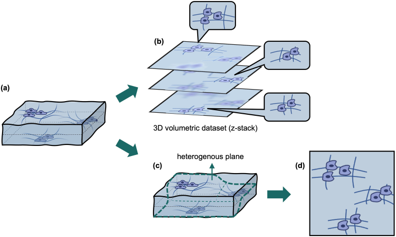
Cytology is the study of whole cells in diagnostic pathology. Unlike standard histologic thinly sliced specimens, cytologic preparations consist of preparations of whole cells where cells commonly cluster and aggregate. As such, cytology preparations are generally much thicker than histologic slides, resulting in large patches of defocus when examined under the microscope. A diagnostic aggregate of cells often cannot be viewed in focus together, requiring pathologists to continually manipulate the focal plane, complicating the task of accurately assessing the entire cellular aggregate and thus in making a diagnosis. Further, it is extremely difficult to acquire useful uniformly in-focus digital images of cytology preparations for applications such as remote diagnostic evaluations and artificial intelligence models. The predominant current method to address this issue is to acquire digital images at multiple focal planes of the entire slide, which demands long scanning time, complex and expensive scanning systems, and huge storage capacity.
Here we report a unique imaging method that can acquire cytologic images efficiently and computationally render all-in-focus digital images that are highly compact.
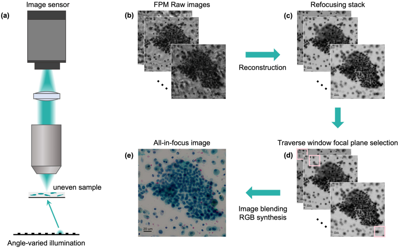
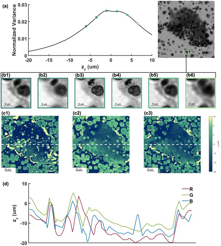
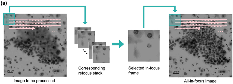
This method applies a metric-based digital refocusing to microscopy data collected with a Fourier ptychographic microscope (FPM). The digitally refocused patches of images are then synthesized into an all-in-focus image.
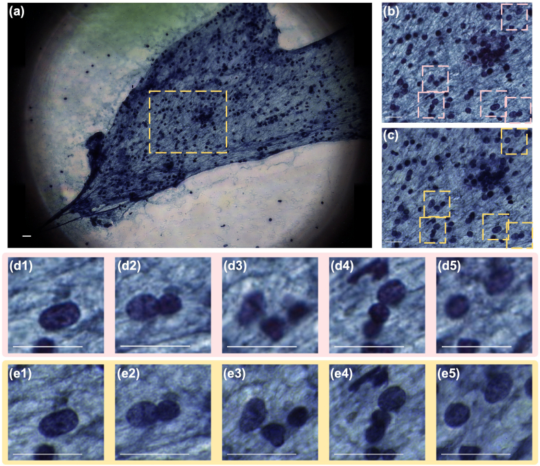
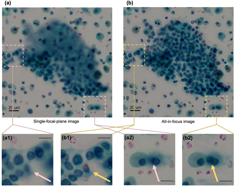
We report all-in-focus FPM results of thyroid fine needle aspiration (FNA) cytology samples, demonstrating our method's ability to overcome the height variance of 30 μm caused by cell aggregation, and rendering images at high resolution (corresponds to a standard microscope with objective NA of 0.75) and that are all-in-focus.
This technology is applicable to standard microscopes, and we believe can have an impact on diagnostic accuracy as well as ease and speed of diagnosing challenging specimens. While we focus on cytology slides here, we anticipate this technology's advantages will translate well for histology applications. This technique also addresses the issue of remote rapid evaluation of cytology preparations. Finally, we believe that by resolving the focus heterogeneity issues in standard digital images, this technique is a critical advance for applying machine learning to cytology specimens.
细胞学是诊断病理学中对完整细胞的研究。与标准组织学薄切片标本不同,细胞学标本由完整细胞的制剂组成,细胞通常会聚集在一起。因此,细胞学标本通常比组织学切片厚得多,在显微镜下检查时会产生大片散焦区域。一个具有诊断意义的细胞聚集体往往无法同时清晰聚焦观察,这就要求病理学家不断调整焦平面,使得准确评估整个细胞聚集体并做出诊断的任务变得复杂。此外,要获取用于远程诊断评估和人工智能模型等应用的细胞学标本的有用的均匀清晰聚焦数字图像极其困难。当前解决这个问题的主要方法是在整个载玻片的多个焦平面获取数字图像,这需要很长的扫描时间、复杂且昂贵的扫描系统以及巨大的存储容量。
在此我们报告一种独特的成像方法,该方法能够高效获取细胞学图像并通过计算生成高度紧凑的全聚焦数字图像。
该方法将基于度量的数字重聚焦应用于用傅里叶叠层显微镜(FPM)收集的显微镜数据。然后将数字重聚焦后的图像块合成成全聚焦图像。
我们报告了甲状腺细针穿刺(FNA)细胞学样本的全聚焦FPM结果,证明了我们的方法能够克服由细胞聚集引起的30μm高度差异,并以高分辨率(相当于物镜数值孔径为0.75的标准显微镜)生成全聚焦图像。
这项技术适用于标准显微镜,我们相信它会对诊断准确性以及诊断具有挑战性标本的便捷性和速度产生影响。虽然我们在此专注于细胞学载玻片,但我们预计这项技术的优势在组织学应用中也能很好地体现。这项技术还解决了细胞学标本的远程快速评估问题。最后,我们认为通过解决标准数字图像中的聚焦异质性问题,这项技术是将机器学习应用于细胞学标本的一项关键进展。