Chor Ana, Takiya Christina Maeda, Dias Marcos Lopes, Gonçalves Raquel Pires, Petithory Tatiana, Cypriano Jefferson, de Andrade Leonardo Rodrigues, Farina Marcos, Anselme Karine
Biomineralization Laboratory, Institute of Biomedical Sciences, Federal University of Rio de Janeiro, Rio de Janeiro 21941-902, Brazil.
Immunopathology Laboratory, Institute of Biophysics Carlos Chagas Filho, Federal University of Rio de Janeiro, Rio de Janeiro 20941-902, Brazil.
Polymers (Basel). 2022 Oct 21;14(20):4460. doi: 10.3390/polym14204460.
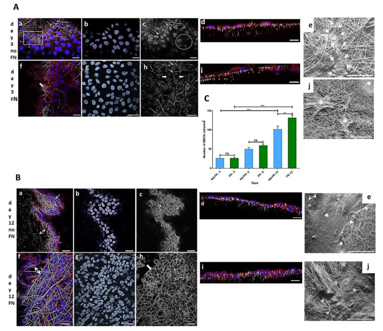
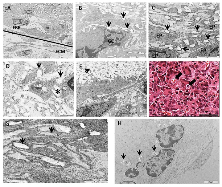
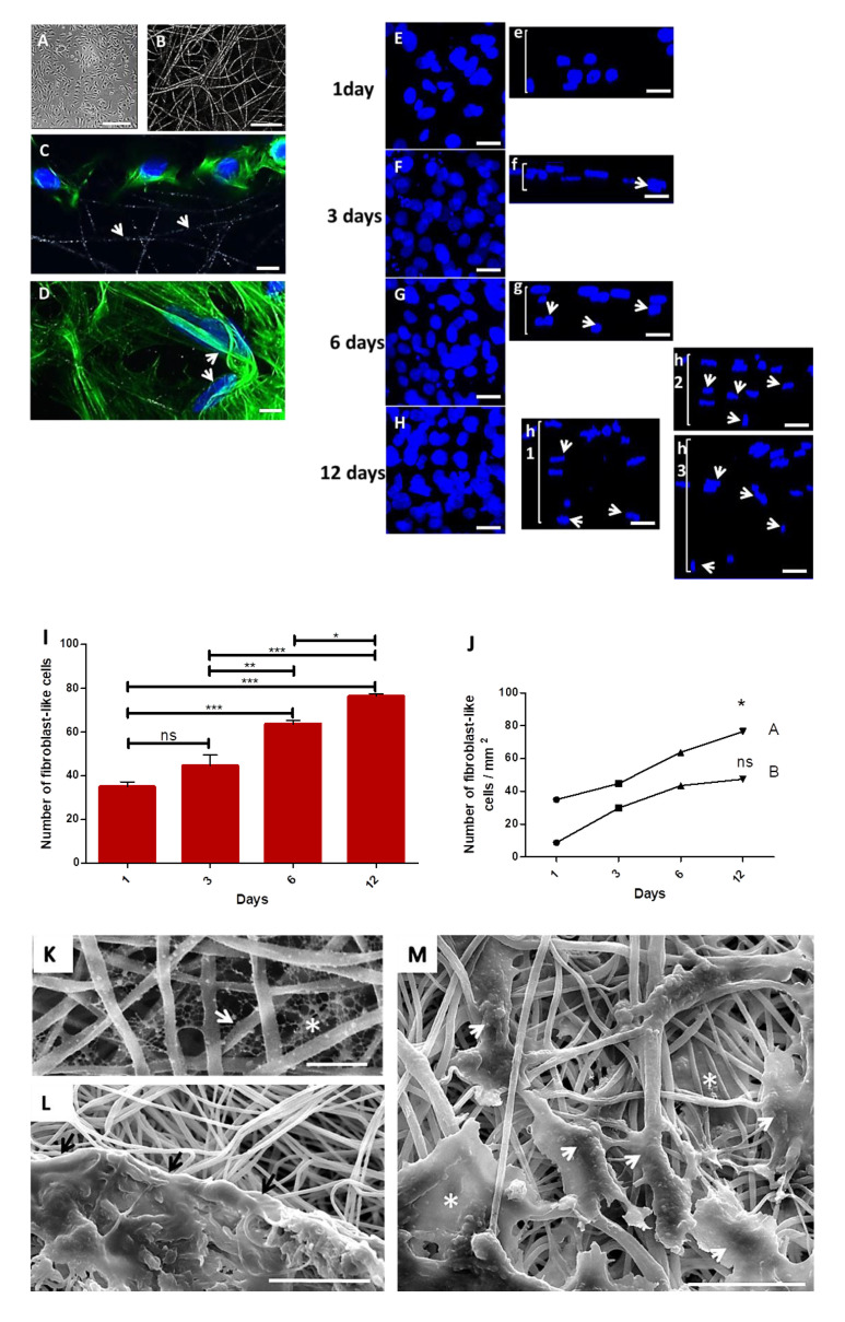
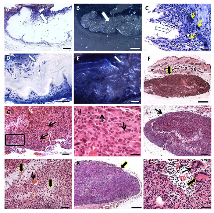
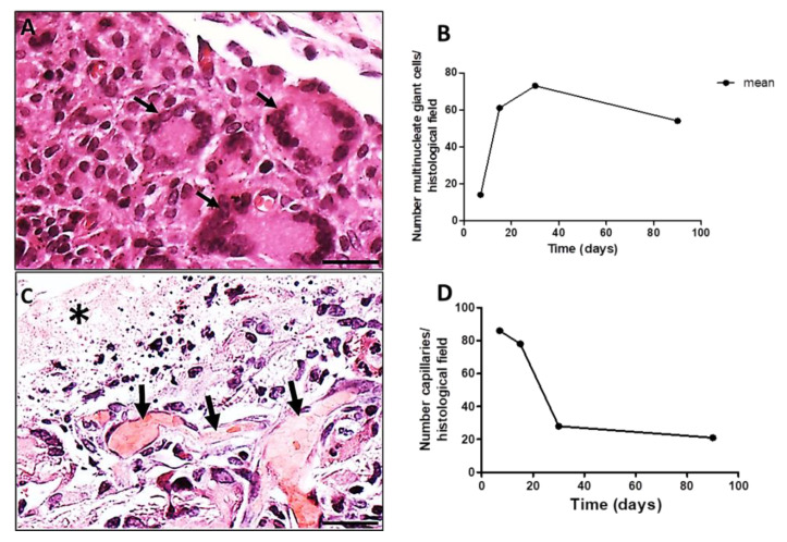
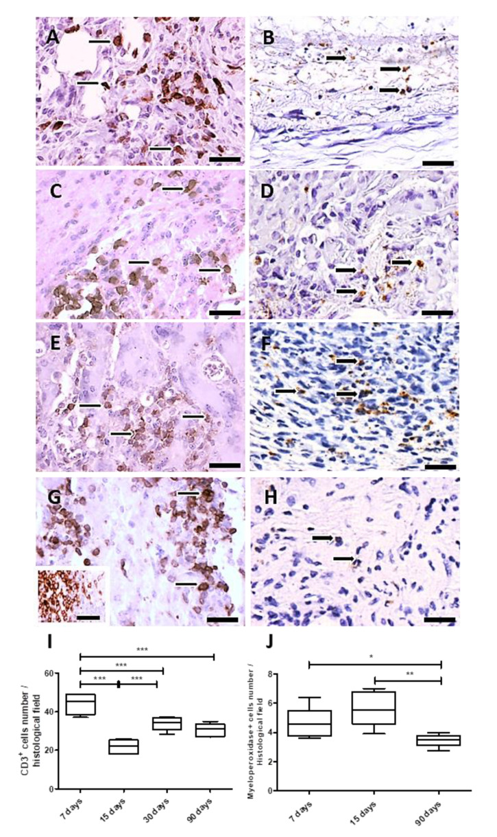
Random electrospun three-dimensional fiber membranes mimic the extracellular matrix and the interfibrillar spaces promotes the flow of nutrients for cells. Electrospun PLGA membranes were analyzed in vitro and in vivo after being sterilized with gamma radiation and bioactivated with fibronectin or collagen. Madin-Darby Canine Kidney (MDCK) epithelial cells and primary fibroblast-like cells from hamster's cheek paunch proliferated over time on these membranes, evidencing their good biocompatibility. Cell-free irradiated PLGA membranes implanted on the back of hamsters resulted in a chronic granulomatous inflammatory response, observed after 7, 15, 30 and 90 days. Morphological analysis of implanted PLGA using light microscopy revealed epithelioid cells, Langhans type of multinucleate giant cells (LCs) and multinucleated giant cells (MNGCs) with internalized biomaterial. Lymphocytes increased along time due to undegraded polymer fragments, inducing the accumulation of cells of the phagocytic lineage, and decreased after 90 days post implantation. Myeloperoxidase cells increased after 15 days and decreased after 90 days. LCs, MNGCs and capillaries decreased after 90 days. Analysis of implanted PLGA after 7, 15, 30 and 90 days using transmission electron microscope (TEM) showed cells exhibiting internalized PLGA fragments and filopodia surrounding PLGA fragments. Over time, TEM analysis showed less PLGA fragments surrounded by cells without fibrous tissue formation. Accordingly, MNGC constituted a granulomatous reaction around the polymer, which resolves with time, probably preventing a fibrous capsule formation. Finally, this study confirms the biocompatibility of electrospun PLGA membranes and their potential to accelerate the healing process of oral ulcerations in hamsters' model in association with autologous cells.
随机电纺三维纤维膜模拟细胞外基质,纤维间空间促进细胞营养物质流动。电纺聚乳酸-羟基乙酸共聚物(PLGA)膜经γ射线灭菌并用纤连蛋白或胶原蛋白进行生物活化后,进行了体外和体内分析。Madin-Darby犬肾(MDCK)上皮细胞和来自仓鼠颊囊的原代成纤维样细胞在这些膜上随时间增殖,证明了它们良好的生物相容性。植入仓鼠背部的无细胞辐照PLGA膜在7天、15天、30天和90天后出现慢性肉芽肿性炎症反应。使用光学显微镜对植入的PLGA进行形态学分析,发现有上皮样细胞、朗汉斯型多核巨细胞(LCs)和内化生物材料的多核巨细胞(MNGCs)。由于未降解的聚合物片段,淋巴细胞随时间增加,诱导吞噬细胞系细胞积累,并在植入后90天减少。髓过氧化物酶细胞在15天后增加,90天后减少。90天后LCs、MNGCs和毛细血管减少。使用透射电子显微镜(TEM)在7天、15天、30天和90天后对植入的PLGA进行分析,显示细胞内有内化的PLGA片段,丝状伪足围绕着PLGA片段。随着时间的推移,TEM分析显示被细胞包围的PLGA片段减少,无纤维组织形成。因此,MNGC在聚合物周围形成肉芽肿反应,随时间消退,可能阻止了纤维囊的形成。最后,本研究证实了电纺PLGA膜的生物相容性及其与自体细胞联合加速仓鼠口腔溃疡愈合过程的潜力。