Division of Engineering in Medicine, Department of Medicine, Harvard Medical School, and Brigham and Women's Hospital, Cambridge, Massachusetts 02139, United States.
Department of Biology, Main Campus, Khalifa University, Abu Dhabi 127788, United Arab Emirates.
ACS Appl Mater Interfaces. 2022 Nov 23;14(46):51602-51618. doi: 10.1021/acsami.2c12585. Epub 2022 Nov 8.
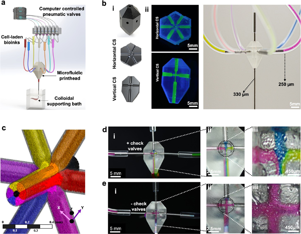
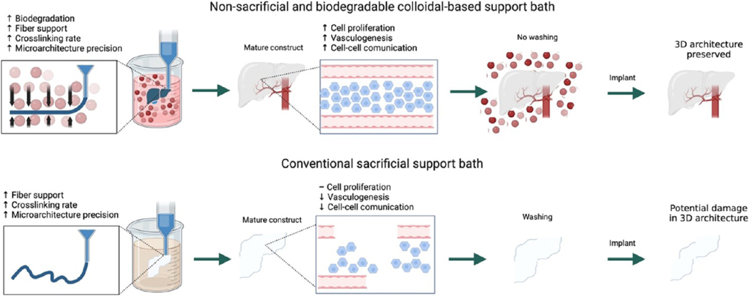
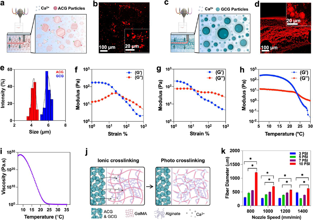
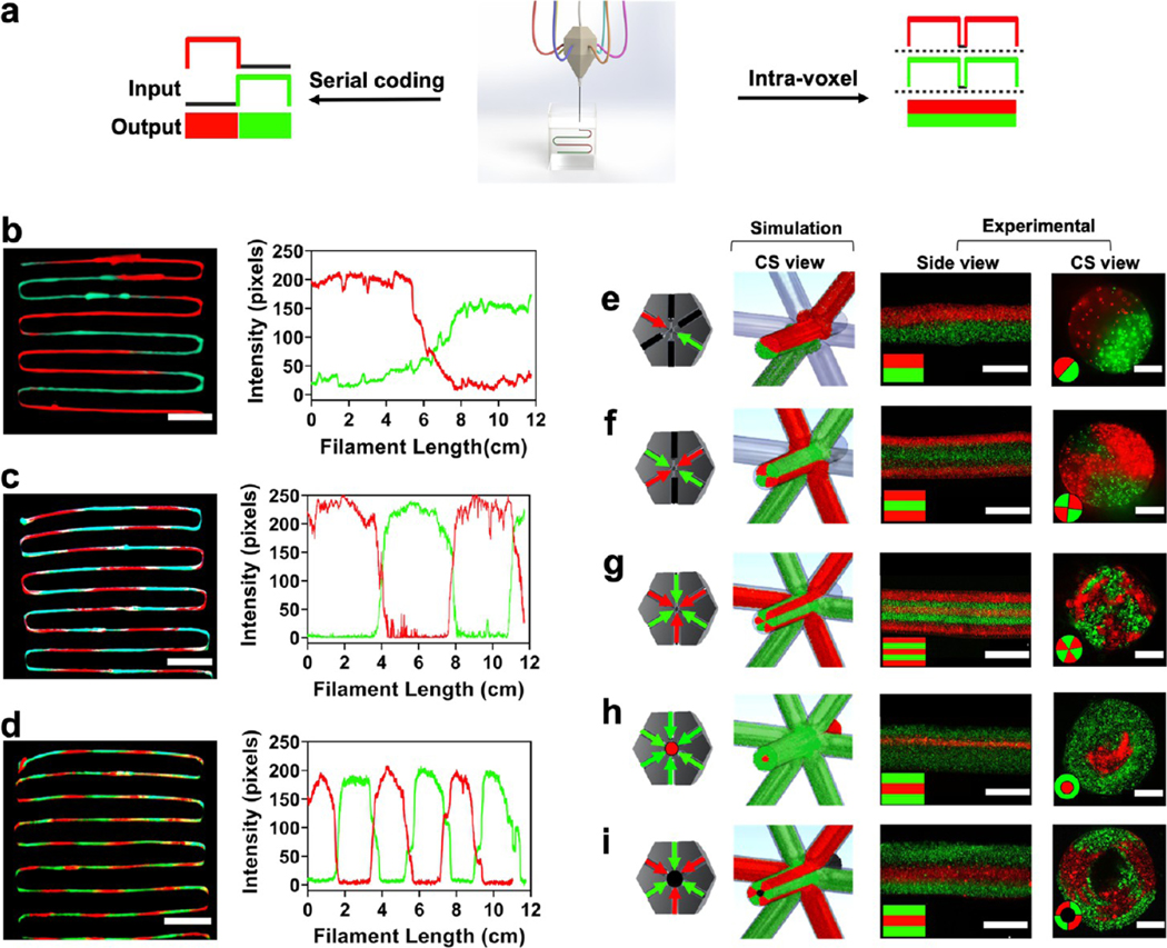
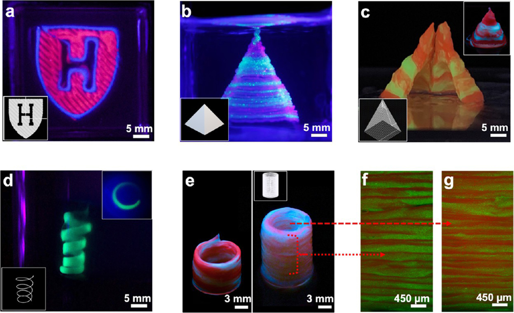
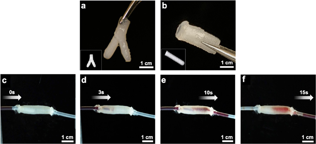
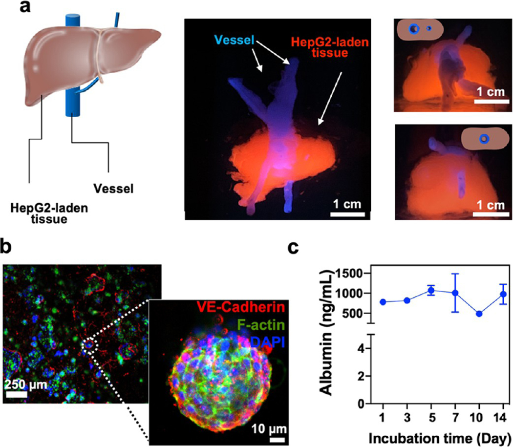
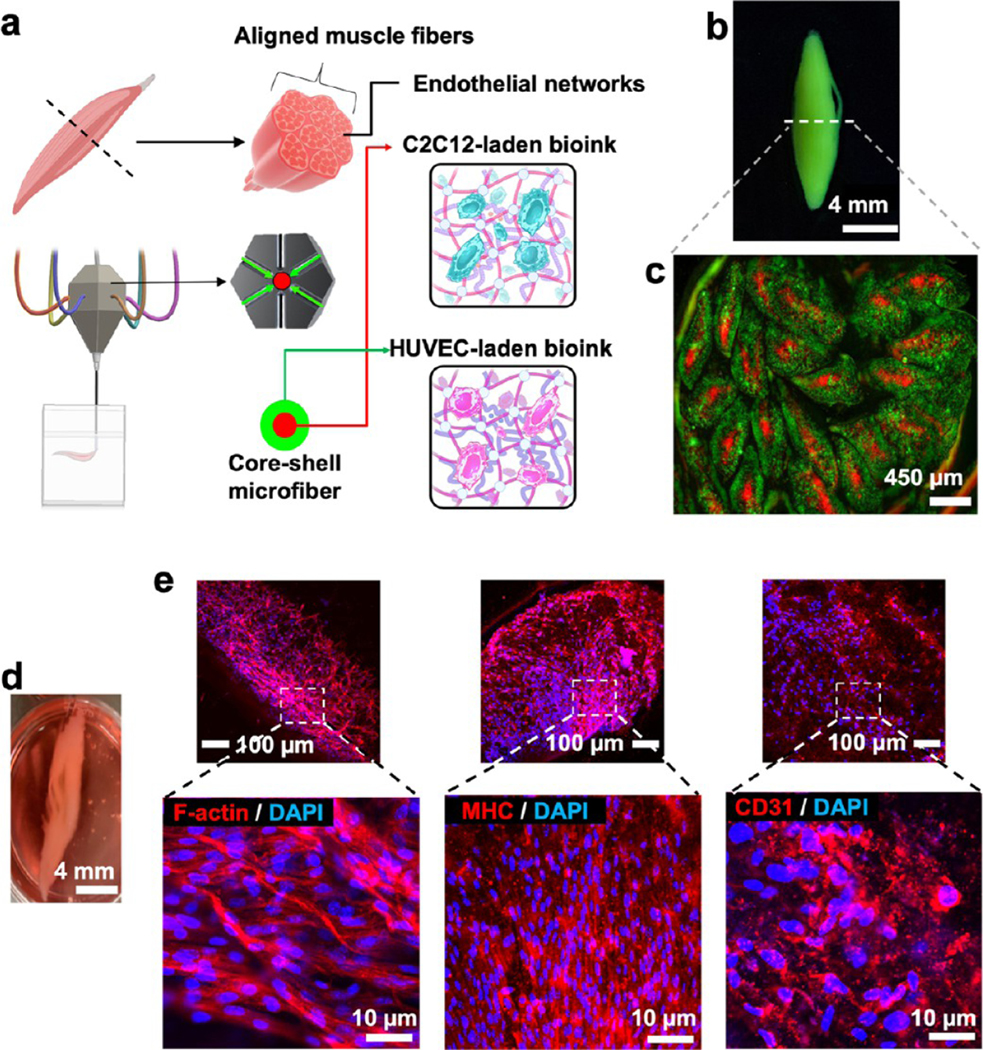
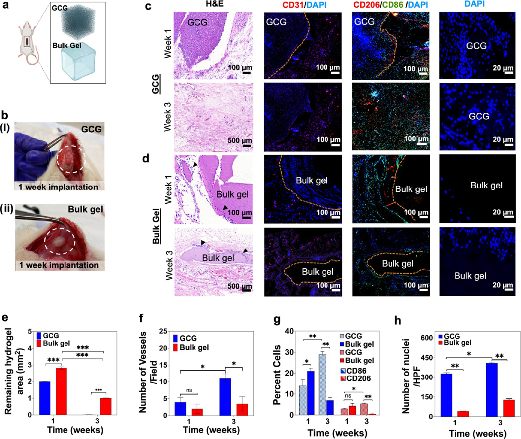
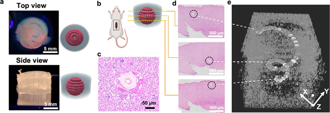
Recapitulating inherent heterogeneity and complex microarchitectures within confined print volumes for developing implantable constructs that could maintain their structure has remained challenging. Here, we present a combinational multimaterial and embedded bioprinting approach to fabricate complex tissue constructs that can be implanted postprinting and retain their three-dimensional (3D) shape . The microfluidics-based single nozzle printhead with computer-controlled pneumatic pressure valves enables laminar flow-based voxelation of up to seven individual bioinks with rapid switching between various bioinks that can solve alignment issues generated during switching multiple nozzles. To improve the spatial organization of various bioinks, printing fidelity with the -direction, and printing speed, self-healing and biodegradable colloidal gels as support baths are introduced to build complex geometries. Furthermore, the colloidal gels provide suitable microenvironments like native extracellular matrices (ECMs) for achieving cell growths and fast host cell invasion via interconnected microporous networks and . Multicompartment microfibers (, solid, core-shell, or donut shape), composed of two different bioink fractions with various lengths or their intravolume space filled by two, four, and six bioink fractions, are successfully printed in the ECM-like support bath. We also print various acellular complex geometries such as pyramids, spirals, and perfusable branched/linear vessels. Successful fabrication of vascularized liver and skeletal muscle tissue constructs show albumin secretion and bundled muscle mimic fibers, respectively. The interconnected microporous networks of colloidal gels result in maintaining printed complex geometries while enabling rapid cell infiltration, .
在受限的打印体积内重现内在异质性和复杂的微观结构,以开发能够维持其结构的可植入构建体,这一直是具有挑战性的。在这里,我们提出了一种组合多材料和嵌入式生物打印方法来制造可在打印后植入并保持其三维 (3D) 形状的复杂组织构建体。基于微流控的单喷嘴打印头带有计算机控制的气动压力阀,可实现多达七种单独生物墨水的层流分块,并且可以在各种生物墨水中快速切换,从而解决在切换多个喷嘴时产生的对准问题。为了提高各种生物墨水的空间组织,提高 - 方向的打印保真度和打印速度,引入自修复和可生物降解的胶体凝胶作为支撑浴来构建复杂的几何形状。此外,胶体凝胶通过相互连接的微孔网络提供类似于天然细胞外基质 (ECM) 的合适微环境,以实现细胞生长和快速宿主细胞入侵。由两种不同生物墨水分数组成的多腔微纤维(实心、核壳或环形),或其体积内用两种、四种和六种生物墨水分数填充的多腔微纤维,在 ECM 样支撑浴中成功打印。我们还打印了各种无细胞复杂几何形状,如金字塔、螺旋和可灌注的分支/线性血管。血管化肝脏和骨骼肌组织构建体的成功制造分别显示出白蛋白分泌和束状肌肉模拟纤维。胶体凝胶的互连微孔网络有助于保持打印的复杂几何形状,同时允许快速细胞渗透。