Department of Computer Science & Information Technology, Hazara University, Mansehra, Pakistan.
Department of Biomedical Engineering, College of Engineering, Princess Nourah bint Abdulrahman University, Riyadh, Saudi Arabia.
PLoS One. 2022 Nov 10;17(11):e0275781. doi: 10.1371/journal.pone.0275781. eCollection 2022.
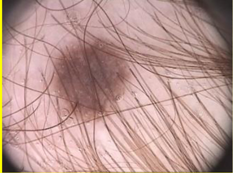
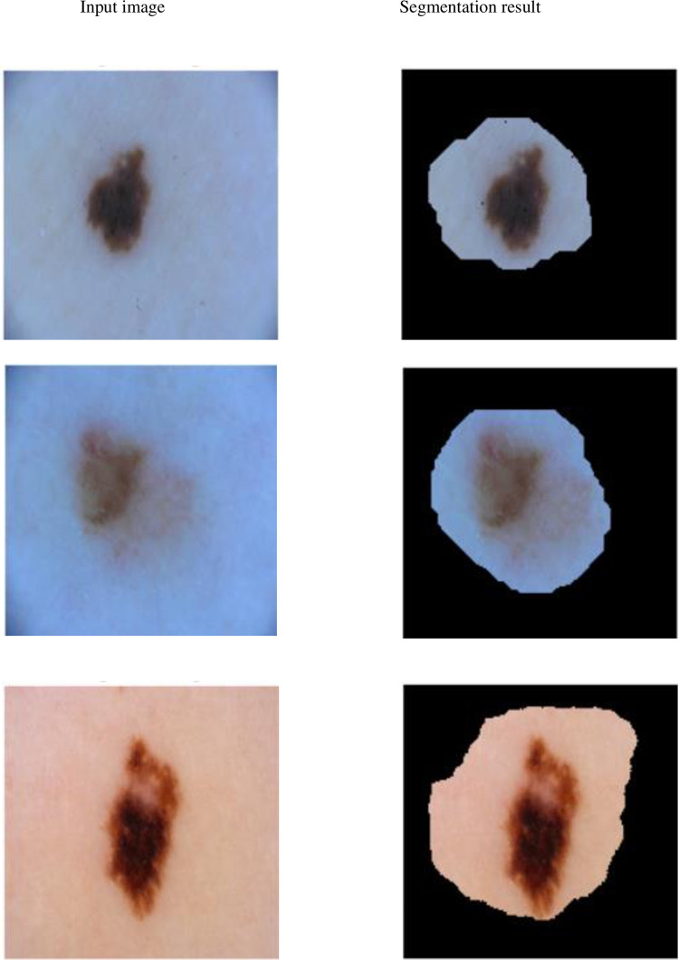
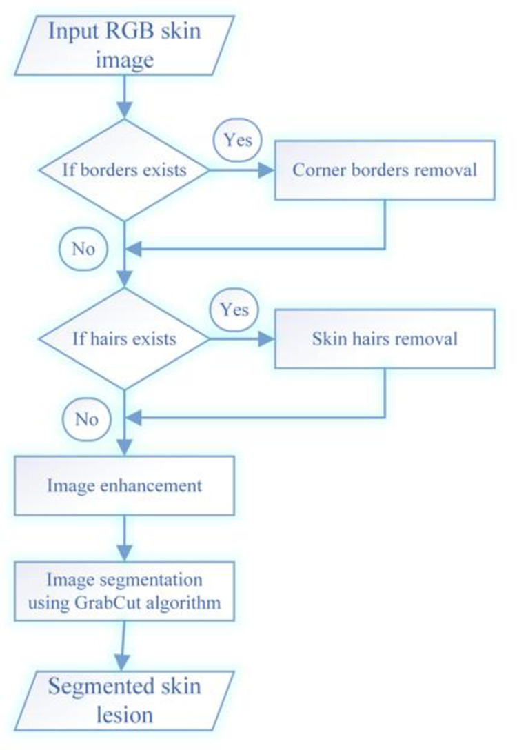
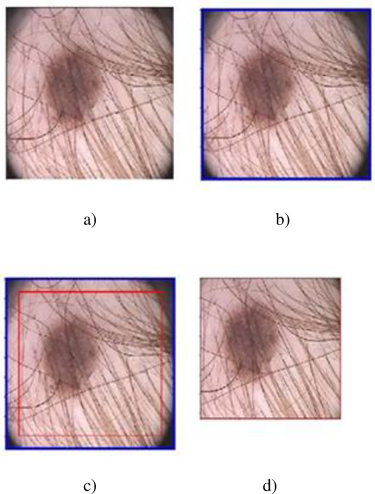
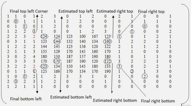
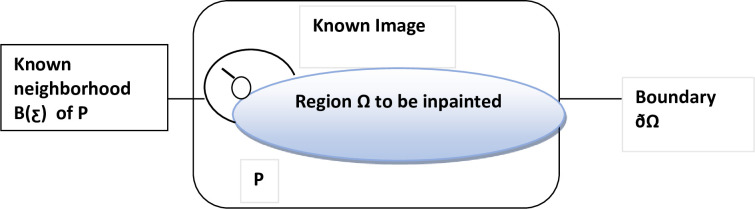
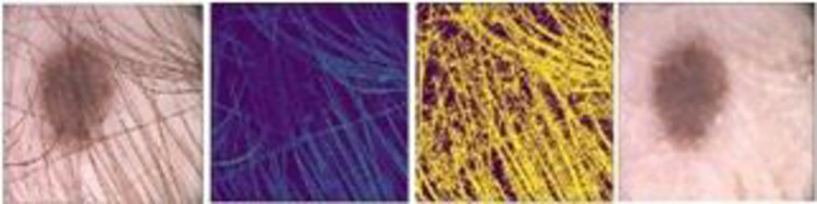
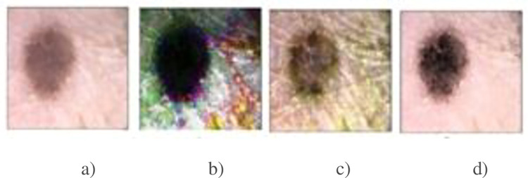
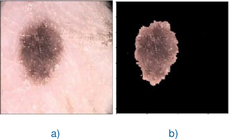
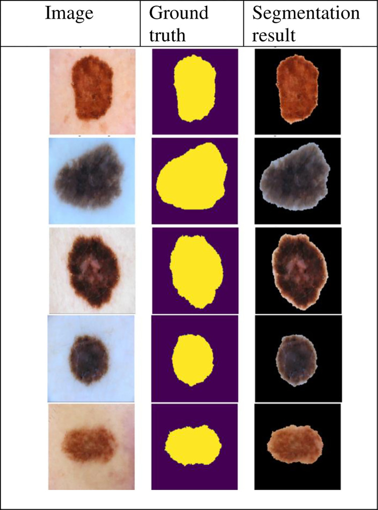
The effective segmentation of lesion(s) from dermoscopic skin images assists the Computer-Aided Diagnosis (CAD) systems in improving the diagnosing rate of skin cancer. The results of the existing skin lesion segmentation techniques are not up to the mark for dermoscopic images with artifacts like varying size corner borders with color similar to lesion(s) and/or hairs having low contrast with surrounding background. To improve the results of the existing skin lesion segmentation techniques for such kinds of dermoscopic images, an effective skin lesion segmentation method is proposed in this research work. The proposed method searches for the presence of corner borders in the given dermoscopc image and removes them if found otherwise it starts searching for the presence of hairs on it and eliminate them if present. Next, it enhances the resultant image using state-of-the-art image enhancement method and segments lesion from it using machine learning technique namely, GrabCut method. The proposed method was tested on PH2 and ISIC 2018 datasets containing 200 images each and its accuracy was measured with two evaluation metrics, i.e., Jaccard index, and Dice index. The evaluation results show that our proposed skin lesion segmentation method obtained Jaccard Index of 0.77, 0.80 and Dice index of 0.87, 0.82 values on PH2, and ISIC2018 datasets, respectively, which are better than state-of-the-art skin lesion segmentation techniques.
病变的有效分割(S)从皮肤镜图像协助计算机辅助诊断(CAD)系统提高皮肤癌的诊断率。现有的皮肤病变分割技术的结果对于具有伪影的皮肤镜图像(如具有与病变相似颜色的不同大小的角边界和/或与周围背景对比度低的毛发)并不理想。为了提高现有的皮肤病变分割技术对于这种类型的皮肤镜图像的结果,本研究提出了一种有效的皮肤病变分割方法。该方法在给定的皮肤镜图像中搜索角边界的存在,如果存在则将其删除,否则开始搜索其上毛发的存在,并将其消除。接下来,它使用最先进的图像增强方法增强所得图像,并使用机器学习技术(即GrabCut 方法)从其中分割病变。该方法在包含 200 张图像的 PH2 和 ISIC 2018 数据集上进行了测试,并使用两种评估指标,即 Jaccard 指数和骰子指数来衡量其准确性。评估结果表明,我们提出的皮肤病变分割方法在 PH2 和 ISIC2018 数据集上分别获得了 0.77、0.80 和 0.87、0.82 的 Jaccard 指数值,优于现有的皮肤病变分割技术。