Facultad de Farmacia y Bioquímica, Universidad de Buenos Aires, Junín 956, Piso 3°, Buenos Aires C1113AAD, Argentina.
Cátedra de Química Analítica Instrumental, Junín 956, Piso 3°, Buenos Aires C1113AAD, Argentina.
Int J Mol Sci. 2022 Nov 2;23(21):13379. doi: 10.3390/ijms232113379.
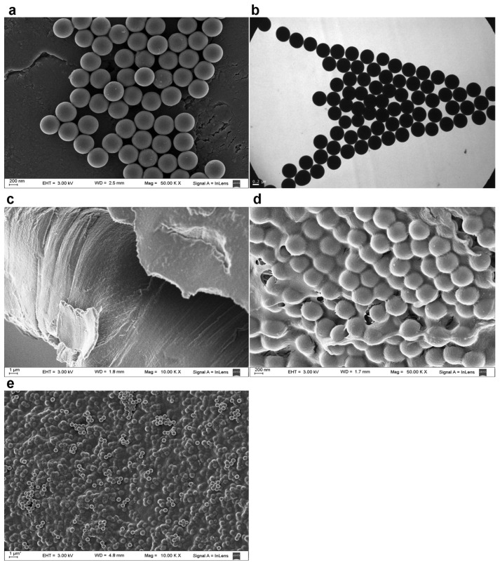
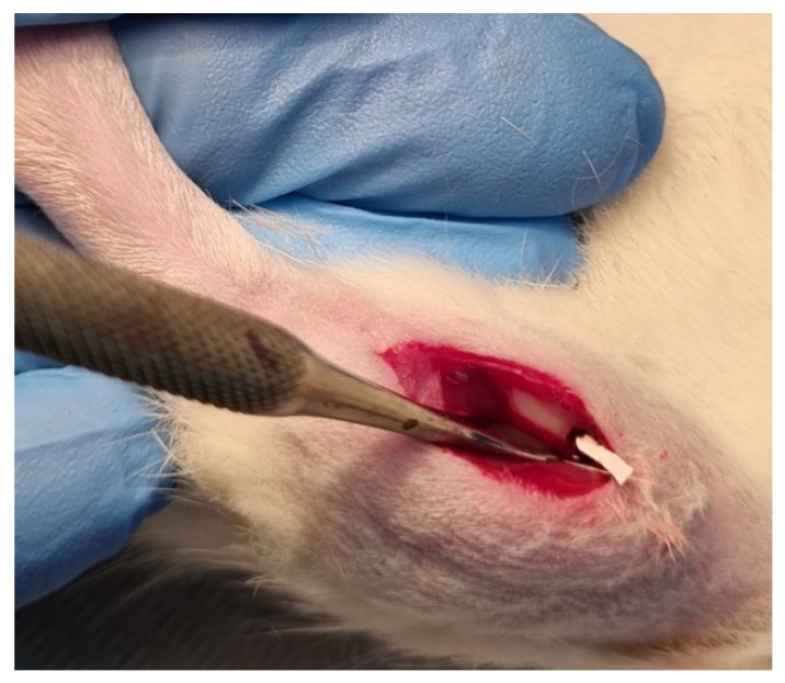
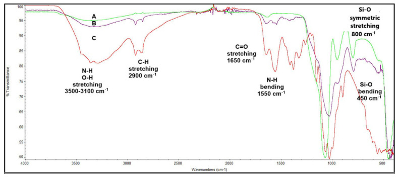
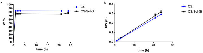
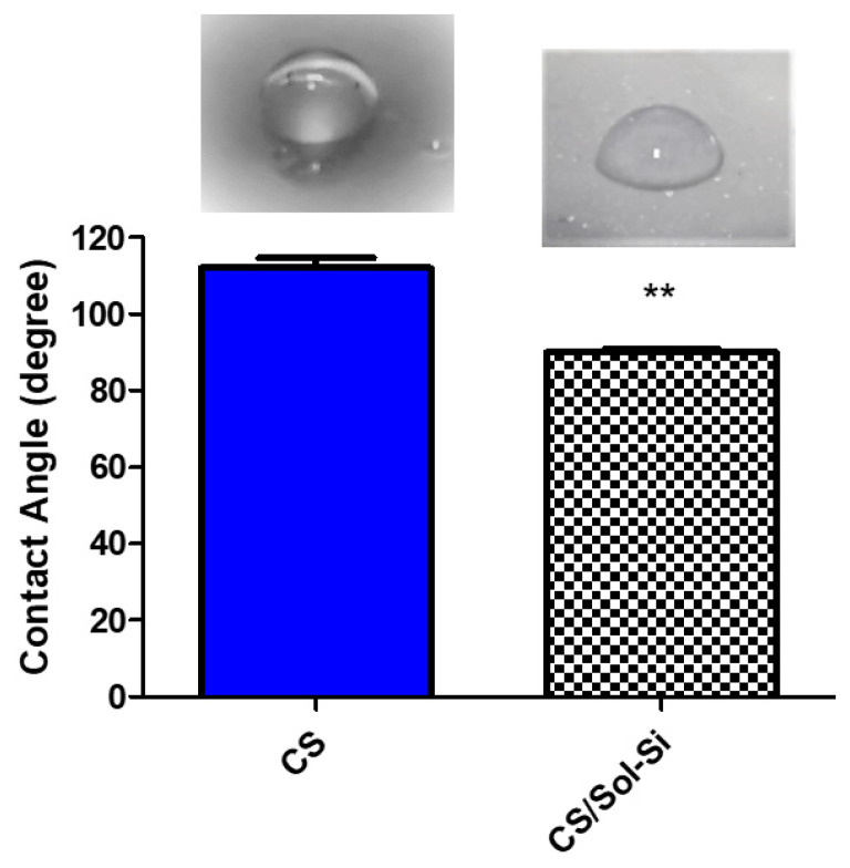
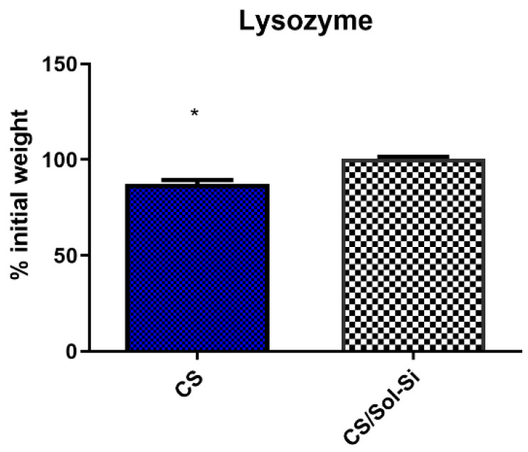
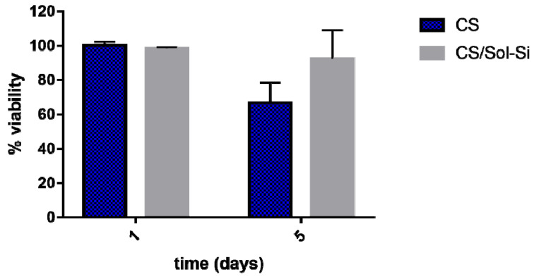
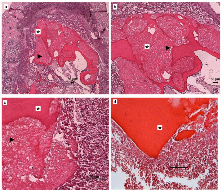
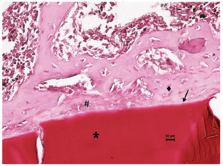
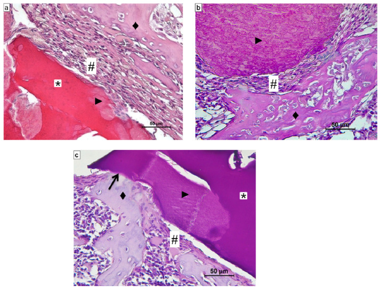
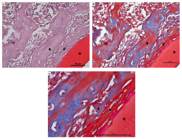
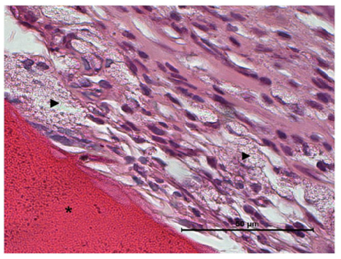
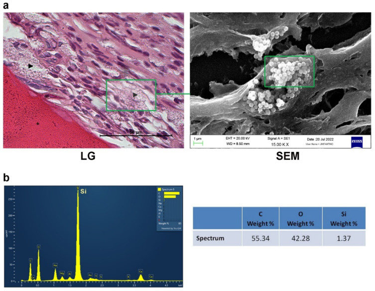
Bone defects have prompted the development of biomaterial-based bone substitutes for restoring the affected tissue completely. Although many biomaterials have been designed and evaluated, the combination of properties required in a biomaterial for bone tissue engineering still poses a challenge. In this study, a chitosan-silica-based biocomposite was synthetized, and its physicochemical characteristics and biocompatibility were characterized, with the aim of exploring the advantages and drawbacks of its use in bone tissue engineering. Dynamic light scattering measurements showed that the mean hydrodynamic size of solid silica particles (Sol-Si) was 482 ± 3 nm. Scanning electron microscopy of the biocomposite showed that Sol-Si were homogenously distributed within the chitosan (CS) matrix. The biocomposite swelled rapidly and was observed to have no cytotoxic effect on the [3T3] cell line within 24 h. Biocompatibility was also analyzed in vivo 14 days post-implant using a murine experimental model (Wistar rats). The biocomposite was implanted in the medullary compartment of both tibiae ( = 12). Histologically, no acute inflammatory infiltrate or multinucleated giant cells associated to the biocomposite were observed, indicating good biocompatibility. At the tissue-biocomposite interface, there was new formation of woven bone tissue in close contact with the biocomposite surface (osseointegration). The new bone formation may be attributed to the action of silica. Free silica particles originating from the biocomposite were observed at the tissue-biocomposite interface. According to our results, the biocomposite may act as a template for cellular interactions and extracellular matrix formation, providing a structural support for new bone tissue formation. The CS/Sol-Si biocomposite may act as a Si reservoir, promoting new bone formation. A scaffold with these properties is essential for cell differentiation and filling a bone defect.
骨缺损促使人们开发基于生物材料的骨替代物来完全恢复受影响的组织。尽管已经设计和评估了许多生物材料,但对于骨组织工程用生物材料所需性能的结合仍然是一个挑战。在这项研究中,合成了一种壳聚糖-二氧化硅基生物复合材料,并对其理化特性和生物相容性进行了表征,旨在探索其在骨组织工程中的优缺点。动态光散射测量表明,固体二氧化硅颗粒(Sol-Si)的平均水动力粒径为 482±3nm。生物复合材料的扫描电子显微镜观察表明,Sol-Si 均匀分布在壳聚糖(CS)基质中。该生物复合材料迅速溶胀,在 24 小时内观察到对[3T3]细胞系无细胞毒性作用。在植入后 14 天使用小鼠实验模型(Wistar 大鼠)还进行了体内生物相容性分析。将生物复合材料植入双侧胫骨骨髓腔(n=12)。组织学观察结果表明,在生物复合材料界面处未观察到与生物复合材料相关的急性炎症浸润或多核巨细胞,表明具有良好的生物相容性。在组织-生物复合材料界面处,有新形成的编织骨组织与生物复合材料表面紧密接触(骨整合)。新骨形成可能归因于二氧化硅的作用。在组织-生物复合材料界面处观察到源自生物复合材料的游离二氧化硅颗粒。根据我们的结果,生物复合材料可能作为细胞相互作用和细胞外基质形成的模板,为新骨组织形成提供结构支撑。CS/Sol-Si 生物复合材料可以作为硅的储存库,促进新骨形成。具有这些特性的支架对于细胞分化和填充骨缺损至关重要。