• 文献检索
  • 文档翻译
  • 深度研究
  • 学术资讯
  • Suppr Zotero 插件Zotero 插件
  • 邀请有礼
  • 套餐&价格
  • 历史记录
应用&插件
Suppr Zotero 插件Zotero 插件浏览器插件Mac 客户端Windows 客户端微信小程序
定价
高级版会员购买积分包购买API积分包
服务
文献检索文档翻译深度研究API 文档MCP 服务
关于我们
关于 Suppr公司介绍联系我们用户协议隐私条款
关注我们

Suppr 超能文献

核心技术专利:CN118964589B侵权必究
粤ICP备2023148730 号-1Suppr @ 2026

文献检索

告别复杂PubMed语法,用中文像聊天一样搜索,搜遍4000万医学文献。AI智能推荐,让科研检索更轻松。

立即免费搜索

文件翻译

保留排版,准确专业,支持PDF/Word/PPT等文件格式,支持 12+语言互译。

免费翻译文档

深度研究

AI帮你快速写综述,25分钟生成高质量综述,智能提取关键信息,辅助科研写作。

立即免费体验

使用通用CZT系统通过快速全身Tc-HMDP SPECT/CT检测前列腺癌骨转移

Detection of prostate cancer bone metastases with fast whole-body Tc-HMDP SPECT/CT using a general-purpose CZT system.

作者信息

Arvola Samuli, Seppänen Marko, Timonen Kirsi L, Rautio Pentti, Ettala Otto, Anttinen Mikael, Boström Peter J, Noponen Tommi

机构信息

Department of Clinical Physiology, Nuclear Medicine and Turku PET Centre, Turku University Hospital and University of Turku, Kiinamyllynkatu 4-8, P.O. Box 52, 20521, Turku, Finland.

Department of Clinical Physiology and Nuclear Medicine, Hospital Nova of Central Finland, Jyväskylä, Finland.

出版信息

EJNMMI Phys. 2022 Dec 12;9(1):85. doi: 10.1186/s40658-022-00517-4.

DOI:10.1186/s40658-022-00517-4
PMID:36508016
原文链接:https://pmc.ncbi.nlm.nih.gov/articles/PMC9743860/
Abstract

BACKGROUND

We evaluated the effects of acquisition time, energy window width, and matrix size on the image quality, quantitation, and diagnostic performance of whole-body Tc-HMDP SPECT/CT in the primary metastasis staging of prostate cancer.

METHODS

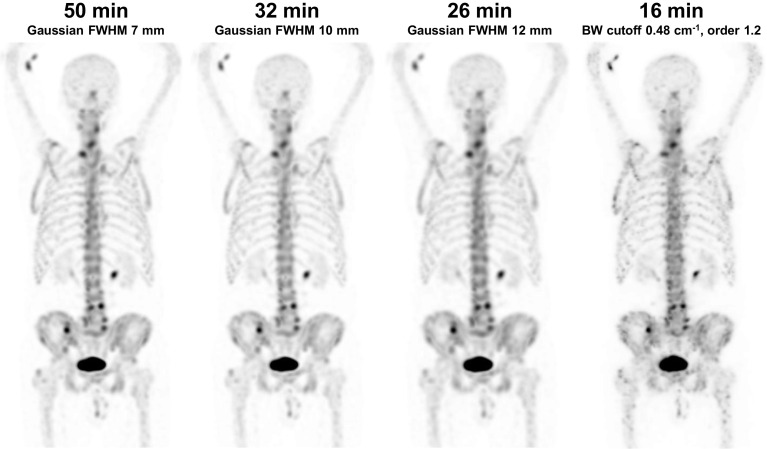
Thirty prostate cancer patients underwent Tc-HMDP SPECT/CT from the top of the head to the mid-thigh using a Discovery NM/CT 670 CZT system with list-mode acquisition, 50-min acquisition time, 15% energy window width, and 128 × 128 matrix size. The acquired list-mode data were resampled to produce data sets with shorter acquisition times of 41, 38, 32, 26, 20, and 16 min, narrower energy windows of 10, 8, 6, and 4%, and a larger matrix size of 256 × 256. Images were qualitatively evaluated by three experienced nuclear medicine physicians and quantitatively evaluated by noise, lesion contrast and SUV measurements. Diagnostic performance was evaluated from the readings of two experienced nuclear medicine physicians in terms of patient-, region-, and lesion-level sensitivity and specificity.

RESULTS

The originally acquired images had the best qualitative image quality and lowest noise. However, the acquisition time could be reduced to 38 min, the energy window narrowed to 8%, and the matrix size increased to 256 × 256 with still acceptable qualitative image quality. Lesion contrast and SUVs were not affected by changes in acquisition parameters. Acquisition time reduction had no effect on the diagnostic performance, as sensitivity, specificity, accuracy, and area under the receiver-operating characteristic curve were not significantly different between the 50-min and reduced acquisition time images. The average patient-level sensitivities of the two readers were 88, 92, 100, and 96% for the 50-, 32-, 26-, and 16-min images, respectively, and the corresponding specificities were 78, 84, 84, and 78%. The average region-level sensitivities of the two readers were 55, 58, 59, and 56% for the 50-, 32-, 26-, and 16-min images, respectively, and the corresponding specificities were 95, 98, 96, and 95%. The number of equivocal lesions tended to increase as the acquisition time decreased.

CONCLUSION

Whole-body Tc-HMDP SPECT/CT can be acquired using a general-purpose CZT system in less than 20 min without any loss in diagnostic performance in metastasis staging of high-risk prostate cancer patients.

摘要

背景

我们评估了采集时间、能量窗宽度和矩阵大小对全身Tc-HMDP SPECT/CT在前列腺癌原发转移分期中的图像质量、定量分析及诊断性能的影响。

方法

30例前列腺癌患者使用Discovery NM/CT 670 CZT系统进行从头至大腿中部的Tc-HMDP SPECT/CT检查,采用列表模式采集,采集时间为50分钟,能量窗宽度为15%,矩阵大小为128×128。将采集到的列表模式数据重新采样,以生成采集时间分别为41、38、32、26、20和16分钟的数据集,能量窗更窄,分别为10%、8%、6%和4%,以及矩阵大小为256×256的数据集。由三名经验丰富的核医学医师对图像进行定性评估,并通过噪声、病变对比度和SUV测量进行定量评估。由两名经验丰富的核医学医师根据患者、区域和病变水平的敏感性和特异性对诊断性能进行评估。

结果

最初采集的图像具有最佳的定性图像质量和最低的噪声。然而,采集时间可缩短至38分钟,能量窗可窄至8%,矩阵大小可增至256×256,而定性图像质量仍可接受。病变对比度和SUV不受采集参数变化的影响。采集时间的减少对诊断性能没有影响,因为50分钟和缩短采集时间的图像之间的敏感性、特异性、准确性和受试者操作特征曲线下面积没有显著差异。两位阅片者在50分钟、32分钟、26分钟和16分钟图像上的平均患者水平敏感性分别为88%、92%、100%和96%,相应的特异性分别为78%、84%、84%和78%。两位阅片者在50分钟、32分钟、26分钟和16分钟图像上的平均区域水平敏感性分别为55%、58%、59%和56%,相应的特异性分别为95%、98%、96%和95%。随着采集时间的减少,可疑病变的数量趋于增加。

结论

对于高危前列腺癌患者的转移分期,使用通用CZT系统在不到20分钟内即可完成全身Tc-HMDP SPECT/CT检查,且诊断性能不会有任何损失。

https://cdn.ncbi.nlm.nih.gov/pmc/blobs/2621/9743860/b99861ae858b/40658_2022_517_Fig2_HTML.jpg
https://cdn.ncbi.nlm.nih.gov/pmc/blobs/2621/9743860/1d8887e17ad2/40658_2022_517_Fig1_HTML.jpg
https://cdn.ncbi.nlm.nih.gov/pmc/blobs/2621/9743860/b99861ae858b/40658_2022_517_Fig2_HTML.jpg
https://cdn.ncbi.nlm.nih.gov/pmc/blobs/2621/9743860/1d8887e17ad2/40658_2022_517_Fig1_HTML.jpg
https://cdn.ncbi.nlm.nih.gov/pmc/blobs/2621/9743860/b99861ae858b/40658_2022_517_Fig2_HTML.jpg

相似文献

1
Detection of prostate cancer bone metastases with fast whole-body Tc-HMDP SPECT/CT using a general-purpose CZT system.使用通用CZT系统通过快速全身Tc-HMDP SPECT/CT检测前列腺癌骨转移
EJNMMI Phys. 2022 Dec 12;9(1):85. doi: 10.1186/s40658-022-00517-4.
2
More advantages in detecting bone and soft tissue metastases from prostate cancer using F-PSMA PET/CT.使用F-PSMA PET/CT检测前列腺癌骨和软组织转移方面有更多优势。
Hell J Nucl Med. 2019 Jan-Apr;22(1):6-9. doi: 10.1967/s002449910952. Epub 2019 Mar 7.
3
Evaluation of a new multipurpose whole-body CzT-based camera: comparison with a dual-head Anger camera and first clinical images.一种基于多用途全身CzT的新型相机的评估:与双头Anger相机的比较及首批临床图像
EJNMMI Phys. 2020 Mar 17;7(1):18. doi: 10.1186/s40658-020-0284-5.
4
A Prospective Comparison of F-prostate-specific Membrane Antigen-1007 Positron Emission Tomography Computed Tomography, Whole-body 1.5 T Magnetic Resonance Imaging with Diffusion-weighted Imaging, and Single-photon Emission Computed Tomography/Computed Tomography with Traditional Imaging in Primary Distant Metastasis Staging of Prostate Cancer (PROSTAGE).F-前列腺特异性膜抗原-1007 正电子发射断层扫描计算机断层扫描、全身 1.5T 磁共振成像弥散加权成像与传统成像单光子发射计算机断层扫描/计算机断层扫描在前列腺癌原发远处转移分期(PROSTAGE)中的前瞻性比较
Eur Urol Oncol. 2021 Aug;4(4):635-644. doi: 10.1016/j.euo.2020.06.012. Epub 2020 Jul 13.
5
Comparison of hybrid Ga-PSMA-PET/CT and Tc-DPD-SPECT/CT for the detection of bone metastases in prostate cancer patients: Additional value of morphologic information from low dose CT.镓-PSMA-PET/CT 与 Tc-DPD-SPECT/CT 比较在前列腺癌患者骨转移检测中的应用:低剂量 CT 形态学信息的附加价值。
Eur Radiol. 2018 Feb;28(2):610-619. doi: 10.1007/s00330-017-4994-6. Epub 2017 Aug 4.
6
(89)Sr bremsstrahlung single photon emission computed tomography using a gamma camera for bone metastases.(89)Sr 轫致辐射单光子发射计算机断层扫描,使用伽马相机检测骨转移。
Ann Nucl Med. 2014 Feb;28(2):112-9. doi: 10.1007/s12149-013-0788-3. Epub 2013 Dec 3.
7
Head-to-head comparison of Tc-PSMA and Tc-MDP SPECT/CT in diagnosing prostate cancer bone metastasis: a prospective, comparative imaging trial.Tc-PSMA 与 Tc-MDP SPECT/CT 对头对头比较诊断前列腺癌骨转移:一项前瞻性、对比成像试验。
Sci Rep. 2022 Sep 26;12(1):15993. doi: 10.1038/s41598-022-20280-x.
8
Skeletal standardized uptake values obtained by quantitative SPECT/CT as an osteoblastic biomarker for the discrimination of active bone metastasis in prostate cancer.通过定量SPECT/CT获得的骨骼标准化摄取值作为鉴别前列腺癌中活跃骨转移的成骨生物标志物。
Eur J Hybrid Imaging. 2017;1(1):2. doi: 10.1186/s41824-017-0006-y. Epub 2017 Oct 12.
9
Prospective evaluation of planar bone scintigraphy, SPECT, SPECT/CT, 18F-NaF PET/CT and whole body 1.5T MRI, including DWI, for the detection of bone metastases in high risk breast and prostate cancer patients: SKELETA clinical trial.对平面骨闪烁显像、单光子发射计算机断层扫描(SPECT)、SPECT/CT、18F-氟化钠正电子发射断层显像/计算机断层扫描(PET/CT)以及全身1.5T磁共振成像(MRI,包括扩散加权成像(DWI))用于检测高危乳腺癌和前列腺癌患者骨转移的前瞻性评估:SKELETA临床试验。
Acta Oncol. 2016;55(1):59-67. doi: 10.3109/0284186X.2015.1027411. Epub 2015 Apr 2.
10
Optimization of Tc whole-body SPECT/CT image quality: A phantom study.Tc 全身 SPECT/CT 图像质量优化:一项体模研究。
J Appl Clin Med Phys. 2022 Apr;23(4):e13528. doi: 10.1002/acm2.13528. Epub 2022 Jan 20.

引用本文的文献

1
A critical phantom study of the energy window used for Tc quantitative explorations with a ring CZT SPECT system.使用环形CZT SPECT系统对用于Tc定量探测的能量窗进行的关键体模研究。
EJNMMI Phys. 2025 Aug 20;12(1):79. doi: 10.1186/s40658-025-00793-w.
2
Quantitative capabilities of commercial CZT SPECT-CT cameras with Tc.配备锝的商用碲锌镉单光子发射计算机断层扫描-计算机断层扫描(CZT SPECT-CT)相机的定量能力。
EJNMMI Phys. 2025 May 10;12(1):44. doi: 10.1186/s40658-025-00754-3.
3
The SwiftScan step-and-shoot continuous mode improves SPECT scanning efficiency: a preliminary phantom and clinical test.

本文引用的文献

1
Ultra high speed SPECT bone imaging enabled by a deep learning enhancement method: a proof of concept.基于深度学习增强方法的超高速单光子发射计算机断层扫描骨成像:概念验证
EJNMMI Phys. 2022 Jun 13;9(1):43. doi: 10.1186/s40658-022-00472-0.
2
Comparison of reprojected bone SPECT/CT and planar bone scintigraphy for the detection of bone metastases in breast and prostate cancer.骨 SPECT/CT 与平面骨闪烁显像术在乳腺癌和前列腺癌骨转移检测中的比较。
Nucl Med Commun. 2022 May 1;43(5):510-517. doi: 10.1097/MNM.0000000000001536.
3
Optimization of Tc whole-body SPECT/CT image quality: A phantom study.
SwiftScan 步进式连续模式可提高单光子发射计算机断层扫描(SPECT)扫描效率:初步体模和临床测试
EJNMMI Phys. 2025 Jan 2;12(1):1. doi: 10.1186/s40658-024-00709-0.
4
Diagnostic Ability of Quantitative Parameters of Whole-Body Bone SPECT/CT Using a Full-Ring 360° Cadmium-Zinc-Telluride Camera for Detecting Bone Metastasis in Patients with Prostate Cancer.使用全环360°碲化镉锌相机的全身骨SPECT/CT定量参数对前列腺癌患者骨转移的诊断能力
Diagnostics (Basel). 2024 Dec 2;14(23):2714. doi: 10.3390/diagnostics14232714.
5
Letter to the Editor: EANM position paper on challenges and opportunities of full-ring 360° CZT bone imaging: it's time to let go of planar whole-body bone imaging.致编辑的信:欧洲核医学与分子影像学会(EANM)关于全环360°碲锌镉(CZT)骨显像的挑战与机遇的立场文件:是时候摒弃平面全身骨显像了。
Eur J Nucl Med Mol Imaging. 2025 Feb;52(3):806-807. doi: 10.1007/s00259-024-06984-4. Epub 2024 Nov 26.
6
Whole-Body SPECT/CT: Protocol Variation and Technical Consideration-A Narrative Review.全身SPECT/CT:协议差异与技术考量——一篇叙述性综述
Diagnostics (Basel). 2024 Aug 21;14(16):1827. doi: 10.3390/diagnostics14161827.
Tc 全身 SPECT/CT 图像质量优化:一项体模研究。
J Appl Clin Med Phys. 2022 Apr;23(4):e13528. doi: 10.1002/acm2.13528. Epub 2022 Jan 20.
4
Satisfied quantitative value can be acquired by short-time bone SPECT/CT using a whole-body cadmium-zinc-telluride gamma camera.使用全身碲化镉锌γ相机进行短时间骨SPECT/CT检查可获得满意的定量值。
Sci Rep. 2021 Dec 21;11(1):24320. doi: 10.1038/s41598-021-03853-0.
5
Feasibility of ultra-high-speed acquisition in xSPECT bone algorithm: a phantom study with advanced bone SPECT-specific phantom.超高速度采集在 xSPECT 骨算法中的可行性:使用先进的骨 SPECT 专用体模的体模研究。
Ann Nucl Med. 2022 Feb;36(2):183-190. doi: 10.1007/s12149-021-01689-2. Epub 2021 Nov 3.
6
Reduced acquisition time for thallium myocardial perfusion imaging with large field cadmium-zinc-telluride SPECT/CT cameras: An equivalence study.大视野镉锌碲硫(CdZnTe)SPECT/CT 相机进行铊心肌灌注成像的采集时间缩短:一项等效性研究。
J Nucl Cardiol. 2022 Aug;29(4):1933-1941. doi: 10.1007/s12350-021-02611-z. Epub 2021 Apr 22.
7
Experimental evaluation of the GE NM/CT 870 CZT clinical SPECT system equipped with WEHR and MEHRS collimator.配备 WEHR 和 MEHRS 准直器的 GE NM/CT 870 CZT 临床 SPECT 系统的实验评估。
J Appl Clin Med Phys. 2021 Feb;22(2):165-177. doi: 10.1002/acm2.13158. Epub 2021 Jan 10.
8
Quantitative bone SPECT/CT reconstruction utilizing anatomical information.利用解剖学信息的定量骨SPECT/CT重建。
EJNMMI Phys. 2021 Jan 6;8(1):2. doi: 10.1186/s40658-020-00348-1.
9
Diagnostic performance of whole-body SPECT/CT in bone metastasis detection using Tc-labelled diphosphate: a systematic review and meta-analysis.全身 SPECT/CT 探测 Tc 标记二膦酸盐骨转移的诊断性能:系统评价和荟萃分析。
Clin Radiol. 2020 Dec;75(12):961.e11-961.e24. doi: 10.1016/j.crad.2020.07.026. Epub 2020 Aug 28.
10
Inter- and intraobserver agreement in standard and ultra-fast single-photon emission computed tomography/computed tomography for the assessment of bone metastases.标准和超快速单光子发射计算机断层扫描/计算机断层扫描评估骨转移的观察者间和观察者内一致性。
Nucl Med Commun. 2020 Oct;41(10):1005-1009. doi: 10.1097/MNM.0000000000001252.