MOE Key Laboratory for Analytical Science of Food Safety and Biology, College of Chemistry, Fuzhou University, Fuzhou, 350108, P. R. China.
Department of Nuclear Medicine, Han Dan Central Hospital, Handan, Hebei, 056001, P. R. China.
Adv Sci (Weinh). 2023 Mar;10(8):e2202051. doi: 10.1002/advs.202202051. Epub 2023 Jan 22.
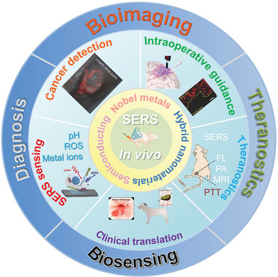
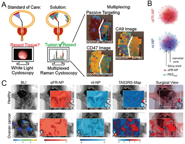
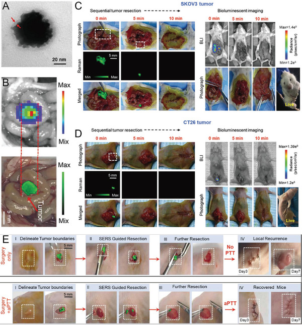
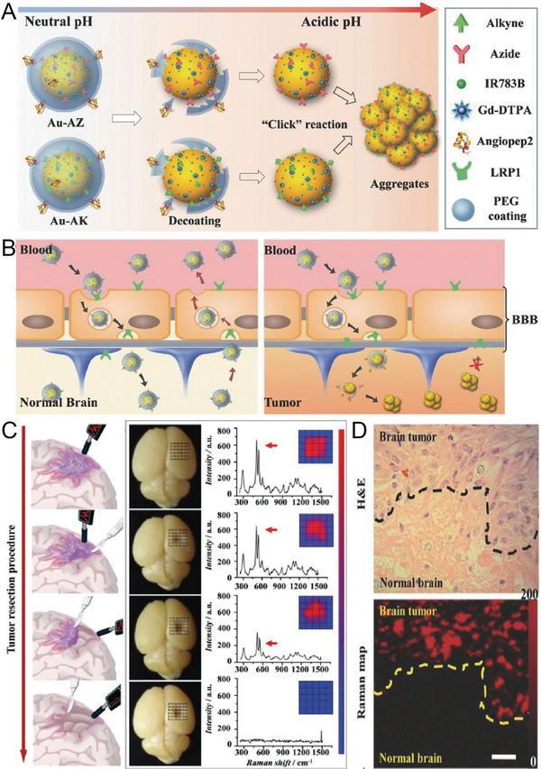
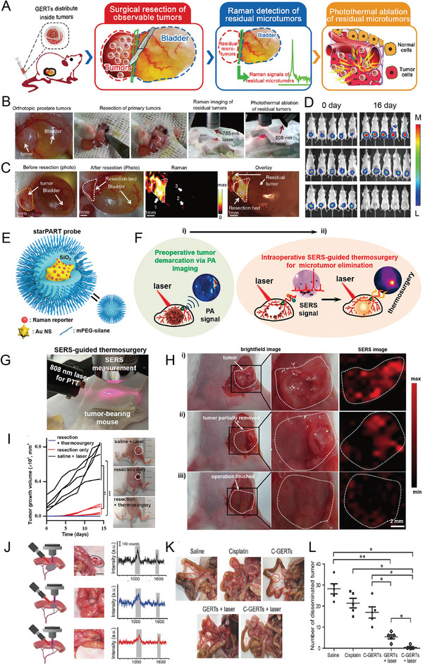
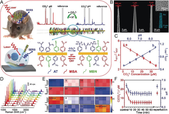
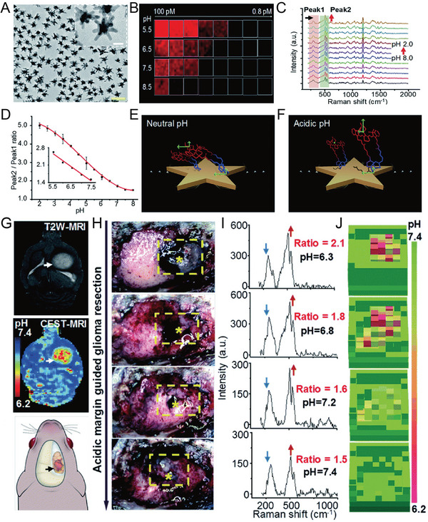
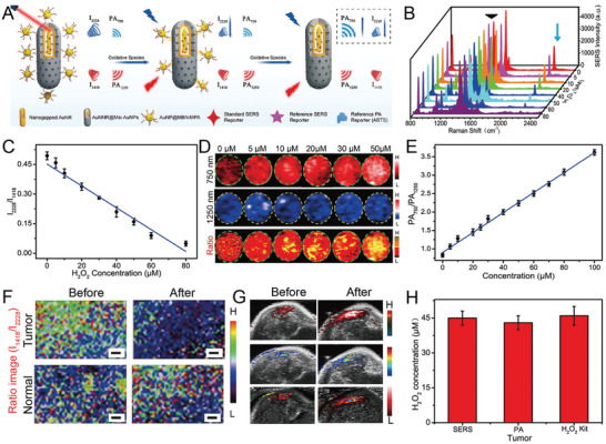
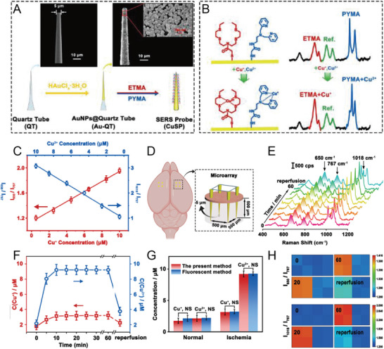
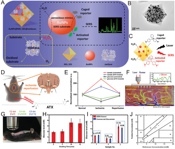
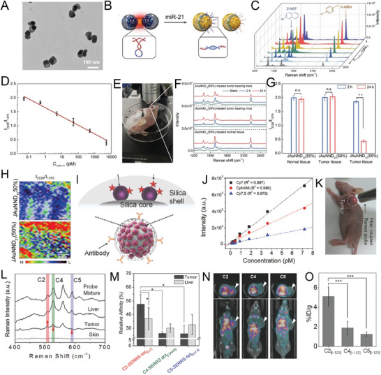
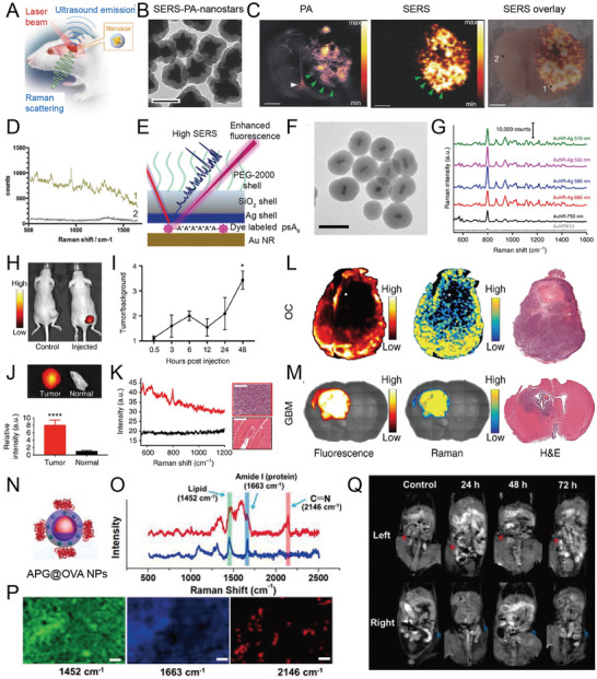
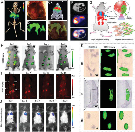
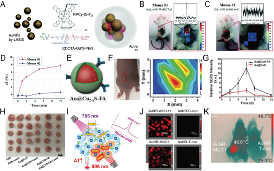
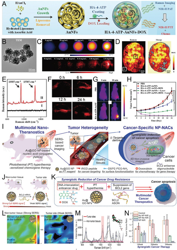
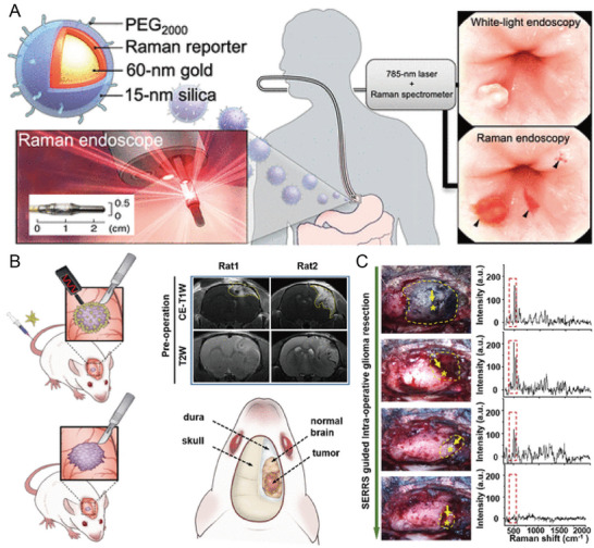
Surface-enhanced Raman scattering (SERS) is a feasible and ultra-sensitive method for biomedical imaging and disease diagnosis. SERS is widely applied to in vivo imaging due to the development of functional nanoparticles encoded by Raman active molecules (SERS nanoprobes) and improvements in instruments. Herein, the recent developments in SERS active materials and their in vivo imaging and biosensing applications are overviewed. Various SERS substrates that have been successfully used for in vivo imaging are described. Then, the applications of SERS imaging in cancer detection and in vivo intraoperative guidance are summarized. The role of highly sensitive SERS biosensors in guiding the detection and prevention of diseases is discussed in detail. Moreover, its role in the identification and resection of microtumors and as a diagnostic and therapeutic platform is also reviewed. Finally, the progress and challenges associated with SERS active materials, equipment, and clinical translation are described. The present evidence suggests that SERS could be applied in clinical practice in the future.
表面增强拉曼散射(SERS)是一种用于生物医学成像和疾病诊断的可行且超灵敏的方法。由于拉曼活性分子编码的功能纳米粒子(SERS 纳米探针)和仪器的改进,SERS 得到了广泛应用于活体成像。本文综述了 SERS 活性材料及其在活体成像和生物传感应用中的最新进展。描述了各种已成功用于活体成像的 SERS 基底。然后,总结了 SERS 成像在癌症检测和术中实时指导中的应用。详细讨论了高灵敏度 SERS 生物传感器在指导疾病检测和预防中的作用。此外,还回顾了其在识别和切除微肿瘤以及作为诊断和治疗平台方面的作用。最后,描述了 SERS 活性材料、设备和临床转化相关的进展和挑战。目前的证据表明,SERS 将来可能会应用于临床实践。