Department of Radiology, Baylor College of Medicine, One Baylor Plaza BCM360, TX, Houston, USA.
Department of Radiology, University of Utah, Salt Lake City, UT, USA.
J Digit Imaging. 2023 Jun;36(3):1279-1284. doi: 10.1007/s10278-023-00784-2. Epub 2023 Jan 30.
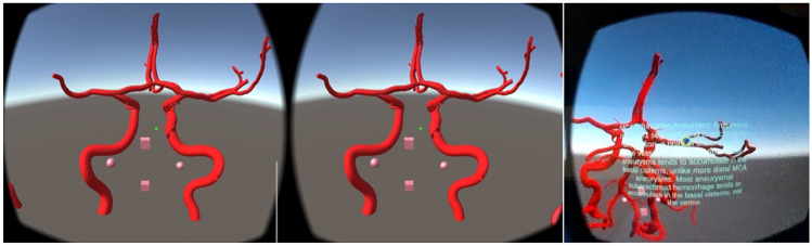
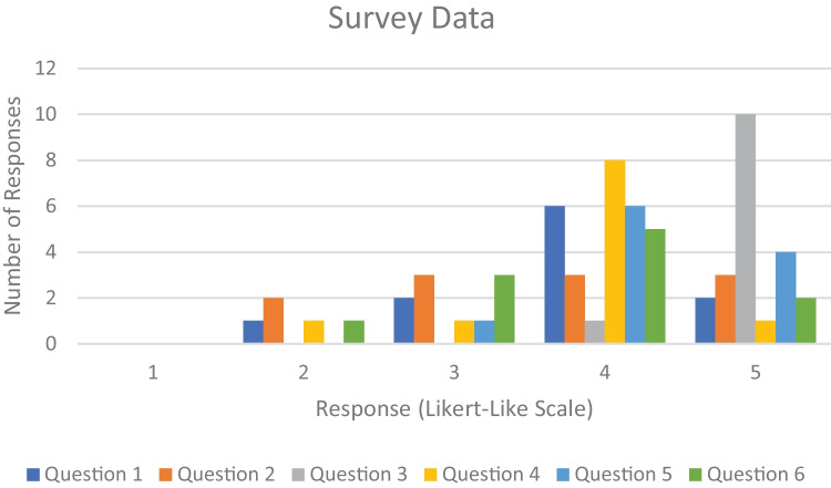
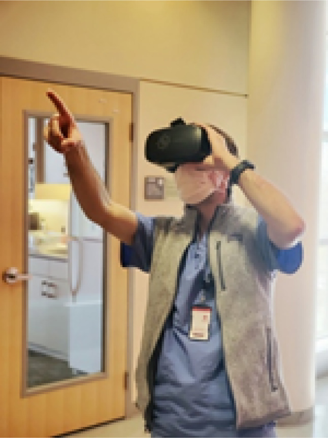
While radiological imaging is presented as two-dimensional images either on radiography or cross-sectional imaging, it is important for interpreters to understand three-dimensional anatomy and pathology. We hypothesized that virtual reality (VR) may serve as an engaging and effective way for trainees to learn to extrapolate from two-dimensional images to an understanding of these three-dimensional structures. We created a Google Cardboard Virtual Reality application that depicts intracranial vasculature and aneurysms. We then recruited 12 medical students to voluntarily participate in our study. The performance of the students in identifying intracranial aneurysms before and after the virtual reality training was evaluated and compared to a control group. While the experimental group's performance in correctly identifying aneurysms after virtual reality educational intervention was better than the control's (experimental increased by 5.3%, control decreased by 2.1%), the difference was not statistically significant (p-value of 0.06). Significantly, survey data from the medical students was very positive with students noting they preferred the immersive virtual reality training over conventional education and believed that VR would be a helpful educational tool for them in the future. We believe virtual reality can serve as an important tool to help radiology trainees better understand three-dimensional anatomy and pathology.
虽然放射影像学以二维图像的形式呈现,无论是放射摄影还是横截面成像,但对于解释器来说,理解三维解剖结构和病理学很重要。我们假设虚拟现实(VR)可能是一种吸引人且有效的方法,可以帮助学员从二维图像中推断出这些三维结构。我们创建了一个 Google Cardboard VR 应用程序,用于描绘颅内血管和动脉瘤。然后,我们招募了 12 名医学生自愿参加我们的研究。评估了学生在虚拟现实培训前后识别颅内动脉瘤的表现,并与对照组进行了比较。虽然实验组在虚拟现实教育干预后正确识别动脉瘤的表现优于对照组(实验组增加了 5.3%,对照组减少了 2.1%),但差异无统计学意义(p 值为 0.06)。值得注意的是,医学生的调查数据非常积极,学生们表示他们更喜欢沉浸式虚拟现实培训而不是传统教育,并认为 VR 将成为他们未来有帮助的教育工具。我们相信虚拟现实可以作为帮助放射科学员更好地理解三维解剖结构和病理学的重要工具。