• 文献检索
  • 文档翻译
  • 深度研究
  • 学术资讯
  • Suppr Zotero 插件Zotero 插件
  • 邀请有礼
  • 套餐&价格
  • 历史记录
应用&插件
Suppr Zotero 插件Zotero 插件浏览器插件Mac 客户端Windows 客户端微信小程序
定价
高级版会员购买积分包购买API积分包
服务
文献检索文档翻译深度研究API 文档MCP 服务
关于我们
关于 Suppr公司介绍联系我们用户协议隐私条款
关注我们

Suppr 超能文献

核心技术专利:CN118964589B侵权必究
粤ICP备2023148730 号-1Suppr @ 2026

文献检索

告别复杂PubMed语法,用中文像聊天一样搜索,搜遍4000万医学文献。AI智能推荐,让科研检索更轻松。

立即免费搜索

文件翻译

保留排版,准确专业,支持PDF/Word/PPT等文件格式,支持 12+语言互译。

免费翻译文档

深度研究

AI帮你快速写综述,25分钟生成高质量综述,智能提取关键信息,辅助科研写作。

立即免费体验

使用基于深度学习的不规则网络分割来改进黑色素瘤自动诊断

Improving Automatic Melanoma Diagnosis Using Deep Learning-Based Segmentation of Irregular Networks.

作者信息

Nambisan Anand K, Maurya Akanksha, Lama Norsang, Phan Thanh, Patel Gehana, Miller Keith, Lama Binita, Hagerty Jason, Stanley Ronald, Stoecker William V

机构信息

Electrical and Computer Engineering Department, Missouri University of Science and Technology, Rolla, MO 65409, USA.

Department of Biological Sciences, College of Arts, Sciences, and Education, Missouri University of Science and Technology, Rolla, MO 65409, USA.

出版信息

Cancers (Basel). 2023 Feb 16;15(4):1259. doi: 10.3390/cancers15041259.

DOI:10.3390/cancers15041259
PMID:36831599
原文链接:https://pmc.ncbi.nlm.nih.gov/articles/PMC9953766/
Abstract

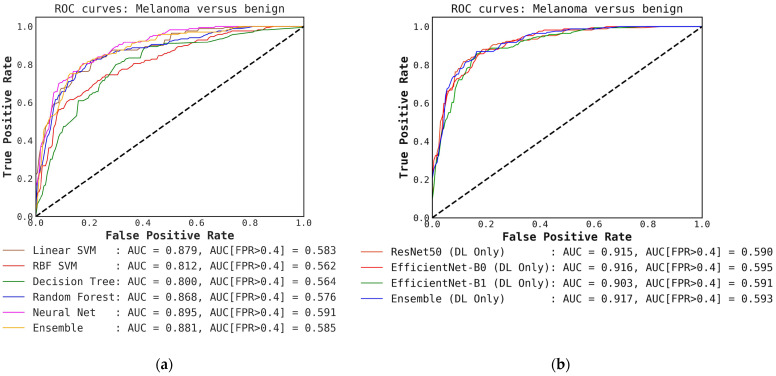
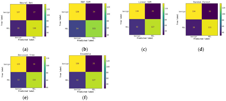
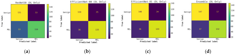
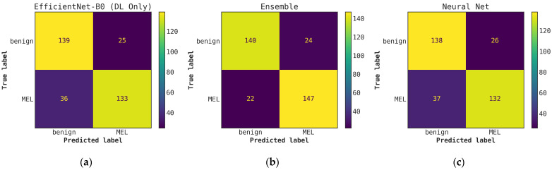
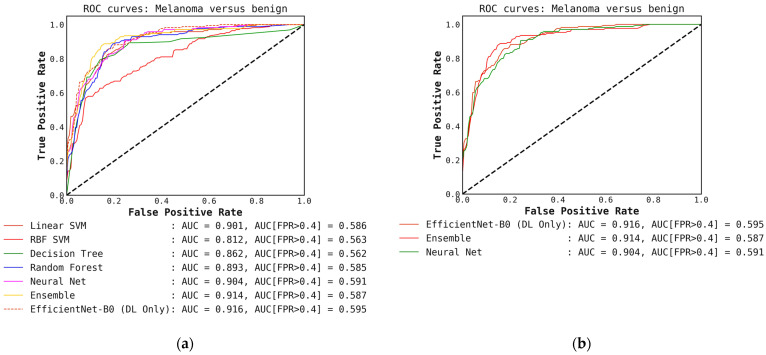
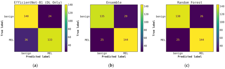
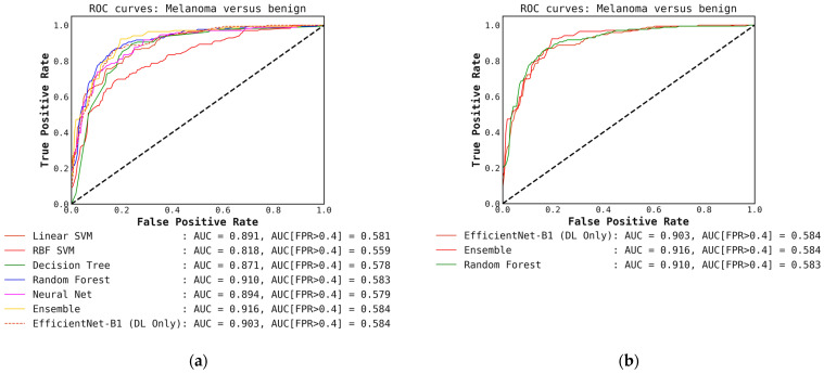
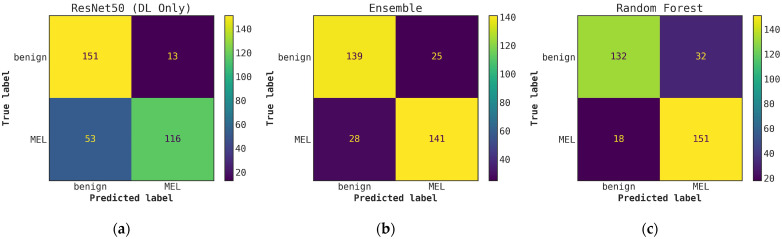
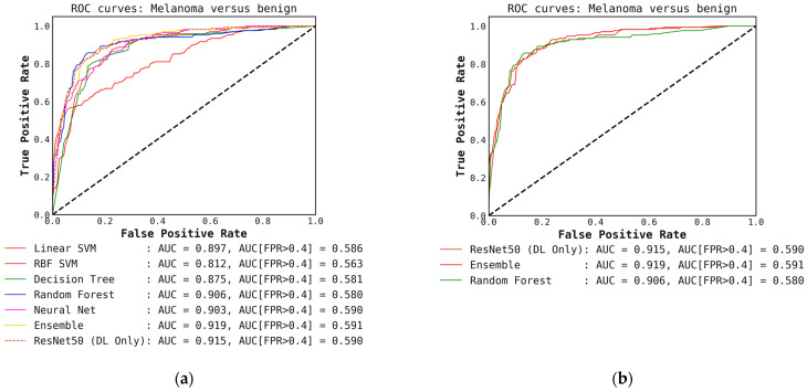
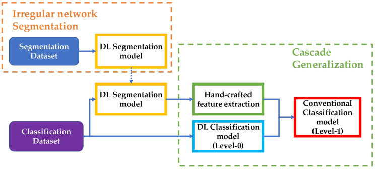
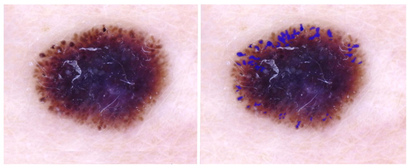
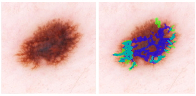
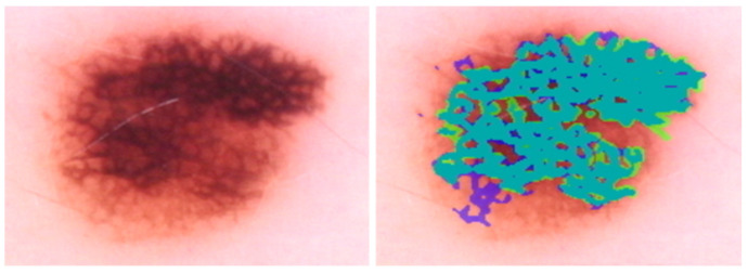
Deep learning has achieved significant success in malignant melanoma diagnosis. These diagnostic models are undergoing a transition into clinical use. However, with melanoma diagnostic accuracy in the range of ninety percent, a significant minority of melanomas are missed by deep learning. Many of the melanomas missed have irregular pigment networks visible using dermoscopy. This research presents an annotated irregular network database and develops a classification pipeline that fuses deep learning image-level results with conventional hand-crafted features from irregular pigment networks. We identified and annotated 487 unique dermoscopic melanoma lesions from images in the ISIC 2019 dermoscopic dataset to create a ground-truth irregular pigment network dataset. We trained multiple transfer learned segmentation models to detect irregular networks in this training set. A separate, mutually exclusive subset of the International Skin Imaging Collaboration (ISIC) 2019 dataset with 500 melanomas and 500 benign lesions was used for training and testing deep learning models for the binary classification of melanoma versus benign. The best segmentation model, U-Net++, generated irregular network masks on the 1000-image dataset. Other classical color, texture, and shape features were calculated for the irregular network areas. We achieved an increase in the recall of melanoma versus benign of 11% and in accuracy of 2% over DL-only models using conventional classifiers in a sequential pipeline based on the cascade generalization framework, with the highest increase in recall accompanying the use of the random forest algorithm. The proposed approach facilitates leveraging the strengths of both deep learning and conventional image processing techniques to improve the accuracy of melanoma diagnosis. Further research combining deep learning with conventional image processing on automatically detected dermoscopic features is warranted.

摘要

深度学习在恶性黑色素瘤诊断方面取得了显著成功。这些诊断模型正在向临床应用过渡。然而,由于黑色素瘤诊断准确率在90%左右,深度学习仍会遗漏相当一部分黑色素瘤。许多被遗漏的黑色素瘤在皮肤镜检查下可见不规则色素网络。本研究提出了一个带注释的不规则网络数据库,并开发了一种分类流程,将深度学习图像级结果与来自不规则色素网络的传统手工特征相融合。我们从国际皮肤影像协作组织(ISIC)2019皮肤镜数据集的图像中识别并注释了487个独特的皮肤镜下黑色素瘤病变,以创建一个真实的不规则色素网络数据集。我们训练了多个迁移学习分割模型,以在这个训练集中检测不规则网络。国际皮肤影像协作组织(ISIC)2019数据集的一个单独的、相互排斥的子集,包含500个黑色素瘤和500个良性病变,用于训练和测试深度学习模型,以进行黑色素瘤与良性病变的二元分类。最佳分割模型U-Net++在1000图像数据集上生成了不规则网络掩码。针对不规则网络区域计算了其他经典的颜色、纹理和形状特征。在基于级联泛化框架的顺序流程中,使用传统分类器,我们相对于仅使用深度学习的模型,黑色素瘤与良性病变的召回率提高了11%,准确率提高了2%,其中召回率的最大提高伴随着随机森林算法的使用。所提出的方法有助于利用深度学习和传统图像处理技术的优势,提高黑色素瘤诊断的准确性。有必要进一步开展将深度学习与传统图像处理相结合的研究,以自动检测皮肤镜特征。

https://cdn.ncbi.nlm.nih.gov/pmc/blobs/e8e2/9953766/9a18b44411eb/cancers-15-01259-g006.jpg
https://cdn.ncbi.nlm.nih.gov/pmc/blobs/e8e2/9953766/0ec6e0890098/cancers-15-01259-g0A1.jpg
https://cdn.ncbi.nlm.nih.gov/pmc/blobs/e8e2/9953766/c0ce8fa20abb/cancers-15-01259-g0A2.jpg
https://cdn.ncbi.nlm.nih.gov/pmc/blobs/e8e2/9953766/f638d1447c8e/cancers-15-01259-g0A3.jpg
https://cdn.ncbi.nlm.nih.gov/pmc/blobs/e8e2/9953766/8e9b41654fb0/cancers-15-01259-g0A4.jpg
https://cdn.ncbi.nlm.nih.gov/pmc/blobs/e8e2/9953766/a6809a4be7a1/cancers-15-01259-g0A5.jpg
https://cdn.ncbi.nlm.nih.gov/pmc/blobs/e8e2/9953766/f74e988d73f8/cancers-15-01259-g0A6.jpg
https://cdn.ncbi.nlm.nih.gov/pmc/blobs/e8e2/9953766/3beae42b7258/cancers-15-01259-g0A7.jpg
https://cdn.ncbi.nlm.nih.gov/pmc/blobs/e8e2/9953766/77a85db67447/cancers-15-01259-g0A8.jpg
https://cdn.ncbi.nlm.nih.gov/pmc/blobs/e8e2/9953766/387262d36e39/cancers-15-01259-g0A9.jpg
https://cdn.ncbi.nlm.nih.gov/pmc/blobs/e8e2/9953766/eae1882e830b/cancers-15-01259-g001.jpg
https://cdn.ncbi.nlm.nih.gov/pmc/blobs/e8e2/9953766/1d9d75e4a5ca/cancers-15-01259-g002.jpg
https://cdn.ncbi.nlm.nih.gov/pmc/blobs/e8e2/9953766/eb142976a461/cancers-15-01259-g003.jpg
https://cdn.ncbi.nlm.nih.gov/pmc/blobs/e8e2/9953766/5df19fc5c0ae/cancers-15-01259-g004.jpg
https://cdn.ncbi.nlm.nih.gov/pmc/blobs/e8e2/9953766/5619f94a10aa/cancers-15-01259-g005.jpg
https://cdn.ncbi.nlm.nih.gov/pmc/blobs/e8e2/9953766/9a18b44411eb/cancers-15-01259-g006.jpg

相似文献

1
Improving Automatic Melanoma Diagnosis Using Deep Learning-Based Segmentation of Irregular Networks.使用基于深度学习的不规则网络分割来改进黑色素瘤自动诊断
Cancers (Basel). 2023 Feb 16;15(4):1259. doi: 10.3390/cancers15041259.
2
Deep Learning Approaches Towards Skin Lesion Segmentation and Classification from Dermoscopic Images - A Review.深度学习方法在皮肤镜图像的皮肤损伤分割和分类中的应用综述。
Curr Med Imaging. 2020;16(5):513-533. doi: 10.2174/1573405615666190129120449.
3
Deep Learning Based Skin Lesion Segmentation and Classification of Melanoma Using Support Vector Machine (SVM).基于深度学习的皮肤病变分割以及使用支持向量机(SVM)对黑色素瘤进行分类
Asian Pac J Cancer Prev. 2019 May 25;20(5):1555-1561. doi: 10.31557/APJCP.2019.20.5.1555.
4
Automatic lesion segmentation using atrous convolutional deep neural networks in dermoscopic skin cancer images.基于空洞卷积深度神经网络的皮肤镜图像中自动病变分割。
BMC Med Imaging. 2022 May 29;22(1):103. doi: 10.1186/s12880-022-00829-y.
5
Melanoma Classification Using a Novel Deep Convolutional Neural Network with Dermoscopic Images.基于皮肤镜图像的新型深度卷积神经网络的黑色素瘤分类。
Sensors (Basel). 2022 Feb 2;22(3):1134. doi: 10.3390/s22031134.
6
Melanoma segmentation using deep learning with test-time augmentations and conditional random fields.使用带有测试时增强和条件随机场的深度学习进行黑色素瘤分割。
Sci Rep. 2022 Mar 10;12(1):3948. doi: 10.1038/s41598-022-07885-y.
7
HMA-Net: A deep U-shaped network combined with HarDNet and multi-attention mechanism for medical image segmentation.HMA-Net:一种结合 HarDNet 和多注意力机制的深度 U 形网络,用于医学图像分割。
Med Phys. 2023 Mar;50(3):1635-1646. doi: 10.1002/mp.16065. Epub 2022 Nov 3.
8
Multiple skin lesions diagnostics via integrated deep convolutional networks for segmentation and classification.基于集成深度卷积网络的多皮肤损伤诊断,用于分割和分类。
Comput Methods Programs Biomed. 2020 Jul;190:105351. doi: 10.1016/j.cmpb.2020.105351. Epub 2020 Jan 23.
9
Reducing the Impact of Confounding Factors on Skin Cancer Classification via Image Segmentation: Technical Model Study.通过图像分割减少混杂因素对皮肤癌分类的影响:技术模型研究。
J Med Internet Res. 2021 Mar 25;23(3):e21695. doi: 10.2196/21695.
10
Dermoscopy, with and without visual inspection, for diagnosing melanoma in adults.使用或不使用肉眼检查的皮肤镜检查在成人黑色素瘤诊断中的应用
Cochrane Database Syst Rev. 2018 Dec 4;12(12):CD011902. doi: 10.1002/14651858.CD011902.pub2.

引用本文的文献

1
Artificial Intelligence in the Non-Invasive Detection of Melanoma.人工智能在黑色素瘤的非侵入性检测中的应用
Life (Basel). 2024 Dec 4;14(12):1602. doi: 10.3390/life14121602.
2
A novel deep learning framework for accurate melanoma diagnosis integrating imaging and genomic data for improved patient outcomes.一种新颖的深度学习框架,用于准确诊断黑色素瘤,整合成像和基因组数据,以改善患者的治疗效果。
Skin Res Technol. 2024 Jun;30(6):e13770. doi: 10.1111/srt.13770.
3
Hybrid Topological Data Analysis and Deep Learning for Basal Cell Carcinoma Diagnosis.

本文引用的文献

1
BCN20000: Dermoscopic Lesions in the Wild.BCN20000:野外的皮肤镜病变。
Sci Data. 2024 Jun 17;11(1):641. doi: 10.1038/s41597-024-03387-w.
2
Cancer statistics, 2023.癌症统计数据,2023 年。
CA Cancer J Clin. 2023 Jan;73(1):17-48. doi: 10.3322/caac.21763.
3
ChimeraNet: U-Net for Hair Detection in Dermoscopic Skin Lesion Images.ChimeraNet:用于皮肤镜皮肤病变图像中毛发检测的 U-Net。
混合拓扑数据分析和深度学习在基底细胞癌诊断中的应用。
J Imaging Inform Med. 2024 Feb;37(1):92-106. doi: 10.1007/s10278-023-00924-8. Epub 2024 Jan 12.
4
DDCNN-F: double decker convolutional neural network 'F' feature fusion as a medical image classification framework.DDCNN-F:作为医学图像分类框架的双层卷积神经网络“F”特征融合
Sci Rep. 2024 Jan 5;14(1):676. doi: 10.1038/s41598-023-49721-x.
5
Leveraging Machine Learning for Accurate Detection and Diagnosis of Melanoma and Nevi: An Interdisciplinary Study in Dermatology.利用机器学习实现黑色素瘤和痣的准确检测与诊断:皮肤病学的跨学科研究
Cureus. 2023 Aug 25;15(8):e44120. doi: 10.7759/cureus.44120. eCollection 2023 Aug.
J Digit Imaging. 2023 Apr;36(2):526-535. doi: 10.1007/s10278-022-00740-6. Epub 2022 Nov 16.
4
An Updated Algorithm Integrated With Patient Data for the Differentiation of Atypical Nevi From Early Melanomas: the idScore 2021.一种结合患者数据用于鉴别非典型痣与早期黑色素瘤的更新算法:idScore 2021
Dermatol Pract Concept. 2022 Jul 1;12(3):e2022134. doi: 10.5826/dpc.1203a134. eCollection 2022 Jul.
5
Skin Cancer Classification With Deep Learning: A Systematic Review.基于深度学习的皮肤癌分类:一项系统综述。
Front Oncol. 2022 Jul 13;12:893972. doi: 10.3389/fonc.2022.893972. eCollection 2022.
6
BreastScreening-AI: Evaluating medical intelligent agents for human-AI interactions.BreastScreening-AI:评估用于人机交互的医学智能体。
Artif Intell Med. 2022 May;127:102285. doi: 10.1016/j.artmed.2022.102285. Epub 2022 Mar 29.
7
Cancer statistics, 2022.癌症统计数据,2022 年。
CA Cancer J Clin. 2022 Jan;72(1):7-33. doi: 10.3322/caac.21708. Epub 2022 Jan 12.
8
Analysis of the ISIC image datasets: Usage, benchmarks and recommendations.国际皮肤影像协作组(ISIC)图像数据集分析:用途、基准和建议。
Med Image Anal. 2022 Jan;75:102305. doi: 10.1016/j.media.2021.102305. Epub 2021 Nov 16.
9
Generalised Dice Overlap as a Deep Learning Loss Function for Highly Unbalanced Segmentations.广义骰子重叠作为高度不平衡分割的深度学习损失函数
Deep Learn Med Image Anal Multimodal Learn Clin Decis Support (2017). 2017;2017:240-248. doi: 10.1007/978-3-319-67558-9_28. Epub 2017 Sep 9.
10
Skin Cancer Detection: A Review Using Deep Learning Techniques.皮肤癌检测:深度学习技术的综述。
Int J Environ Res Public Health. 2021 May 20;18(10):5479. doi: 10.3390/ijerph18105479.