Department of Diagnostic and Interventional Radiology, Hannover Medical School, Hannover, Germany.
Department of Trauma Surgery, Hannover Medical School, Hannover, Germany.
Eur Radiol Exp. 2023 Mar 27;7(1):15. doi: 10.1186/s41747-023-00329-w.
Photon-counting detector computed tomography (PCD-CT) has the potential to provide superior image quality compared to energy-integrating detector computed tomography (EID-CT). We compared the two systems for elbow imaging in off-center arm positioning, 90° flexion, and cast fixation in a simulated post-trauma setting.
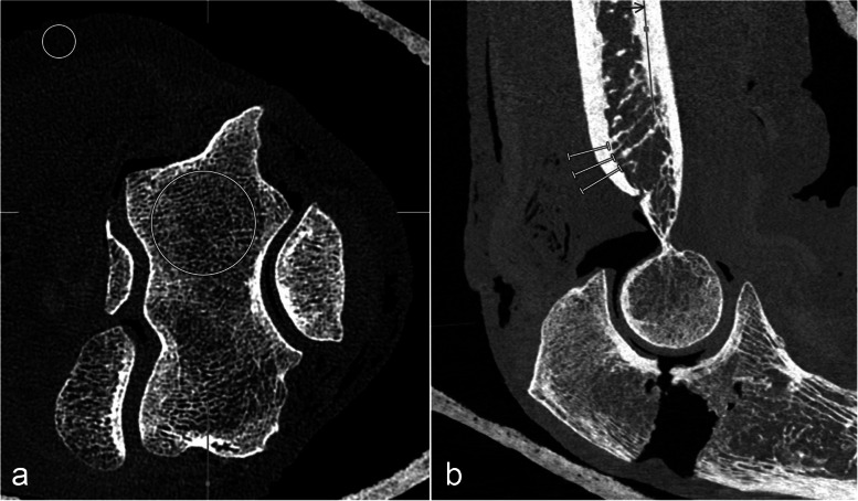
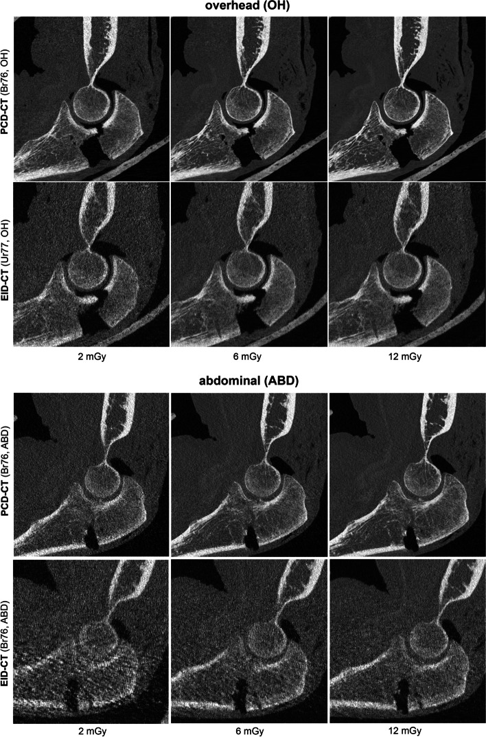
The institutional review board approved the study protocol. In a cadaver study, an olecranon fracture was artificially created in ten whole arm specimens. Two different scanning positions were evaluated: (a) arm overhead; and (b) arm on top of the abdomen of a whole-body phantom. The ultra-high resolution mode with three dose protocols and two reconstruction kernels was applied. Two blinded radiologists independently evaluated fracture and trabecular bone delineation. Signal-to-noise ratio (SNR), contrast-to-noise ratio (CNR), and cortical sharpness measurements were performed. Cohen κ correlations, Mann-Whitney U and Wilcoxon signed rank tests were used. A p value lower than 0.05 was considered statistically significant.
Dose-equivalent PCD-CT scans were rated better for fracture and trabecular bone evaluation (p < 0.001). SNR, CNR, and cortical sharpness were higher for all diagnostic (Br76) PCD-CT images (p < 0.001). The arm position had less effect on image quality in the PCD-CT compared to the EID-CT. The use of a sharp bone kernel (Br89) improved image quality ratings for PCD-CT. In the low-dose scan mode, PCD-CT resulted in more diagnostic scans (75%) compared to EID-CT (19%).
PCD-CT provided superior objective and subjective image quality for fracture and trabecular bone structures delineation of the elbow compared to EID-CT in a typical post-trauma setting.
• Photon-counting detector computed tomography (PCD-CT) preserved high image quality in elbow imaging with off-center positions. • PCD-CT was advantageous for bone evaluation in trauma elbows. • PCD-CT ultra-high-resolution mode and very sharp reconstruction kernels facilitated higher image quality.
与能量积分探测器计算机断层扫描(EID-CT)相比,光子计数探测器计算机断层扫描(PCD-CT)具有提供更好图像质量的潜力。我们比较了两种系统在模拟创伤后环境下的偏心手臂定位、90°弯曲和石膏固定的肘部成像。
机构审查委员会批准了研究方案。在一项尸体研究中,在十个完整手臂标本中人工创建尺骨鹰嘴骨折。评估了两种不同的扫描位置:(a)手臂上方;(b)全身体模腹部上方的手臂。应用超高分辨率模式和三种剂量方案及两种重建核。两位盲法放射科医生独立评估骨折和小梁骨勾画。进行了信噪比(SNR)、对比噪声比(CNR)和皮质锐度测量。使用 Cohen κ 相关、Mann-Whitney U 和 Wilcoxon 符号秩检验。p 值低于 0.05 被认为具有统计学意义。
剂量等效的 PCD-CT 扫描在骨折和小梁骨评估方面的评分更好(p<0.001)。所有诊断性(Br76)PCD-CT 图像的 SNR、CNR 和皮质锐度均较高(p<0.001)。与 EID-CT 相比,PCD-CT 中手臂位置对图像质量的影响较小。使用锐利骨核(Br89)可改善 PCD-CT 的图像质量评分。在低剂量扫描模式下,PCD-CT 产生的诊断性扫描(75%)多于 EID-CT(19%)。
在典型的创伤后环境中,与 EID-CT 相比,PCD-CT 为肘部骨折和小梁骨结构的勾画提供了更好的客观和主观图像质量。
• 光子计数探测器计算机断层扫描(PCD-CT)在偏心位置的肘部成像中保持了高图像质量。• PCD-CT 对创伤性肘部的骨评估有利。• PCD-CT 超高分辨率模式和非常锐利的重建核有助于提高图像质量。