• 文献检索
  • 文档翻译
  • 深度研究
  • 学术资讯
  • Suppr Zotero 插件Zotero 插件
  • 邀请有礼
  • 套餐&价格
  • 历史记录
应用&插件
Suppr Zotero 插件Zotero 插件浏览器插件Mac 客户端Windows 客户端微信小程序
定价
高级版会员购买积分包购买API积分包
服务
文献检索文档翻译深度研究API 文档MCP 服务
关于我们
关于 Suppr公司介绍联系我们用户协议隐私条款
关注我们

Suppr 超能文献

核心技术专利:CN118964589B侵权必究
粤ICP备2023148730 号-1Suppr @ 2026

文献检索

告别复杂PubMed语法,用中文像聊天一样搜索,搜遍4000万医学文献。AI智能推荐,让科研检索更轻松。

立即免费搜索

文件翻译

保留排版,准确专业,支持PDF/Word/PPT等文件格式,支持 12+语言互译。

免费翻译文档

深度研究

AI帮你快速写综述,25分钟生成高质量综述,智能提取关键信息,辅助科研写作。

立即免费体验

暴露磷脂酰丝氨酸的膜联蛋白A1阳性细胞外囊泡:潜在的癌症生物标志物

Phosphatidylserine-Exposing Annexin A1-Positive Extracellular Vesicles: Potential Cancer Biomarkers.

作者信息

Perez Gloria I, Bernard Matthew P, Vocelle Daniel, Zarea Ahmed A, Saleh Najla A, Gagea Matthew A, Schneider Doug, Bauzon Maxine, Hermiston Terry, Kanada Masamitsu

机构信息

Institute for Quantitative Health Science and Engineering (IQ), Michigan State University, East Lansing, MI 48824, USA.

College of Osteopathic Medicine, Michigan State University, East Lansing, MI 48824, USA.

出版信息

Vaccines (Basel). 2023 Mar 13;11(3):639. doi: 10.3390/vaccines11030639.

DOI:10.3390/vaccines11030639
PMID:36992223
原文链接:https://pmc.ncbi.nlm.nih.gov/articles/PMC10059271/
Abstract

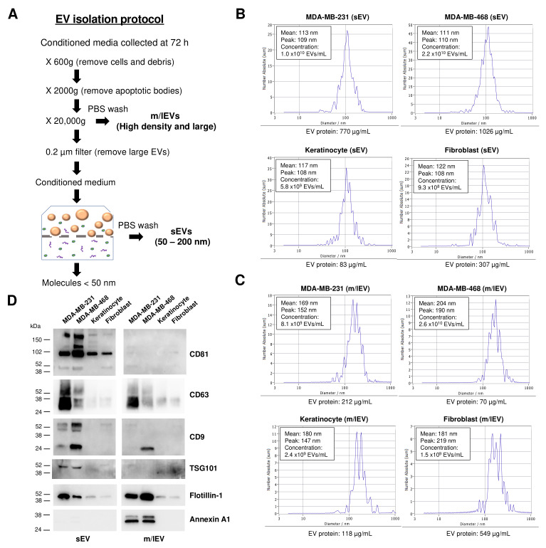
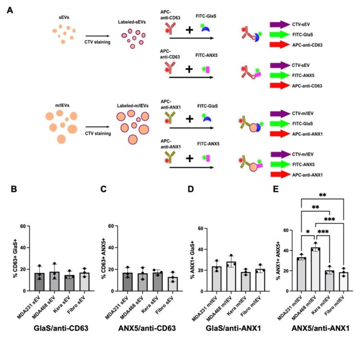
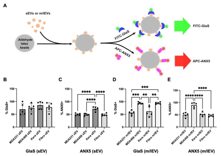
Under physiological conditions, phosphatidylserine (PS) predominantly localizes to the cytosolic leaflet of the plasma membrane of cells. During apoptosis, PS is exposed on the cell surface and serves as an "eat-me" signal for macrophages to prevent releasing self-immunogenic cellular components from dying cells which could potentially lead to autoimmunity. However, increasing evidence indicates that viable cells can also expose PS on their surface. Interestingly, tumor cell-derived extracellular vesicles (EVs) externalize PS. Recent studies have proposed PS-exposing EVs as a potential biomarker for the early detection of cancer and other diseases. However, there are confounding results regarding subtypes of PS-positive EVs, and knowledge of PS exposure on the EV surface requires further elucidation. In this study, we enriched small EVs (sEVs) and medium/large EVs (m/lEVs) from conditioned media of breast cancer cells (MDA-MB-231, MDA-MB-468) and non-cancerous cells (keratinocytes, fibroblasts). Since several PS-binding molecules are available to date, we compared recombinant proteins of annexin A5 and the carboxylated glutamic acid domain of Protein S (GlaS), also specific for PS, to detect PS-exposing EVs. Firstly, PS externalization in each EV fraction was analyzed using a bead-based EV assay, which combines EV capture using microbeads and analysis of PS-exposing EVs by flow cytometry. The bulk EV assay showed higher PS externalization in m/lEVs derived from MDA-MB-468 cells but not from MDA-MB-231 cells, while higher binding of GlaS was also observed in m/lEVs from fibroblasts. Second, using single EV flow cytometry, PS externalization was also analyzed on individual sEVs and m/lEVs. Significantly higher PS externalization was detected in m/lEVs (annexin A1) derived from cancer cells compared to m/lEVs (annexin A1) from non-cancerous cells. These results emphasize the significance of PS-exposing m/lEVs (annexin A1) as an undervalued EV subtype for early cancer detection and provide a better understanding of PS externalization in disease-associated EV subtypes.

摘要

在生理条件下,磷脂酰丝氨酸(PS)主要定位于细胞质膜的胞质小叶。在细胞凋亡过程中,PS暴露于细胞表面,并作为巨噬细胞的“吃掉我”信号,以防止死亡细胞释放可能导致自身免疫的自身免疫原性细胞成分。然而,越来越多的证据表明,活细胞也可以在其表面暴露PS。有趣的是,肿瘤细胞衍生的细胞外囊泡(EVs)会使PS外化。最近的研究提出,暴露PS的EVs作为癌症和其他疾病早期检测的潜在生物标志物。然而,关于PS阳性EVs亚型的结果存在混淆,并且对EV表面PS暴露的了解需要进一步阐明。在本研究中,我们从乳腺癌细胞(MDA-MB-231、MDA-MB-468)和非癌细胞(角质形成细胞、成纤维细胞)的条件培养基中富集了小EVs(sEVs)和中/大EVs(m/lEVs)。由于目前有几种PS结合分子,我们比较了膜联蛋白A5的重组蛋白和同样对PS具有特异性的蛋白S的羧化谷氨酸结构域(GlaS),以检测暴露PS的EVs。首先,使用基于磁珠的EV检测法分析每个EV组分中的PS外化,该方法结合了使用微珠捕获EV和通过流式细胞术分析暴露PS的EVs。大量EV检测显示,来自MDA-MB-468细胞而非MDA-MB-231细胞的m/lEVs中PS外化程度更高,同时在成纤维细胞的m/lEVs中也观察到GlaS的结合更高。其次,使用单个EV流式细胞术,还对单个sEVs和m/lEVs的PS外化进行了分析。与来自非癌细胞的m/lEVs(膜联蛋白A1)相比,在来自癌细胞的m/lEVs(膜联蛋白A1)中检测到显著更高的PS外化。这些结果强调了暴露PS的m/lEVs(膜联蛋白A1)作为早期癌症检测中被低估的EV亚型的重要性,并为更好地理解疾病相关EV亚型中的PS外化提供了依据。

https://cdn.ncbi.nlm.nih.gov/pmc/blobs/4943/10059271/e53290b4b47a/vaccines-11-00639-g004.jpg
https://cdn.ncbi.nlm.nih.gov/pmc/blobs/4943/10059271/179721e2528d/vaccines-11-00639-g001.jpg
https://cdn.ncbi.nlm.nih.gov/pmc/blobs/4943/10059271/69cf327b70ba/vaccines-11-00639-g002.jpg
https://cdn.ncbi.nlm.nih.gov/pmc/blobs/4943/10059271/4c3b52ccbf9c/vaccines-11-00639-g003.jpg
https://cdn.ncbi.nlm.nih.gov/pmc/blobs/4943/10059271/e53290b4b47a/vaccines-11-00639-g004.jpg

相似文献

1
Phosphatidylserine-Exposing Annexin A1-Positive Extracellular Vesicles: Potential Cancer Biomarkers.暴露磷脂酰丝氨酸的膜联蛋白A1阳性细胞外囊泡:潜在的癌症生物标志物
Vaccines (Basel). 2023 Mar 13;11(3):639. doi: 10.3390/vaccines11030639.
2
Circulating extracellular vesicles as novel biomarkers for pulmonary arterial hypertension in patients with systemic lupus erythematosus.循环细胞外囊泡作为系统性红斑狼疮肺动脉高压患者的新型生物标志物。
Front Immunol. 2024 Sep 19;15:1374100. doi: 10.3389/fimmu.2024.1374100. eCollection 2024.
3
Characterisation of adipocyte-derived extracellular vesicle subtypes identifies distinct protein and lipid signatures for large and small extracellular vesicles.脂肪细胞衍生的细胞外囊泡亚型的表征确定了大、小细胞外囊泡不同的蛋白质和脂质特征。
J Extracell Vesicles. 2017 Apr 10;6(1):1305677. doi: 10.1080/20013078.2017.1305677. eCollection 2017.
4
Extracellular Vesicle Subtypes Released From Activated or Apoptotic T-Lymphocytes Carry a Specific and Stimulus-Dependent Protein Cargo.从活化或凋亡的 T 淋巴细胞释放的细胞外囊泡亚型携带特定的、刺激依赖性的蛋白质货物。
Front Immunol. 2018 Mar 15;9:534. doi: 10.3389/fimmu.2018.00534. eCollection 2018.
5
A simple flow cytometry method improves the detection of phosphatidylserine-exposing extracellular vesicles.一种简单的流式细胞术方法可提高对暴露磷脂酰丝氨酸的细胞外囊泡的检测。
J Thromb Haemost. 2015 Feb;13(2):237-47. doi: 10.1111/jth.12767. Epub 2014 Nov 29.
6
Tissue factor activity of small and large extracellular vesicles in different diseases.不同疾病中小细胞外囊泡和大细胞外囊泡的组织因子活性
Res Pract Thromb Haemost. 2023 Mar;7(3):100124. doi: 10.1016/j.rpth.2023.100124. Epub 2023 Mar 15.
7
Comparative proteomic analysis of three major extracellular vesicle classes secreted from human primary and metastatic colorectal cancer cells: Exosomes, microparticles, and shed midbody remnants.三种主要的细胞外囊泡(外泌体、微粒体和脱落中体残片)在人原发性和转移性结直肠癌细胞中的分泌的比较蛋白质组学分析。
Proteomics. 2024 Jun;24(11):e2300057. doi: 10.1002/pmic.202300057. Epub 2023 Jul 28.
8
Assessment of technical and clinical utility of a bead-based flow cytometry platform for multiparametric phenotyping of CNS-derived extracellular vesicles.评估基于珠粒的流式细胞仪平台在中枢神经系统来源细胞外囊泡的多参数表型分析中的技术和临床实用性。
Cell Commun Signal. 2023 Oct 6;21(1):276. doi: 10.1186/s12964-023-01308-9.
9
Fluorescence triggering: A general strategy for enumerating and phenotyping extracellular vesicles by flow cytometry.荧光触发:一种通过流式细胞术计数和表型分析细胞外囊泡的通用策略。
Cytometry A. 2016 Feb;89(2):184-95. doi: 10.1002/cyto.a.22669. Epub 2015 Apr 9.
10
Cooperative binding of Annexin A5 to phosphatidylserine on apoptotic cell membranes.膜联蛋白 A5 与凋亡细胞膜上的磷脂酰丝氨酸的协同结合。
Phys Biol. 2013 Dec;10(6):065006. doi: 10.1088/1478-3975/10/6/065006. Epub 2013 Dec 4.

引用本文的文献

1
Emerging Biomimetic Drug Delivery Nanoparticles Inspired by Extracellular Vesicles.受细胞外囊泡启发的新型仿生药物递送纳米颗粒
Wiley Interdiscip Rev Nanomed Nanobiotechnol. 2025 Jul-Aug;17(4):e70025. doi: 10.1002/wnan.70025.
2
Bioinspired Nanoplatforms: Polydopamine and Exosomes for Targeted Antimicrobial Therapy.仿生纳米平台:用于靶向抗菌治疗的聚多巴胺和外泌体
Polymers (Basel). 2025 Jun 16;17(12):1670. doi: 10.3390/polym17121670.
3
Extracellular Vesicle Lipids and Their Role in Delivery.细胞外囊泡脂质及其在递送中的作用。

本文引用的文献

1
Gla-domain mediated targeting of externalized phosphatidylserine for intracellular delivery.Gla 结构域介导的靶向细胞内化的磷脂酰丝氨酸
FASEB J. 2023 Aug;37(8):e23113. doi: 10.1096/fj.202201250RRR.
2
In Vitro and In Vivo Analysis of Extracellular Vesicle-Mediated Metastasis Using a Bright, Red-Shifted Bioluminescent Reporter Protein.使用一种明亮的、红移生物发光报告蛋白对细胞外囊泡介导的转移进行体外和体内分析。
Adv Genet (Hoboken). 2022 Jan 19;3(1):2100055. doi: 10.1002/ggn2.202100055. eCollection 2022 Mar.
3
Advances in the analysis of single extracellular vesicles: A critical review.
J Extracell Biol. 2025 Jun 23;4(6):e70064. doi: 10.1002/jex2.70064. eCollection 2025 Jun.
4
TGF-β Induces the Secretion of Extracellular Vesicles Enriched with CD39 and CD73 from Cervical Cancer Cells.转化生长因子-β诱导宫颈癌细胞分泌富含CD39和CD73的细胞外囊泡。
Int J Mol Sci. 2025 Mar 7;26(6):2413. doi: 10.3390/ijms26062413.
5
EV-Elute: A universal platform for the enrichment of functional surface marker-defined extracellular vesicle subpopulations.EV-Elute:用于富集功能性表面标志物定义的细胞外囊泡亚群的通用平台。
J Extracell Vesicles. 2024 Dec;13(12):e70017. doi: 10.1002/jev2.70017.
6
Dynamic reorganization of multivesicular bodies and exosome production impacted by sonoporation.声孔作用对多泡体和外泌体产生的动态重组的影响。
Sci Rep. 2024 Nov 9;14(1):27432. doi: 10.1038/s41598-024-79042-6.
7
Extracellular vesicle mimetics as delivery vehicles for oligonucleotide-based therapeutics and plasmid DNA.细胞外囊泡模拟物作为基于寡核苷酸的治疗药物和质粒DNA的递送载体。
Front Bioeng Biotechnol. 2024 Oct 17;12:1437817. doi: 10.3389/fbioe.2024.1437817. eCollection 2024.
8
Monocyte derived large extracellular vesicles in polytrauma.多发伤中单核细胞衍生的大型细胞外囊泡
J Extracell Biol. 2024 Sep 2;3(9):e70005. doi: 10.1002/jex2.70005. eCollection 2024 Sep.
9
Tailoring of apoptotic bodies for diagnostic and therapeutic applications:advances, challenges, and prospects.为诊断和治疗应用定制凋亡小体:进展、挑战和前景。
J Transl Med. 2024 Sep 1;22(1):810. doi: 10.1186/s12967-024-05451-w.
10
Impacts of tissue context on extracellular vesicles-mediated cancer-host cell communications.组织微环境对细胞外囊泡介导的肿瘤-宿主细胞通讯的影响。
Cancer Sci. 2024 Jun;115(6):1726-1737. doi: 10.1111/cas.16161. Epub 2024 Mar 26.
单细胞外囊泡分析的进展:批判性综述
Sens Actuators Rep. 2021 Nov;3. doi: 10.1016/j.snr.2021.100052. Epub 2021 Oct 10.
4
Supermeres are functional extracellular nanoparticles replete with disease biomarkers and therapeutic targets.超级膜是充满疾病生物标志物和治疗靶点的功能性细胞外纳米颗粒。
Nat Cell Biol. 2021 Dec;23(12):1240-1254. doi: 10.1038/s41556-021-00805-8. Epub 2021 Dec 9.
5
Binding of phosphatidylserine-positive microparticles by PBMCs classifies disease severity in COVID-19 patients.血小板结合 PS 阳性微粒可对 COVID-19 患者的疾病严重程度进行分类。
J Extracell Vesicles. 2021 Dec;10(14):e12173. doi: 10.1002/jev2.12173.
6
Phosphatidylserine-deficient small extracellular vesicle is a major somatic cell-derived sEV subpopulation in blood.磷脂酰丝氨酸缺陷型小细胞外囊泡是血液中主要的体细胞来源的小细胞外囊泡亚群。
iScience. 2021 Jul 10;24(8):102839. doi: 10.1016/j.isci.2021.102839. eCollection 2021 Aug 20.
7
Specificities of exosome versus small ectosome secretion revealed by live intracellular tracking of CD63 and CD9.通过活细胞内追踪 CD63 和 CD9 揭示外泌体与小细胞外囊泡分泌的特异性。
Nat Commun. 2021 Jul 19;12(1):4389. doi: 10.1038/s41467-021-24384-2.
8
Chemokines act as phosphatidylserine-bound "find-me" signals in apoptotic cell clearance.趋化因子作为磷脂酰丝氨酸结合的“找我”信号在凋亡细胞清除中发挥作用。
PLoS Biol. 2021 May 26;19(5):e3001259. doi: 10.1371/journal.pbio.3001259. eCollection 2021 May.
9
Extracellular vesicles from human plasma and serum are carriers of extravesicular cargo-Implications for biomarker discovery.人血浆和血清中的细胞外囊泡是细胞外囊泡货物的载体——对生物标志物发现的启示。
PLoS One. 2020 Aug 19;15(8):e0236439. doi: 10.1371/journal.pone.0236439. eCollection 2020.
10
Annexin A5 as an immune checkpoint inhibitor and tumor-homing molecule for cancer treatment.膜联蛋白 A5 作为免疫检查点抑制剂和肿瘤归巢分子用于癌症治疗。
Nat Commun. 2020 Feb 28;11(1):1137. doi: 10.1038/s41467-020-14821-z.