Department of Radiology, The University of Tokyo Hospital, Tokyo, Japan.
Br J Radiol. 2023 Oct;96(1150):20220685. doi: 10.1259/bjr.20220685. Epub 2023 Apr 22.
To investigate the effectiveness of a deep learning model in helping radiologists or radiology residents detect esophageal cancer on contrast-enhanced CT images.
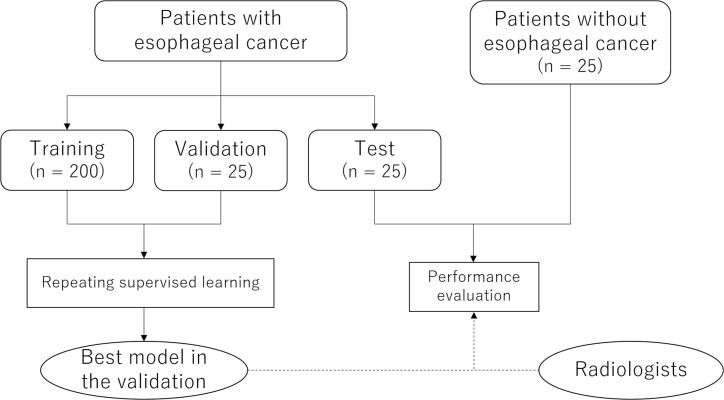
This retrospective study included 250 and 25 patients with and without esophageal cancer, respectively, who underwent contrast-enhanced CT between December 2014 and May 2021 (mean age, 67.9 ± 10.3 years; 233 men). A deep learning model was developed using data from 200 and 25 patients with esophageal cancer as training and validation data sets, respectively. The model was then applied to the test data set, consisting of additional 25 and 25 patients with and without esophageal cancer, respectively. Four readers (one radiologist and three radiology residents) independently registered the likelihood of malignant lesions using a 3-point scale in the test data set. After the scorings were completed, the readers were allowed to reference to the deep learning model results and modify their scores, when necessary.
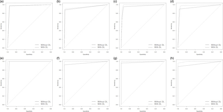
The area under the curve (AUC) of the deep learning model was 0.95 and 0.98 in the image- and patient-based analyses, respectively. By referencing to the deep learning model results, the AUCs for the readers were improved from 0.96/0.93/0.96/0.93 to 0.97/0.95/0.99/0.96 ( = 0.100/0.006/<0.001/<0.001, DeLong's test) in the image-based analysis, with statistically significant differences noted for the three less-experienced readers. Furthermore, the AUCs for the readers tended to improve from 0.98/0.96/0.98/0.94 to 1.00/1.00/1.00/1.00 ( = 0.317/0.149/0.317/0.073, DeLong's test) in the patient-based analysis.
The deep learning model mainly helped less-experienced readers improve their performance in detecting esophageal cancer on contrast-enhanced CT.
A deep learning model could mainly help less-experienced readers to detect esophageal cancer by improving their diagnostic confidence and diagnostic performance.
探究深度学习模型在帮助放射科医生或放射科住院医师检测对比增强 CT 图像中的食管癌中的有效性。
本回顾性研究纳入了 250 例和 25 例分别患有和不患有食管癌的患者,他们在 2014 年 12 月至 2021 年 5 月间进行了对比增强 CT 检查(平均年龄,67.9 ± 10.3 岁;233 名男性)。使用 200 例和 25 例食管癌患者的数据分别作为训练和验证数据集来开发深度学习模型。然后,将模型应用于包含另外 25 例和 25 例患有和不患有食管癌的患者的测试数据集。4 位读者(一位放射科医生和三位放射科住院医师)在测试数据集中独立使用 3 分制对恶性病变的可能性进行评分。评分完成后,当需要时,读者可以参考深度学习模型的结果并修改其评分。
深度学习模型的曲线下面积(AUC)在图像和患者基础分析中分别为 0.95 和 0.98。通过参考深度学习模型的结果,读者的 AUC 从 0.96/0.93/0.96/0.93 提高到 0.97/0.95/0.99/0.96(=0.100/0.006/<0.001/<0.001,DeLong 检验),在图像基础分析中,三位经验较少的读者的差异具有统计学意义。此外,读者的 AUC 从 0.98/0.96/0.98/0.94 提高到 1.00/1.00/1.00/1.00(=0.317/0.149/0.317/0.073,DeLong 检验),在患者基础分析中呈现出上升趋势。
深度学习模型主要帮助经验较少的读者提高在对比增强 CT 上检测食管癌的性能。
深度学习模型可以通过提高诊断信心和诊断性能,主要帮助经验较少的读者检测食管癌。