Department of Radiology, The Second Affiliated Hospital, Zhejiang University School of Medicine, No 88 Jiefang Road, Hangzhou, Zhejiang, China.
Department of Neurology, The Second Affiliated Hospital, Zhejiang University School of Medicine, No 88 Jiefang Road, Hangzhou, Zhejiang, China.
BMC Med. 2023 Apr 7;21(1):136. doi: 10.1186/s12916-023-02855-1.
Migraine is one of the world's most prevalent and disabling diseases. Despite huge advances in neuroimaging research, more valuable neuroimaging markers are still urgently needed to provide important insights into the brain mechanisms that underlie migraine symptoms. We therefore aim to investigate the regional iron deposition in subcortical nuclei of migraineurs as compared to controls and its association with migraine-related pathophysiological assessments.
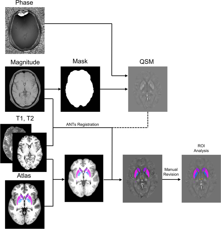
A total of 200 migraineurs (56 chronic migraine [CM], 144 episodic migraine [EM]) and 41 matched controls were recruited. All subjects underwent MRI and clinical variables including frequency/duration of migraine, intensity of migraine, 6-item Headache Impact Test (HIT-6), Migraine Disability Assessment (MIDAS), and Pittsburgh Sleep Quality Index (PSQI) were recorded. Quantitative susceptibility mapping was employed to quantify the regional iron content in subcortical regions. Associations between clinical variables and regional iron deposition were studied as well.
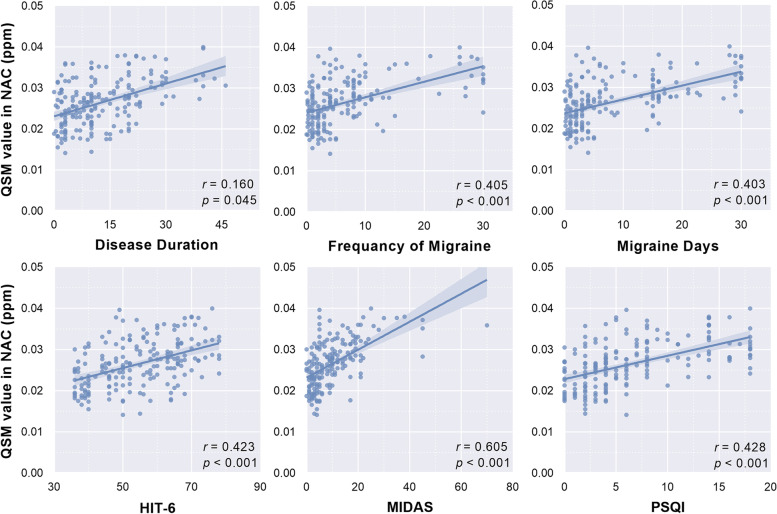
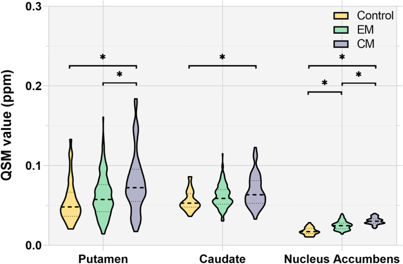
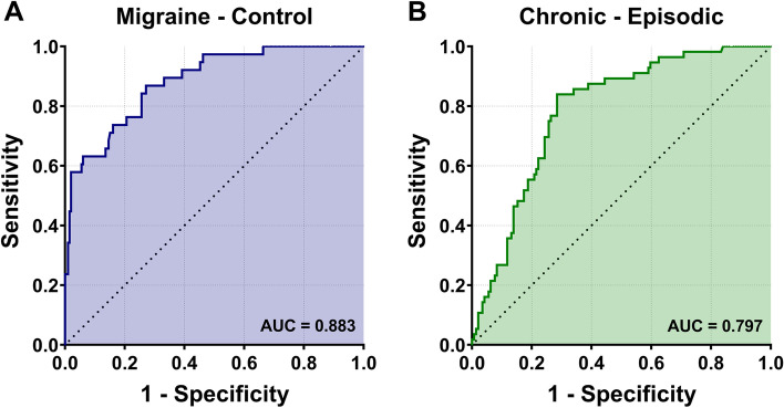
Increased iron deposition in the putamen, caudate, and nucleus accumbens (NAC) was observed in migraineurs more than controls. Meanwhile, patients with CM had a significantly higher volume of iron deposits compared to EM in multiple subcortical nuclei, especially in NAC. Volume of iron in NAC can be used to distinguish patients with CM from EM with a sensitivity of 85.45% and specificity of 71.53%. As the most valuable neuroimaging markers in all of the subcortical nuclei, higher iron deposition in NAC was significantly associated with disease progression, and higher HIT-6, MIDAS, and PSQI.
These findings provide evidence that iron deposition in NAC may be a biomarker for migraine chronicity and migraine-related dysfunctions, thus may help to understand the underlying vascular and neural mechanisms of migraine.
ClinicalTrials.gov, number NCT04939922.
偏头痛是世界上最普遍和致残的疾病之一。尽管神经影像学研究取得了巨大进展,但仍迫切需要更有价值的神经影像学标志物,以提供对偏头痛症状背后的大脑机制的重要见解。因此,我们旨在研究偏头痛患者与对照组相比,皮质下核团的局部铁沉积及其与偏头痛相关病理生理评估的关系。
共招募了 200 名偏头痛患者(56 名慢性偏头痛 [CM],144 名发作性偏头痛 [EM])和 41 名匹配的对照者。所有受试者均接受 MRI 检查,并记录了包括偏头痛发作频率/持续时间、偏头痛强度、6 项头痛影响测试(HIT-6)、偏头痛残疾评估(MIDAS)和匹兹堡睡眠质量指数(PSQI)在内的临床变量。采用定量磁化率成像来量化皮质下区域的局部铁含量。还研究了临床变量与局部铁沉积之间的关系。
与对照组相比,偏头痛患者的壳核、尾状核和伏隔核(NAC)的铁沉积增加。同时,CM 患者的多个皮质下核中铁沉积体积明显高于 EM 患者,尤其是 NAC。NAC 中的铁沉积量可用于区分 CM 和 EM 患者,其敏感性为 85.45%,特异性为 71.53%。作为所有皮质下核中最有价值的神经影像学标志物,NAC 中的铁沉积与疾病进展以及更高的 HIT-6、MIDAS 和 PSQI 显著相关。
这些发现为 NAC 中的铁沉积可能是偏头痛慢性和偏头痛相关功能障碍的生物标志物提供了证据,因此可能有助于理解偏头痛的血管和神经机制。
ClinicalTrials.gov,编号 NCT04939922。