III. Department of Medicine, University Medical Center Hamburg-Eppendorf, Hamburg, Germany.
Hamburg Center for Kidney Health (HCKH), University Medical Center Hamburg-Eppendorf, Hamburg, Germany.
Nat Nanotechnol. 2023 Apr;18(4):336-342. doi: 10.1038/s41565-023-01328-z. Epub 2023 Apr 10.
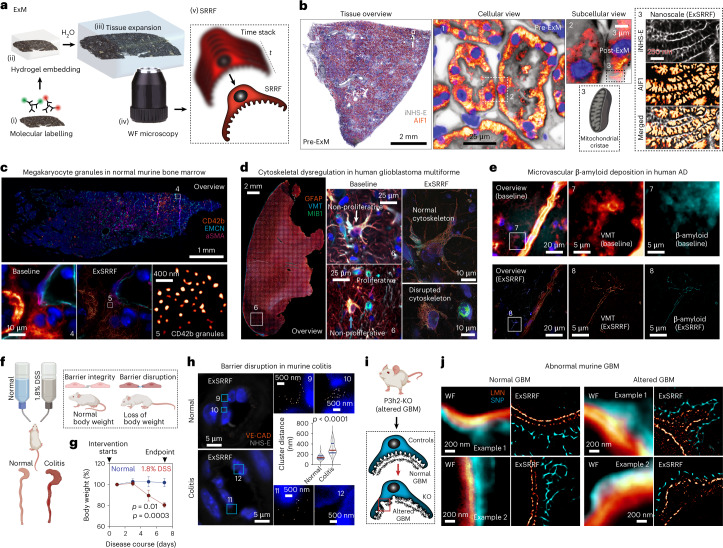
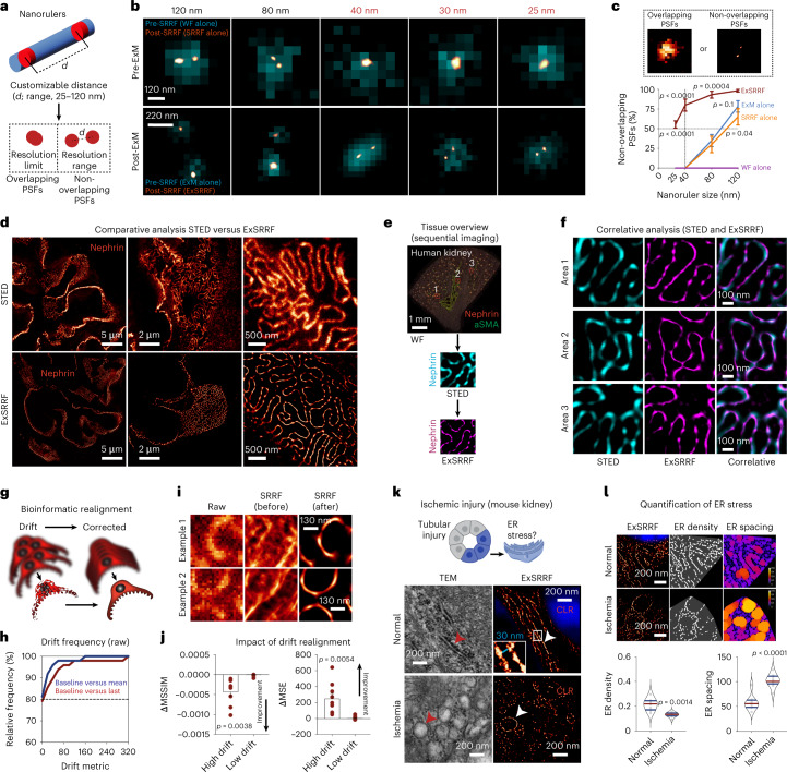
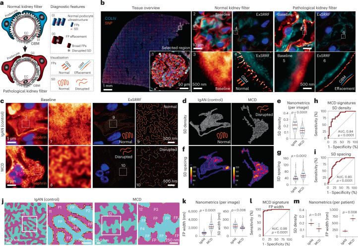
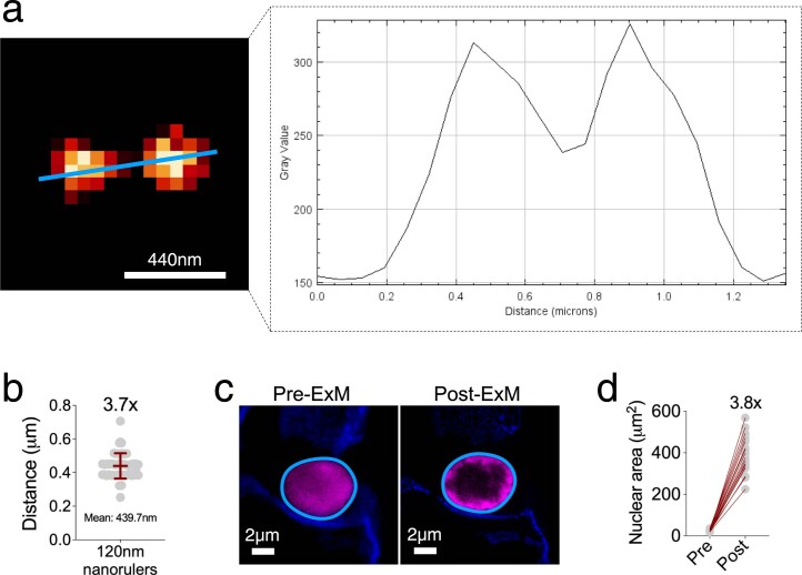
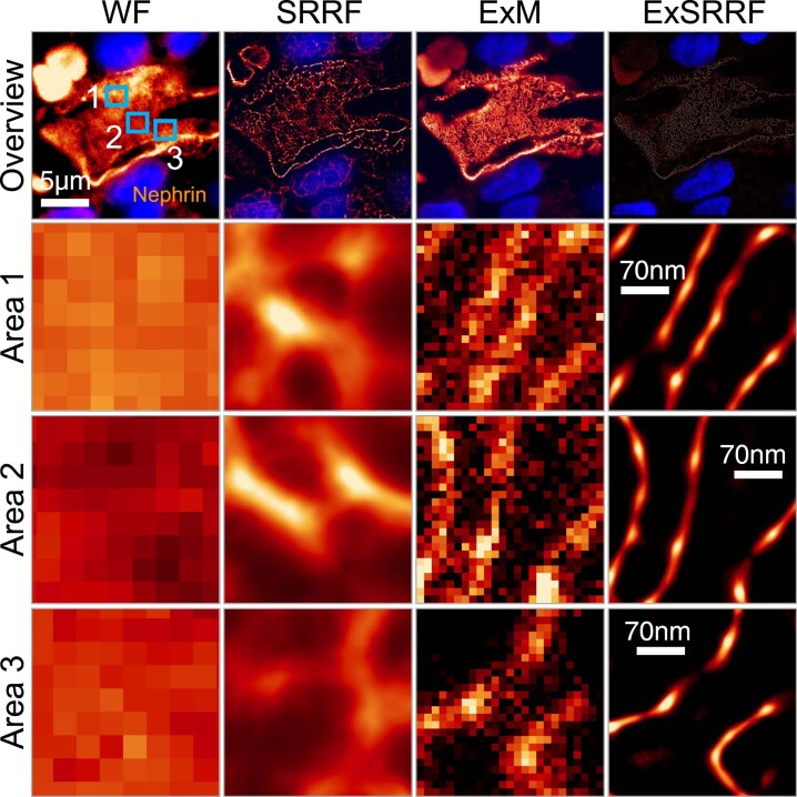
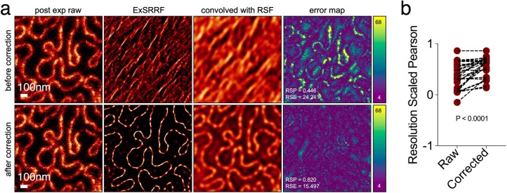
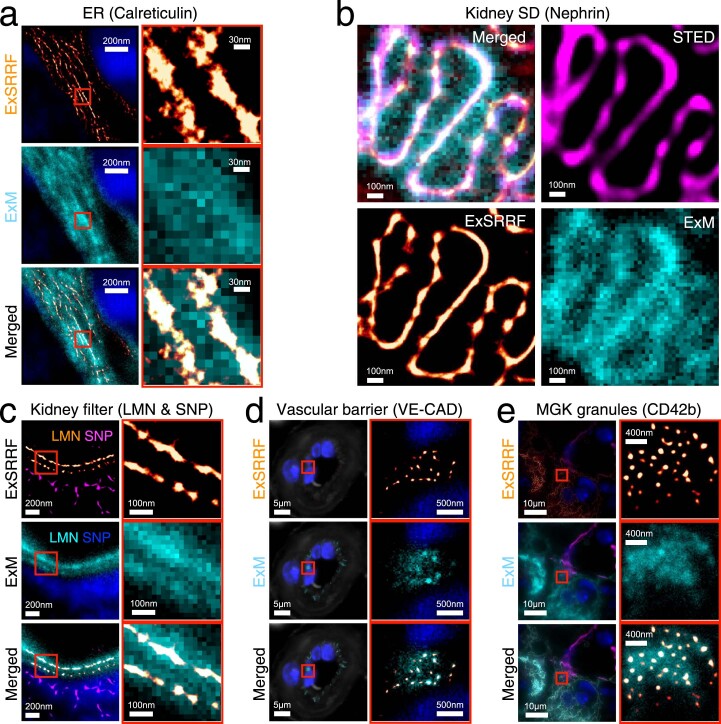
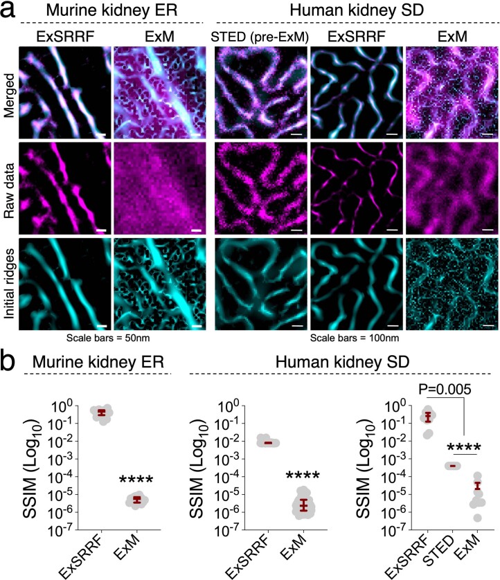
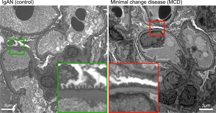
Expansion microscopy physically enlarges biological specimens to achieve nanoscale resolution using diffraction-limited microscopy systems. However, optimal performance is usually reached using laser-based systems (for example, confocal microscopy), restricting its broad applicability in clinical pathology, as most centres have access only to light-emitting diode (LED)-based widefield systems. As a possible alternative, a computational method for image resolution enhancement, namely, super-resolution radial fluctuations (SRRF), has recently been developed. However, this method has not been explored in pathology specimens to date, because on its own, it does not achieve sufficient resolution for routine clinical use. Here, we report expansion-enhanced super-resolution radial fluctuations (ExSRRF), a simple, robust, scalable and accessible workflow that provides a resolution of up to 25 nm using LED-based widefield microscopy. ExSRRF enables molecular profiling of subcellular structures from archival formalin-fixed paraffin-embedded tissues in complex clinical and experimental specimens, including ischaemic, degenerative, neoplastic, genetic and immune-mediated disorders. Furthermore, as examples of its potential application to experimental and clinical pathology, we show that ExSRRF can be used to identify and quantify classical features of endoplasmic reticulum stress in the murine ischaemic kidney and diagnostic ultrastructural features in human kidney biopsies.
扩展显微镜通过物理放大生物样本,在使用衍射受限显微镜系统的情况下实现纳米级分辨率。然而,其最佳性能通常是在基于激光的系统(例如共聚焦显微镜)中实现的,这限制了其在临床病理学中的广泛适用性,因为大多数中心仅能获得基于发光二极管(LED)的宽场系统。作为一种可能的替代方法,最近开发了一种用于图像分辨率增强的计算方法,即超分辨率径向波动(SRRF)。然而,迄今为止,该方法尚未在病理标本中进行探索,因为仅凭自身,它无法达到常规临床应用所需的足够分辨率。在这里,我们报告了扩展增强的超分辨率径向波动(ExSRRF),这是一种简单、稳健、可扩展且易于访问的工作流程,可在基于 LED 的宽场显微镜下提供高达 25nm 的分辨率。ExSRRF 能够对包括缺血性、退行性、肿瘤性、遗传性和免疫介导性疾病在内的复杂临床和实验标本中的固定石蜡包埋组织中的亚细胞结构进行分子分析。此外,作为其在实验和临床病理学中潜在应用的示例,我们表明 ExSRRF 可用于识别和定量小鼠缺血性肾脏中的内质网应激的经典特征以及人类肾活检中的诊断超微结构特征。