• 文献检索
  • 文档翻译
  • 深度研究
  • 学术资讯
  • Suppr Zotero 插件Zotero 插件
  • 邀请有礼
  • 套餐&价格
  • 历史记录
应用&插件
Suppr Zotero 插件Zotero 插件浏览器插件Mac 客户端Windows 客户端微信小程序
定价
高级版会员购买积分包购买API积分包
服务
文献检索文档翻译深度研究API 文档MCP 服务
关于我们
关于 Suppr公司介绍联系我们用户协议隐私条款
关注我们

Suppr 超能文献

核心技术专利:CN118964589B侵权必究
粤ICP备2023148730 号-1Suppr @ 2026

文献检索

告别复杂PubMed语法,用中文像聊天一样搜索,搜遍4000万医学文献。AI智能推荐,让科研检索更轻松。

立即免费搜索

文件翻译

保留排版,准确专业,支持PDF/Word/PPT等文件格式,支持 12+语言互译。

免费翻译文档

深度研究

AI帮你快速写综述,25分钟生成高质量综述,智能提取关键信息,辅助科研写作。

立即免费体验

基于对比学习的预训练可提高糖尿病视网膜病变分类模型的表示能力和迁移能力。

Contrastive learning-based pretraining improves representation and transferability of diabetic retinopathy classification models.

机构信息

Department of Biomedical Data Science, Stanford University School of Medicine, 1265 Welch Road, Stanford, CA, 94305, USA.

Department of Electrical and Computer Engineering, University of North Carolina at Charlotte, 9201 University City Boulevard, Charlotte, NC, 28223, USA.

出版信息

Sci Rep. 2023 Apr 13;13(1):6047. doi: 10.1038/s41598-023-33365-y.

DOI:10.1038/s41598-023-33365-y
PMID:37055475
原文链接:https://pmc.ncbi.nlm.nih.gov/articles/PMC10102012/
Abstract

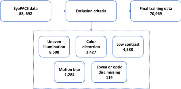
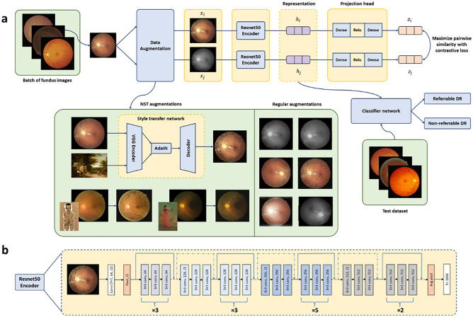
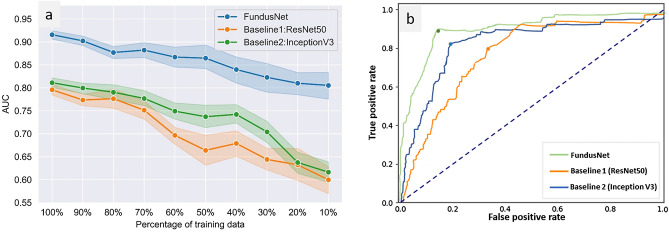
Diabetic retinopathy (DR) is a major cause of vision impairment in diabetic patients worldwide. Due to its prevalence, early clinical diagnosis is essential to improve treatment management of DR patients. Despite recent demonstration of successful machine learning (ML) models for automated DR detection, there is a significant clinical need for robust models that can be trained with smaller cohorts of dataset and still perform with high diagnostic accuracy in independent clinical datasets (i.e., high model generalizability). Towards this need, we have developed a self-supervised contrastive learning (CL) based pipeline for classification of referable vs non-referable DR. Self-supervised CL based pretraining allows enhanced data representation, therefore, the development of robust and generalized deep learning (DL) models, even with small, labeled datasets. We have integrated a neural style transfer (NST) augmentation in the CL pipeline to produce models with better representations and initializations for the detection of DR in color fundus images. We compare our CL pretrained model performance with two state of the art baseline models pretrained with Imagenet weights. We further investigate the model performance with reduced labeled training data (down to 10 percent) to test the robustness of the model when trained with small, labeled datasets. The model is trained and validated on the EyePACS dataset and tested independently on clinical datasets from the University of Illinois, Chicago (UIC). Compared to baseline models, our CL pretrained FundusNet model had higher area under the receiver operating characteristics (ROC) curve (AUC) (CI) values (0.91 (0.898 to 0.930) vs 0.80 (0.783 to 0.820) and 0.83 (0.801 to 0.853) on UIC data). At 10 percent labeled training data, the FundusNet AUC was 0.81 (0.78 to 0.84) vs 0.58 (0.56 to 0.64) and 0.63 (0.60 to 0.66) in baseline models, when tested on the UIC dataset. CL based pretraining with NST significantly improves DL classification performance, helps the model generalize well (transferable from EyePACS to UIC data), and allows training with small, annotated datasets, therefore reducing ground truth annotation burden of the clinicians.

摘要

糖尿病性视网膜病变(DR)是全球糖尿病患者视力损害的主要原因。由于其普遍性,早期临床诊断对于改善 DR 患者的治疗管理至关重要。尽管最近已经证明了用于自动 DR 检测的机器学习(ML)模型的成功,但仍然需要稳健的模型,这些模型可以用较小的数据集进行训练,并在独立的临床数据集(即高模型通用性)中仍然具有高诊断准确性。针对这一需求,我们开发了一种基于自监督对比学习(CL)的分类管道,用于分类可转诊与不可转诊的 DR。基于自监督 CL 的预训练允许增强数据表示,因此,即使使用较小的标记数据集,也可以开发稳健且通用的深度学习(DL)模型。我们在 CL 管道中集成了神经风格转换(NST)增强功能,以生成具有更好表示和初始化的模型,用于检测彩色眼底图像中的 DR。我们将我们的 CL 预训练模型的性能与两个使用 Imagenet 权重预训练的最先进基线模型的性能进行了比较。我们还使用减少的标记训练数据(低至 10%)进一步研究了模型性能,以测试模型在使用小的标记数据集进行训练时的鲁棒性。该模型在 EyePACS 数据集上进行训练和验证,并在伊利诺伊大学芝加哥分校(UIC)的临床数据集上独立进行测试。与基线模型相比,我们的 CL 预训练的 FundusNet 模型在 UIC 数据上具有更高的接收器操作特征(ROC)曲线下面积(AUC)(CI)值(0.91(0.898 至 0.930)比 0.80(0.783 至 0.820)和 0.83(0.801 至 0.853))。在标记训练数据低至 10%时,当在 UIC 数据集上进行测试时,FundusNet 的 AUC 为 0.81(0.78 至 0.84),而基线模型为 0.58(0.56 至 0.64)和 0.63(0.60 至 0.66)。基于 NST 的 CL 预训练显著提高了 DL 分类性能,有助于模型很好地泛化(从 EyePACS 转移到 UIC 数据),并且允许使用小的、有注释的数据集进行训练,从而减少了临床医生的真实注释负担。

https://cdn.ncbi.nlm.nih.gov/pmc/blobs/6de7/10102012/06553dd41456/41598_2023_33365_Fig4_HTML.jpg
https://cdn.ncbi.nlm.nih.gov/pmc/blobs/6de7/10102012/6343096b946e/41598_2023_33365_Fig1_HTML.jpg
https://cdn.ncbi.nlm.nih.gov/pmc/blobs/6de7/10102012/b39b3a6a57eb/41598_2023_33365_Fig2_HTML.jpg
https://cdn.ncbi.nlm.nih.gov/pmc/blobs/6de7/10102012/03eeec46541d/41598_2023_33365_Fig3_HTML.jpg
https://cdn.ncbi.nlm.nih.gov/pmc/blobs/6de7/10102012/06553dd41456/41598_2023_33365_Fig4_HTML.jpg

相似文献

1
Contrastive learning-based pretraining improves representation and transferability of diabetic retinopathy classification models.基于对比学习的预训练可提高糖尿病视网膜病变分类模型的表示能力和迁移能力。
Sci Rep. 2023 Apr 13;13(1):6047. doi: 10.1038/s41598-023-33365-y.
2
Contrastive self-supervised learning for diabetic retinopathy early detection.对比自监督学习在糖尿病视网膜病变早期检测中的应用。
Med Biol Eng Comput. 2023 Sep;61(9):2441-2452. doi: 10.1007/s11517-023-02810-5. Epub 2023 Apr 29.
3
MediDRNet: Tackling category imbalance in diabetic retinopathy classification with dual-branch learning and prototypical contrastive learning.MediDRNet:使用双分支学习和原型对比学习解决糖尿病视网膜病变分类中的类别不平衡问题。
Comput Methods Programs Biomed. 2024 Aug;253:108230. doi: 10.1016/j.cmpb.2024.108230. Epub 2024 May 17.
4
Vision transformer with masked autoencoders for referable diabetic retinopathy classification based on large-size retina image.基于大尺寸视网膜图像的可引用糖尿病视网膜病变分类的掩蔽自动编码器视觉转换器。
PLoS One. 2024 Mar 6;19(3):e0299265. doi: 10.1371/journal.pone.0299265. eCollection 2024.
5
Development and Validation of a Deep Learning Algorithm for Detection of Diabetic Retinopathy in Retinal Fundus Photographs.深度学习算法在视网膜眼底照片糖尿病视网膜病变检测中的开发与验证。
JAMA. 2016 Dec 13;316(22):2402-2410. doi: 10.1001/jama.2016.17216.
6
Performance and Limitation of Machine Learning Algorithms for Diabetic Retinopathy Screening: Meta-analysis.机器学习算法在糖尿病视网膜病变筛查中的性能和局限性:荟萃分析。
J Med Internet Res. 2021 Jul 3;23(7):e23863. doi: 10.2196/23863.
7
Explainable Diabetic Retinopathy using EfficientNET.使用EfficientNET的可解释性糖尿病视网膜病变
Annu Int Conf IEEE Eng Med Biol Soc. 2020 Jul;2020:1966-1969. doi: 10.1109/EMBC44109.2020.9175664.
8
Leveraging Multimodal Deep Learning Architecture with Retina Lesion Information to Detect Diabetic Retinopathy.利用具有视网膜病变信息的多模态深度学习架构来检测糖尿病性视网膜病变。
Transl Vis Sci Technol. 2020 Jul 16;9(2):41. doi: 10.1167/tvst.9.2.41. eCollection 2020 Jul.
9
A reliable diabetic retinopathy grading via transfer learning and ensemble learning with quadratic weighted kappa metric.基于迁移学习和集成学习的二次加权 Kappa 度量在糖尿病性视网膜病变分级中的应用
BMC Med Inform Decis Mak. 2024 Feb 6;24(1):37. doi: 10.1186/s12911-024-02446-x.
10
Development and Validation of a Deep Learning System for Diabetic Retinopathy and Related Eye Diseases Using Retinal Images From Multiethnic Populations With Diabetes.使用来自多民族糖尿病患者群体的视网膜图像开发并验证用于糖尿病视网膜病变及相关眼病的深度学习系统
JAMA. 2017 Dec 12;318(22):2211-2223. doi: 10.1001/jama.2017.18152.

引用本文的文献

1
Automated Foveal Avascular Zone Segmentation in Optical Coherence Tomography Angiography Across Multiple Eye Diseases Using Knowledge Distillation.利用知识蒸馏技术在多种眼部疾病的光学相干断层扫描血管造影中实现黄斑无血管区的自动分割
Bioengineering (Basel). 2025 Mar 23;12(4):334. doi: 10.3390/bioengineering12040334.
2
Distributed training of foundation models for ophthalmic diagnosis.用于眼科诊断的基础模型的分布式训练。
Commun Eng. 2025 Jan 22;4(1):6. doi: 10.1038/s44172-025-00341-5.
3
A Self-Supervised Equivariant Refinement Classification Network for Diabetic Retinopathy Classification.

本文引用的文献

1
Prediction of Diabetes through Retinal Images Using Deep Neural Network.基于深度神经网络的视网膜图像糖尿病预测。
Comput Intell Neurosci. 2022 Jun 3;2022:7887908. doi: 10.1155/2022/7887908. eCollection 2022.
2
A deep learning model for identifying diabetic retinopathy using optical coherence tomography angiography.一种利用光相干断层扫描血管造影术识别糖尿病性视网膜病变的深度学习模型。
Sci Rep. 2021 Nov 26;11(1):23024. doi: 10.1038/s41598-021-02479-6.
3
Learning Domain-Agnostic Visual Representation for Computational Pathology Using Medically-Irrelevant Style Transfer Augmentation.
一种用于糖尿病视网膜病变分类的自监督等变细化分类网络。
J Imaging Inform Med. 2025 Jun;38(3):1796-1811. doi: 10.1007/s10278-024-01270-z. Epub 2024 Sep 19.
使用与医学无关的风格迁移增强学习计算病理学的领域不可知视觉表示。
IEEE Trans Med Imaging. 2021 Dec;40(12):3945-3954. doi: 10.1109/TMI.2021.3101985. Epub 2021 Nov 30.
4
A deep learning system for detecting diabetic retinopathy across the disease spectrum.一种用于在疾病谱中检测糖尿病性视网膜病变的深度学习系统。
Nat Commun. 2021 May 28;12(1):3242. doi: 10.1038/s41467-021-23458-5.
5
Artificial intelligence using deep learning to screen for referable and vision-threatening diabetic retinopathy in Africa: a clinical validation study.人工智能利用深度学习在非洲筛查可转诊和威胁视力的糖尿病视网膜病变:一项临床验证研究。
Lancet Digit Health. 2019 May;1(1):e35-e44. doi: 10.1016/S2589-7500(19)30004-4. Epub 2019 May 2.
6
Transfer Learning for Automated OCTA Detection of Diabetic Retinopathy.基于迁移学习的自动 OCTA 糖尿病视网膜病变检测
Transl Vis Sci Technol. 2020 Jul 2;9(2):35. doi: 10.1167/tvst.9.2.35. eCollection 2020 Jul.
7
Diabetic Retinopathy Preferred Practice Pattern®.糖尿病视网膜病变首选诊疗模式®
Ophthalmology. 2020 Jan;127(1):P66-P145. doi: 10.1016/j.ophtha.2019.09.025. Epub 2019 Sep 25.
8
Deep learning based computer-aided diagnosis systems for diabetic retinopathy: A survey.基于深度学习的糖尿病视网膜病变计算机辅助诊断系统:综述。
Artif Intell Med. 2019 Aug;99:101701. doi: 10.1016/j.artmed.2019.07.009. Epub 2019 Aug 7.
9
Pivotal trial of an autonomous AI-based diagnostic system for detection of diabetic retinopathy in primary care offices.在基层医疗诊所中用于检测糖尿病视网膜病变的基于人工智能的自主诊断系统的关键试验。
NPJ Digit Med. 2018 Aug 28;1:39. doi: 10.1038/s41746-018-0040-6. eCollection 2018.
10
Development and Validation of Deep Learning Models for Screening Multiple Abnormal Findings in Retinal Fundus Images.深度学习模型在视网膜眼底图像多种异常发现筛查中的开发与验证。
Ophthalmology. 2020 Jan;127(1):85-94. doi: 10.1016/j.ophtha.2019.05.029. Epub 2019 May 31.