• 文献检索
  • 文档翻译
  • 深度研究
  • 学术资讯
  • Suppr Zotero 插件Zotero 插件
  • 邀请有礼
  • 套餐&价格
  • 历史记录
应用&插件
Suppr Zotero 插件Zotero 插件浏览器插件Mac 客户端Windows 客户端微信小程序
定价
高级版会员购买积分包购买API积分包
服务
文献检索文档翻译深度研究API 文档MCP 服务
关于我们
关于 Suppr公司介绍联系我们用户协议隐私条款
关注我们

Suppr 超能文献

核心技术专利:CN118964589B侵权必究
粤ICP备2023148730 号-1Suppr @ 2026

文献检索

告别复杂PubMed语法,用中文像聊天一样搜索,搜遍4000万医学文献。AI智能推荐,让科研检索更轻松。

立即免费搜索

文件翻译

保留排版,准确专业,支持PDF/Word/PPT等文件格式,支持 12+语言互译。

免费翻译文档

深度研究

AI帮你快速写综述,25分钟生成高质量综述,智能提取关键信息,辅助科研写作。

立即免费体验

多光谱成像在皮肤病评估中的应用:现状与展望。

Multispectral Imaging for Skin Diseases Assessment-State of the Art and Perspectives.

机构信息

Department of Computer Science and Engineering, University Politehnica of Bucharest, 060042 Bucharest, Romania.

出版信息

Sensors (Basel). 2023 Apr 11;23(8):3888. doi: 10.3390/s23083888.

DOI:10.3390/s23083888
PMID:37112229
原文链接:https://pmc.ncbi.nlm.nih.gov/articles/PMC10140977/
Abstract

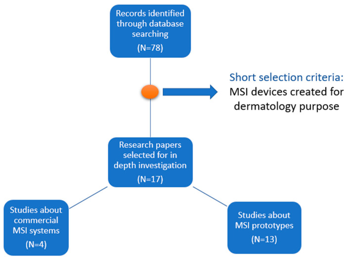
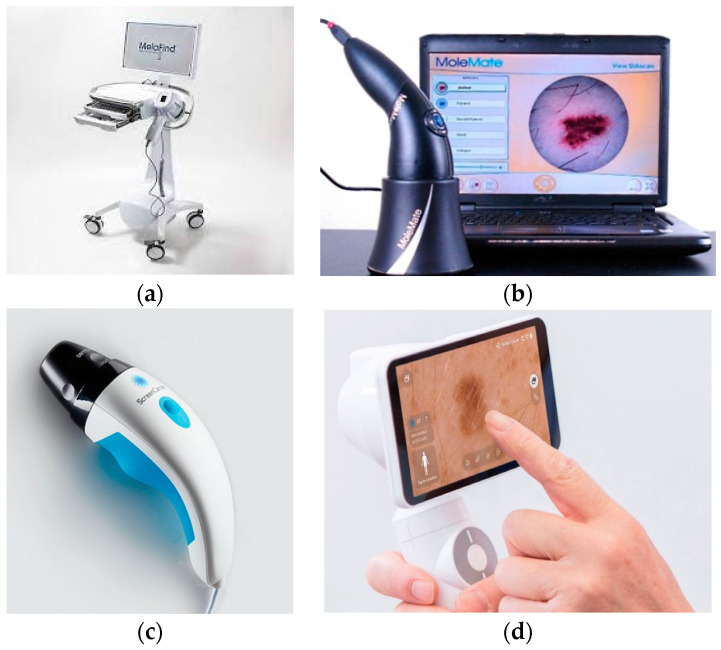
Skin optical inspection is an imperative procedure for a suspicious dermal lesion since very early skin cancer detection can guarantee total recovery. Dermoscopy, confocal laser scanning microscopy, optical coherence tomography, multispectral imaging, multiphoton laser imaging, and 3D topography are the most outstanding optical techniques implemented for skin examination. The accuracy of dermatological diagnoses attained by each of those methods is still debatable, and only dermoscopy is frequently used by all dermatologists. Therefore, a comprehensive method for skin analysis has not yet been established. Multispectral imaging (MSI) is based on light-tissue interaction properties due to radiation wavelength variation. An MSI device collects the reflected radiation after illumination of the lesion with light of different wavelengths and provides a set of spectral images. The concentration maps of the main light-absorbing molecules in the skin, the chromophores, can be retrieved using the intensity values from those images, sometimes even for deeper-located tissues, due to interaction with near-infrared light. Recent studies have shown that portable and cost-efficient MSI systems can be used for extracting skin lesion characteristics useful for early melanoma diagnoses. This review aims to describe the efforts that have been made to develop MSI systems for skin lesions evaluation in the last decade. We examined the hardware characteristics of the produced devices and identified the typical structure of an MSI device for dermatology. The analyzed prototypes showed the possibility of improving the specificity of classification between the melanoma and benign nevi. Currently, however, they are rather adjuvants tools for skin lesion assessment, and efforts are needed towards a fully fledged diagnostic MSI device.

摘要

皮肤光学检查对于可疑的皮肤病变是必要的程序,因为早期发现皮肤癌可以保证完全康复。皮肤镜检查、共焦激光扫描显微镜、光相干断层扫描、多光谱成像、多光子激光成像和 3D topography 是用于皮肤检查的最杰出的光学技术。这些方法中的每一种方法所达到的皮肤科诊断准确性仍存在争议,只有皮肤镜检查经常被所有皮肤科医生使用。因此,尚未建立皮肤分析的综合方法。多光谱成像 (MSI) 基于光与组织相互作用的特性,因为辐射波长的变化。MSI 设备在病变处用不同波长的光照射后收集反射辐射,并提供一组光谱图像。可以从这些图像的强度值中检索到皮肤中主要光吸收分子(发色团)的浓度图,有时甚至可以用于更深层的组织,因为它们与近红外光相互作用。最近的研究表明,便携式和具有成本效益的 MSI 系统可用于提取有助于早期黑色素瘤诊断的皮肤病变特征。本综述旨在描述过去十年中为开发用于皮肤病变评估的 MSI 系统所做的努力。我们检查了所生产设备的硬件特性,并确定了用于皮肤科的 MSI 设备的典型结构。分析的原型表明了改善黑素瘤和良性痣之间分类特异性的可能性。然而,目前它们只是皮肤病变评估的辅助工具,需要努力开发功能齐全的诊断 MSI 设备。

https://cdn.ncbi.nlm.nih.gov/pmc/blobs/1eec/10140977/84602f5f7037/sensors-23-03888-g002.jpg
https://cdn.ncbi.nlm.nih.gov/pmc/blobs/1eec/10140977/6895e4e01b5b/sensors-23-03888-g001.jpg
https://cdn.ncbi.nlm.nih.gov/pmc/blobs/1eec/10140977/84602f5f7037/sensors-23-03888-g002.jpg
https://cdn.ncbi.nlm.nih.gov/pmc/blobs/1eec/10140977/6895e4e01b5b/sensors-23-03888-g001.jpg
https://cdn.ncbi.nlm.nih.gov/pmc/blobs/1eec/10140977/84602f5f7037/sensors-23-03888-g002.jpg

相似文献

1
Multispectral Imaging for Skin Diseases Assessment-State of the Art and Perspectives.多光谱成像在皮肤病评估中的应用:现状与展望。
Sensors (Basel). 2023 Apr 11;23(8):3888. doi: 10.3390/s23083888.
2
Role of In Vivo Reflectance Confocal Microscopy in the Analysis of Melanocytic Lesions.体内反射共聚焦显微镜在黑素细胞性病变分析中的作用
Acta Dermatovenerol Croat. 2018 Apr;26(1):64-67.
3
[The Rise of Artificial Intelligence - High Prediction Accuracy in Early Detection of Pigmented Melanoma].[人工智能的崛起——色素性黑色素瘤早期检测中的高预测准确率]
Laryngorhinootologie. 2023 Jul;102(7):496-503. doi: 10.1055/a-1949-3639. Epub 2022 Dec 29.
4
Noninvasive diagnosis in dermatology.皮肤科的非侵入性诊断。
J Dtsch Dermatol Ges. 2017 Oct;15(10):999-1016. doi: 10.1111/ddg.13347.
5
In vivo assessment of optical properties of melanocytic skin lesions and differentiation of melanoma from non-malignant lesions by high-definition optical coherence tomography.通过高清光学相干断层扫描对黑素细胞性皮肤病变的光学特性进行体内评估以及区分黑色素瘤与非恶性病变。
Arch Dermatol Res. 2016 Jan;308(1):7-20. doi: 10.1007/s00403-015-1608-5. Epub 2015 Nov 13.
6
Deep learning-assisted multispectral imaging for early screening of skin diseases.深度学习辅助多光谱成像在皮肤病早期筛查中的应用。
Photodiagnosis Photodyn Ther. 2024 Aug;48:104292. doi: 10.1016/j.pdpdt.2024.104292. Epub 2024 Jul 26.
7
Non-invasive tools for the diagnosis of cutaneous melanoma.用于诊断皮肤黑色素瘤的非侵入性工具。
Skin Res Technol. 2017 Aug;23(3):261-271. doi: 10.1111/srt.12350. Epub 2016 Nov 22.
8
Optical coherence tomography in dermatology.光学相干断层扫描在皮肤科的应用。
J Biomed Opt. 2013 Jun;18(6):061224. doi: 10.1117/1.JBO.18.6.061224.
9
A Review of Noninvasive Techniques for Skin Cancer Detection in Dermatology.皮肤科中用于皮肤癌检测的无创技术综述
Am J Clin Dermatol. 2020 Aug;21(4):513-524. doi: 10.1007/s40257-020-00517-z.
10
Density-based parallel skin lesion border detection with webCL.基于密度的并行皮肤病变边界检测与WebCL技术
BMC Bioinformatics. 2015;16 Suppl 13(Suppl 13):S5. doi: 10.1186/1471-2105-16-S13-S5. Epub 2015 Sep 25.

引用本文的文献

1
Machine learning-based classification of spatially resolved diffuse reflectance and autofluorescence spectra acquired on human skin for actinic keratoses and skin carcinoma diagnostics aid.基于机器学习对在人体皮肤上采集的空间分辨漫反射和自体荧光光谱进行分类,以辅助光化性角化病和皮肤癌的诊断。
J Biomed Opt. 2025 Mar;30(3):035001. doi: 10.1117/1.JBO.30.3.035001. Epub 2025 Mar 4.
2
Development and performance validation of an affordable and portable high-resolution darkfield polarization-sensitive multispectral imaging microscope for the assessment of radiation dermatitis and fibrosis.用于评估放射性皮炎和纤维化的经济实惠且便携的高分辨率暗场偏振敏感多光谱成像显微镜的开发与性能验证
Biomed Opt Express. 2024 Dec 23;16(1):320-333. doi: 10.1364/BOE.546226. eCollection 2025 Jan 1.
3

本文引用的文献

1
Diagnostics Using Non-Invasive Technologies in Dermatological Oncology.皮肤科肿瘤学中使用非侵入性技术的诊断方法
Cancers (Basel). 2022 Nov 29;14(23):5886. doi: 10.3390/cancers14235886.
2
Hyperspectral and multispectral image processing for gross-level tumor detection in skin lesions: a systematic review.用于皮肤病变中大体水平肿瘤检测的高光谱和多光谱图像处理:系统评价。
J Biomed Opt. 2022 Jun;27(6). doi: 10.1117/1.JBO.27.6.060901.
3
Applications of multispectral and hyperspectral imaging in dermatology.多光谱和高光谱成像在皮肤科中的应用。
A multispectral camera in the VIS-NIR equipped with thermal imaging and environmental sensors for non invasive analysis in precision agriculture.一款配备热成像和环境传感器的可见-近红外多光谱相机,用于精准农业中的非侵入性分析。
HardwareX. 2024 Oct 21;20:e00596. doi: 10.1016/j.ohx.2024.e00596. eCollection 2024 Dec.
4
Skin lesion segmentation using deep learning algorithm with ant colony optimization.基于蚁群算法的深度学习算法在皮肤损伤分割中的应用。
BMC Med Inform Decis Mak. 2024 Sep 27;24(1):265. doi: 10.1186/s12911-024-02686-x.
5
Performance Characterization of an Illumination-Based Low-Cost Multispectral Camera.基于照明的低成本多光谱相机的性能表征
Sensors (Basel). 2024 Aug 13;24(16):5229. doi: 10.3390/s24165229.
6
Classification of melanocytic lesions using direct illumination multispectral imaging.利用直接照明多光谱成像对黑素细胞病变进行分类。
Sci Rep. 2024 Aug 16;14(1):19036. doi: 10.1038/s41598-024-69773-x.
7
Parameter-based transfer learning for severity classification of atopic dermatitis using hyperspectral imaging.基于参数的迁移学习在利用高光谱成像对特应性皮炎严重程度分类中的应用。
Skin Res Technol. 2024 Apr;30(4):e13704. doi: 10.1111/srt.13704.
8
Effect of skin color on optical properties and the implications for medical optical technologies: a review.肤色对光学性质的影响及其对医学光学技术的意义:综述。
J Biomed Opt. 2024 Jan;29(1):010901. doi: 10.1117/1.JBO.29.1.010901. Epub 2024 Jan 24.
9
Optically Guided High-Frequency Ultrasound Shows Superior Efficacy for Preoperative Estimation of Breslow Thickness in Comparison with Multispectral Imaging: A Single-Center Prospective Validation Study.与多光谱成像相比,光学引导高频超声在术前评估Breslow厚度方面显示出更高的疗效:一项单中心前瞻性验证研究。
Cancers (Basel). 2023 Dec 28;16(1):157. doi: 10.3390/cancers16010157.
Exp Dermatol. 2022 Aug;31(8):1128-1135. doi: 10.1111/exd.14624. Epub 2022 Jun 19.
4
Multi-Class CNN for Classification of Multispectral and Autofluorescence Skin Lesion Clinical Images.用于多光谱和自体荧光皮肤病变临床图像分类的多类卷积神经网络
J Clin Med. 2022 May 17;11(10):2833. doi: 10.3390/jcm11102833.
5
Skin cancer detection using non-invasive techniques.使用非侵入性技术进行皮肤癌检测。
RSC Adv. 2018 Aug 6;8(49):28095-28130. doi: 10.1039/c8ra04164d. eCollection 2018 Aug 2.
6
Differentiating Malignant from Benign Pigmented or Non-Pigmented Skin Tumours-A Pilot Study on 3D Hyperspectral Imaging of Complex Skin Surfaces and Convolutional Neural Networks.区分恶性与良性色素沉着或无色素皮肤肿瘤——复杂皮肤表面三维高光谱成像与卷积神经网络的初步研究
J Clin Med. 2022 Mar 30;11(7):1914. doi: 10.3390/jcm11071914.
7
Non-invasive diagnostic techniques in pigmentary skin disorders and skin cancer.非侵入性诊断技术在色素性皮肤疾病和皮肤癌中的应用。
J Cosmet Dermatol. 2022 Feb;21(2):444-450. doi: 10.1111/jocd.14547. Epub 2021 Nov 1.
8
Data-Efficient Sensor Upgrade Path Using Knowledge Distillation.基于知识蒸馏的数据高效传感器升级路径。
Sensors (Basel). 2021 Sep 29;21(19):6523. doi: 10.3390/s21196523.
9
Quantitative Multispectral Imaging Differentiates Melanoma from Seborrheic Keratosis.定量多光谱成像鉴别黑色素瘤与脂溢性角化病。
Diagnostics (Basel). 2021 Jul 22;11(8):1315. doi: 10.3390/diagnostics11081315.
10
Non-invasive optical methods for melanoma diagnosis.用于黑色素瘤诊断的无创性光学方法。
Photodiagnosis Photodyn Ther. 2021 Jun;34:102266. doi: 10.1016/j.pdpdt.2021.102266. Epub 2021 Mar 27.