Materials Science and Engineering Program, University of California, 9500 Gilman Dr. San Diego, 92093, La Jolla, CA, USA.
Department of NanoEngineering, University of California, 9500 Gilman Dr. San Diego, 92093, La Jolla, CA, USA.
Curr Cardiol Rep. 2023 Jun;25(6):505-514. doi: 10.1007/s11886-023-01881-y. Epub 2023 May 2.
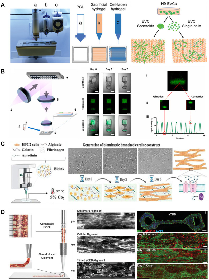
Bioengineering of functional cardiac tissue composed of primary cardiomyocytes has great potential for myocardial regeneration and in vitro tissue modeling. 3D bioprinting was developed to create cardiac tissue in hydrogels that can mimic the structural, physiological, and functional features of native myocardium. Through a detailed review of the 3D printing technologies and bioink materials used in the creation of a heart tissue, this article discusses the potential of engineered heart tissues in biomedical applications.
In this review, we discussed the recent progress in 3D bioprinting strategies for cardiac tissue engineering, including bioink and 3D bioprinting methods as well as examples of engineered cardiac tissue such as in vitro cardiac models and vascular channels. 3D printing is a powerful tool for creating in vitro cardiac tissues that are structurally and functionally similar to real tissues. The use of human-induced pluripotent stem cell-derived cardiomyocytes (iPSC-CM) enables the generation of patient-specific tissues. These tissues have the potential to be used for regenerative therapies, disease modeling, and drug testing.
由原代心肌细胞组成的功能性心脏组织的生物工程在心肌再生和体外组织建模方面具有巨大的潜力。3D 生物打印技术的发展用于在水凝胶中创建可模拟天然心肌结构、生理和功能特征的心脏组织。本文通过详细回顾用于心脏组织构建的 3D 打印技术和生物墨水材料,讨论了工程心脏组织在生物医学应用中的潜力。
在这篇综述中,我们讨论了 3D 生物打印在心脏组织工程中的最新进展,包括生物墨水和 3D 生物打印方法以及工程心脏组织的实例,如体外心脏模型和血管通道。3D 打印是创建结构和功能上与真实组织相似的体外心脏组织的有力工具。使用人诱导多能干细胞衍生的心肌细胞(iPSC-CM)可以生成患者特异性组织。这些组织有可能用于再生疗法、疾病建模和药物测试。