Birmingham Acute Care Research Group, Institute of Inflammation and Ageing, University of Birmingham, Birmingham, United Kingdom.
Institute of Cancer and Genomic Sciences, University of Birmingham, Birmingham, United Kingdom.
Front Immunol. 2023 Apr 27;14:1159831. doi: 10.3389/fimmu.2023.1159831. eCollection 2023.
Acute Respiratory Distress Syndrome (ARDS) is a devastating pulmonary inflammatory disorder, commonly precipitated by sepsis. Glucocorticoids are immunomodulatory steroids that can suppress inflammation. Their anti-inflammatory properties within tissues are influenced by their pre-receptor metabolism and amplification from inactive precursors by 11β-hydroxysteroid dehydrogenase type-1 (HSD-1). We hypothesised that in sepsis-related ARDS, alveolar macrophage (AM) HSD-1 activity and glucocorticoid activation are impaired, and associated with greater inflammatory injury and worse outcomes.
We analysed broncho-alveolar lavage (BAL) and circulating glucocorticoid levels, AM HSD-1 reductase activity and Receptor for Advanced Glycation End-products (RAGE) levels in two cohorts of critically ill sepsis patients, with and without ARDS. AM HSD-1 reductase activity was also measured in lobectomy patients. We assessed inflammatory injury parameters in models of lung injury and sepsis in HSD-1 knockout (KO) and wild type (WT) mice.
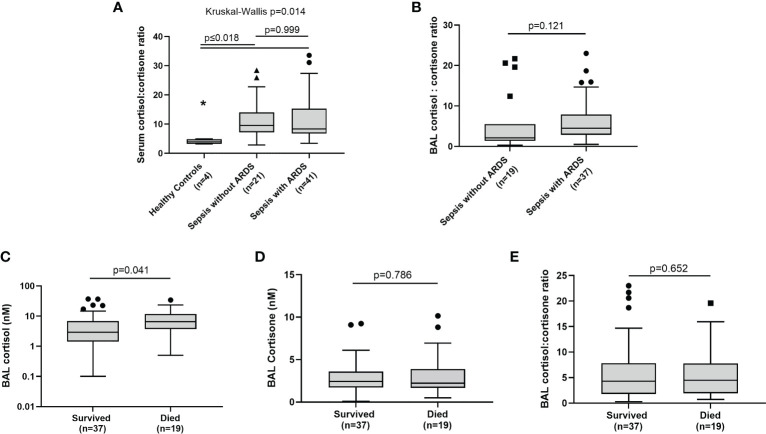
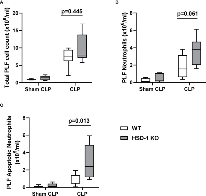
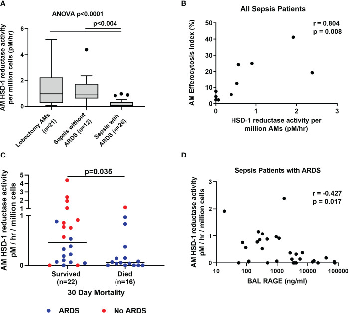
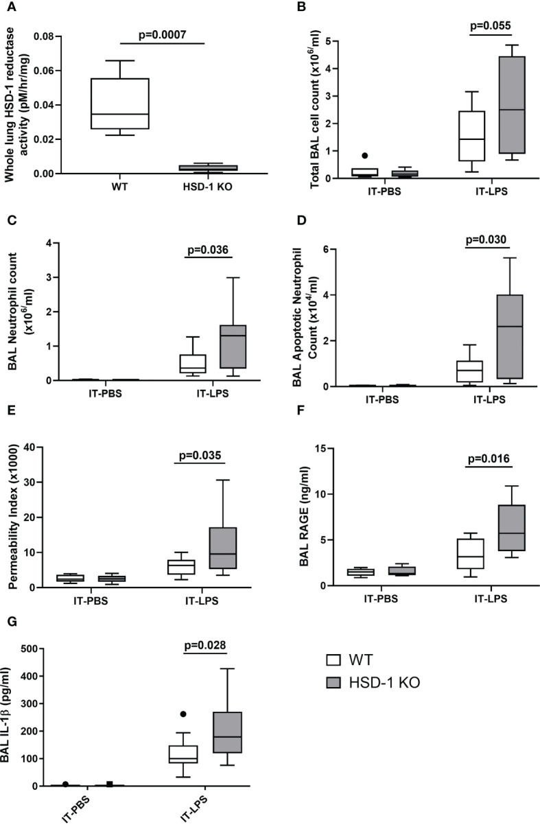
No difference in serum and BAL cortisol: cortisone ratios are shown between sepsis patients with and without ARDS. Across all sepsis patients, there is no association between BAL cortisol: cortisone ratio and 30-day mortality. However, AM HSD-1 reductase activity is impaired in patients with sepsis-related ARDS, compared to sepsis patients without ARDS and lobectomy patients (0.075 v 0.882 v 0.967 pM/hr/10 AMs, p=0.004). Across all sepsis patients (with and without ARDS), impaired AM HSD-1 reductase activity is associated with defective efferocytosis (r=0.804, p=0.008) and increased 30-day mortality. AM HSD-1 reductase activity negatively correlates with BAL RAGE in sepsis patients with ARDS (r=-0.427, p=0.017). Following intra-tracheal lipopolysaccharide (IT-LPS) injury, HSD-1 KO mice demonstrate increased alveolar neutrophil infiltration, apoptotic neutrophil accumulation, alveolar protein permeability and BAL RAGE concentrations compared to WT mice. Caecal Ligation and Puncture (CLP) injury in HSD-1 KO mice results in greater peritoneal apoptotic neutrophil accumulation compared to WT mice.
AM HSD-1 reductase activity does not shape total BAL and serum cortisol: cortisone ratios, however impaired HSD-1 autocrine signalling renders AMs insensitive to the anti-inflammatory effects of local glucocorticoids. This contributes to the decreased efferocytosis, increased BAL RAGE concentrations and mortality seen in sepsis-related ARDS. Upregulation of alveolar HSD-1 activity could restore AM function and improve clinical outcomes in these patients.
急性呼吸窘迫综合征(ARDS)是一种破坏性的肺部炎症性疾病,通常由败血症引发。糖皮质激素是一种具有免疫调节作用的类固醇,可以抑制炎症。它们在组织中的抗炎特性受到其前受体代谢和 11β-羟甾脱氢酶 1 型(HSD-1)将无活性前体转化为有活性形式的影响。我们假设,在与败血症相关的 ARDS 中,肺泡巨噬细胞(AM)的 HSD-1 活性和糖皮质激素激活受损,与更大的炎症损伤和更差的结局相关。
我们分析了两批危重病败血症患者的支气管肺泡灌洗液(BAL)和循环糖皮质激素水平、AM HSD-1 还原酶活性和晚期糖基化终产物受体(RAGE)水平,这些患者有或没有 ARDS。还在肺切除术患者中测量了 AM HSD-1 还原酶活性。我们在 HSD-1 敲除(KO)和野生型(WT)小鼠的肺损伤和败血症模型中评估了炎症损伤参数。
ARDS 与非 ARDS 败血症患者的血清和 BAL 皮质醇:可的松比值无差异。在所有败血症患者中,BAL 皮质醇:可的松比值与 30 天死亡率之间没有关联。然而,与非 ARDS 败血症患者和肺切除术患者相比,与败血症相关的 ARDS 患者的 AM HSD-1 还原酶活性受损(0.075 对 0.882 对 0.967 pM/hr/10 AMs,p=0.004)。在所有败血症患者(有或没有 ARDS)中,受损的 AM HSD-1 还原酶活性与缺陷的胞噬作用相关(r=0.804,p=0.008),并导致 30 天死亡率增加。AM HSD-1 还原酶活性与 ARDS 败血症患者的 BAL RAGE 呈负相关(r=-0.427,p=0.017)。在气管内脂多糖(IT-LPS)损伤后,与 WT 小鼠相比,HSD-1 KO 小鼠的肺泡中性粒细胞浸润、凋亡中性粒细胞积聚、肺泡蛋白通透性和 BAL RAGE 浓度增加。在 HSD-1 KO 小鼠的盲肠结扎和穿孔(CLP)损伤中,与 WT 小鼠相比,腹膜腔中的凋亡中性粒细胞积聚增加。
AM HSD-1 还原酶活性不会影响 BAL 和血清皮质醇:可的松比值,但受损的 HSD-1 自分泌信号会使 AM 对局部糖皮质激素的抗炎作用不敏感。这导致与败血症相关的 ARDS 中胞噬作用减少、BAL RAGE 浓度增加和死亡率增加。肺泡 HSD-1 活性的上调可能恢复 AM 的功能并改善这些患者的临床结局。