• 文献检索
  • 文档翻译
  • 深度研究
  • 学术资讯
  • Suppr Zotero 插件Zotero 插件
  • 邀请有礼
  • 套餐&价格
  • 历史记录
应用&插件
Suppr Zotero 插件Zotero 插件浏览器插件Mac 客户端Windows 客户端微信小程序
定价
高级版会员购买积分包购买API积分包
服务
文献检索文档翻译深度研究API 文档MCP 服务
关于我们
关于 Suppr公司介绍联系我们用户协议隐私条款
关注我们

Suppr 超能文献

核心技术专利:CN118964589B侵权必究
粤ICP备2023148730 号-1Suppr @ 2026

文献检索

告别复杂PubMed语法,用中文像聊天一样搜索,搜遍4000万医学文献。AI智能推荐,让科研检索更轻松。

立即免费搜索

文件翻译

保留排版,准确专业,支持PDF/Word/PPT等文件格式,支持 12+语言互译。

免费翻译文档

深度研究

AI帮你快速写综述,25分钟生成高质量综述,智能提取关键信息,辅助科研写作。

立即免费体验

利用 PHYTOMap 对植物组织进行多重单细胞 3D 空间基因表达分析。

Multiplexed single-cell 3D spatial gene expression analysis in plant tissue using PHYTOMap.

机构信息

Plant Biology Laboratory, The Salk Institute for Biological Studies, La Jolla, CA, USA.

Genomic Analysis Laboratory, The Salk Institute for Biological Studies, La Jolla, CA, USA.

出版信息

Nat Plants. 2023 Jul;9(7):1026-1033. doi: 10.1038/s41477-023-01439-4. Epub 2023 Jun 12.

DOI:10.1038/s41477-023-01439-4
PMID:37308583
原文链接:https://pmc.ncbi.nlm.nih.gov/articles/PMC10356616/
Abstract

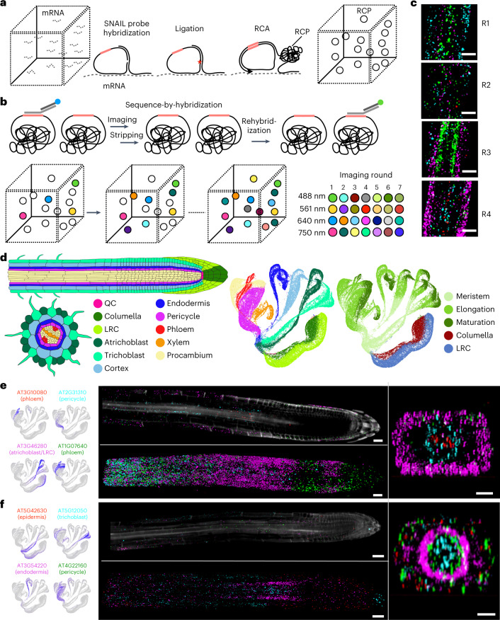
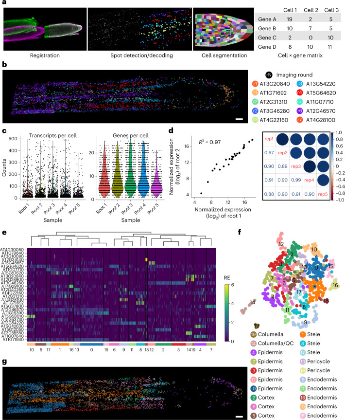
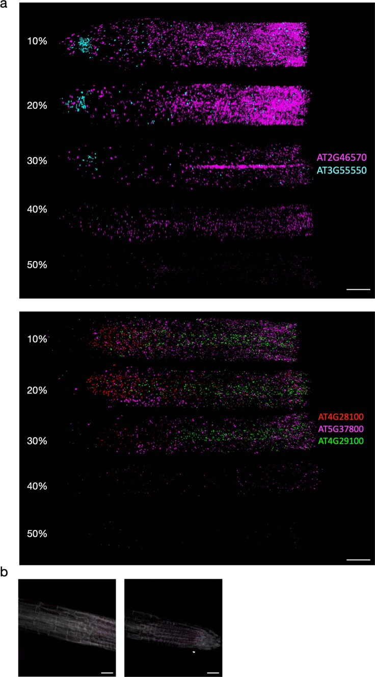
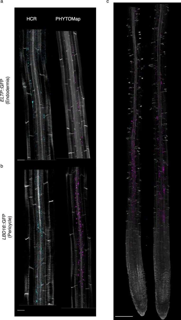
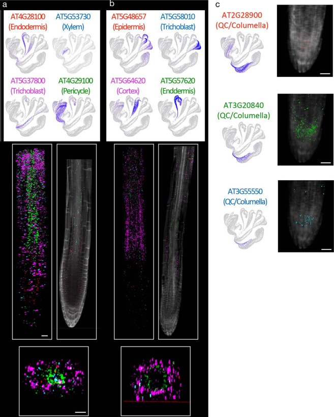
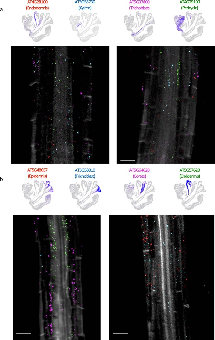
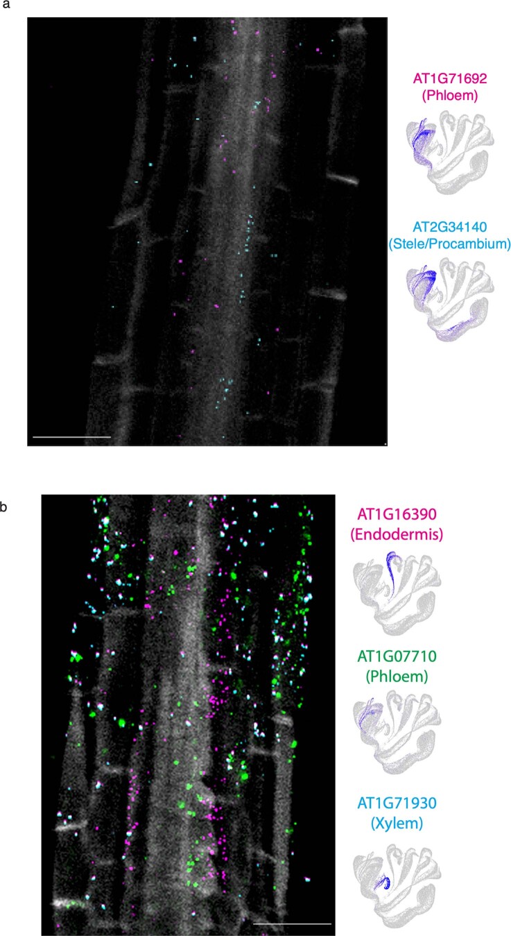
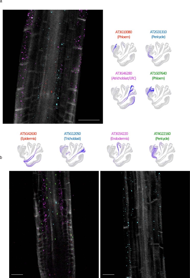
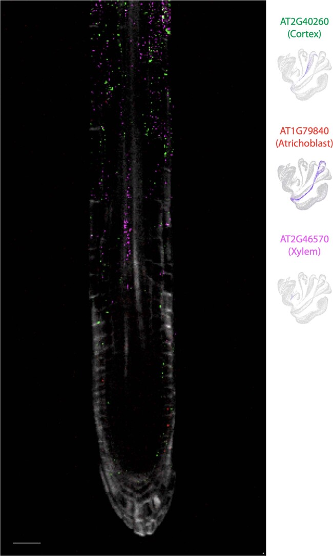
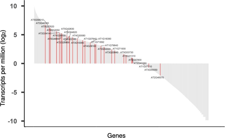
Retrieving the complex responses of individual cells in the native three-dimensional tissue context is crucial for a complete understanding of tissue functions. Here, we present PHYTOMap (plant hybridization-based targeted observation of gene expression map), a multiplexed fluorescence in situ hybridization method that enables single-cell and spatial analysis of gene expression in whole-mount plant tissue in a transgene-free manner and at low cost. We applied PHYTOMap to simultaneously analyse 28 cell-type marker genes in Arabidopsis roots and successfully identified major cell types, demonstrating that our method can substantially accelerate the spatial mapping of marker genes defined in single-cell RNA-sequencing datasets in complex plant tissue.

摘要

在原生的三维组织环境中获取单个细胞的复杂反应对于全面理解组织功能至关重要。在这里,我们介绍了 PHYTOMap(基于植物杂交的靶向观察基因表达图谱),这是一种多重荧光原位杂交方法,可在无转基因且低成本的情况下,对整个植物组织中的单细胞和空间进行基因表达的分析。我们将 PHYTOMap 应用于拟南芥根中 28 个细胞类型标记基因的同时分析,并成功鉴定了主要细胞类型,表明我们的方法可以大大加速在复杂植物组织中单细胞 RNA-seq 数据集定义的标记基因的空间图谱绘制。

https://cdn.ncbi.nlm.nih.gov/pmc/blobs/71fc/10356616/d80ff936698c/41477_2023_1439_Fig12_ESM.jpg
https://cdn.ncbi.nlm.nih.gov/pmc/blobs/71fc/10356616/c7ee89e0734e/41477_2023_1439_Fig1_HTML.jpg
https://cdn.ncbi.nlm.nih.gov/pmc/blobs/71fc/10356616/1131dcdf78e4/41477_2023_1439_Fig2_HTML.jpg
https://cdn.ncbi.nlm.nih.gov/pmc/blobs/71fc/10356616/513a48956f35/41477_2023_1439_Fig3_ESM.jpg
https://cdn.ncbi.nlm.nih.gov/pmc/blobs/71fc/10356616/e8e5b4c497be/41477_2023_1439_Fig4_ESM.jpg
https://cdn.ncbi.nlm.nih.gov/pmc/blobs/71fc/10356616/112e2182a9bb/41477_2023_1439_Fig5_ESM.jpg
https://cdn.ncbi.nlm.nih.gov/pmc/blobs/71fc/10356616/543cf841446b/41477_2023_1439_Fig6_ESM.jpg
https://cdn.ncbi.nlm.nih.gov/pmc/blobs/71fc/10356616/bb8699a6c14d/41477_2023_1439_Fig7_ESM.jpg
https://cdn.ncbi.nlm.nih.gov/pmc/blobs/71fc/10356616/292587e7df48/41477_2023_1439_Fig8_ESM.jpg
https://cdn.ncbi.nlm.nih.gov/pmc/blobs/71fc/10356616/abfdcb7ebefb/41477_2023_1439_Fig9_ESM.jpg
https://cdn.ncbi.nlm.nih.gov/pmc/blobs/71fc/10356616/9d53dad85409/41477_2023_1439_Fig10_ESM.jpg
https://cdn.ncbi.nlm.nih.gov/pmc/blobs/71fc/10356616/dc6772a428b0/41477_2023_1439_Fig11_ESM.jpg
https://cdn.ncbi.nlm.nih.gov/pmc/blobs/71fc/10356616/d80ff936698c/41477_2023_1439_Fig12_ESM.jpg

相似文献

1
Multiplexed single-cell 3D spatial gene expression analysis in plant tissue using PHYTOMap.利用 PHYTOMap 对植物组织进行多重单细胞 3D 空间基因表达分析。
Nat Plants. 2023 Jul;9(7):1026-1033. doi: 10.1038/s41477-023-01439-4. Epub 2023 Jun 12.
2
Analysis of 3D gene expression patterns in plants using whole-mount RNA in situ hybridization.使用整体原位杂交 RNA 分析植物中的 3D 基因表达模式。
Nat Protoc. 2014 Oct;9(10):2464-75. doi: 10.1038/nprot.2014.162. Epub 2014 Sep 25.
3
High-throughput single-cell gene-expression profiling with multiplexed error-robust fluorescence in situ hybridization.利用多重抗误差荧光原位杂交技术进行高通量单细胞基因表达谱分析。
Proc Natl Acad Sci U S A. 2016 Sep 27;113(39):11046-51. doi: 10.1073/pnas.1612826113. Epub 2016 Sep 13.
4
A method for cellular localization of gene expression via quantitative in situ hybridization in plants.一种通过植物定量原位杂交进行基因表达细胞定位的方法。
Plant J. 2007 Apr;50(1):159-75. doi: 10.1111/j.1365-313X.2007.03031.x.
5
A rapid and sensitive, multiplex, whole mount RNA fluorescence in situ hybridization and immunohistochemistry protocol.一种快速灵敏的多重全组织RNA荧光原位杂交和免疫组织化学方法。
Plant Methods. 2023 Nov 22;19(1):131. doi: 10.1186/s13007-023-01108-9.
6
Cell segmentation in imaging-based spatial transcriptomics.基于成像的空间转录组学中的细胞分割
Nat Biotechnol. 2022 Mar;40(3):345-354. doi: 10.1038/s41587-021-01044-w. Epub 2021 Oct 14.
7
Spatial transcriptome profiling by MERFISH reveals subcellular RNA compartmentalization and cell cycle-dependent gene expression.MERFISH 技术进行空间转录组分析揭示了细胞内 RNA 区室化和细胞周期依赖性基因表达。
Proc Natl Acad Sci U S A. 2019 Sep 24;116(39):19490-19499. doi: 10.1073/pnas.1912459116. Epub 2019 Sep 9.
8
Molecularly defined and spatially resolved cell atlas of the whole mouse brain.分子定义和空间分辨的全鼠脑细胞图谱。
Nature. 2023 Dec;624(7991):343-354. doi: 10.1038/s41586-023-06808-9. Epub 2023 Dec 13.
9
High-performance multiplexed fluorescence in situ hybridization in culture and tissue with matrix imprinting and clearing.利用基质印记和透明化技术在培养物和组织中进行的高性能多重荧光原位杂交。
Proc Natl Acad Sci U S A. 2016 Dec 13;113(50):14456-14461. doi: 10.1073/pnas.1617699113. Epub 2016 Nov 22.
10
Characterizing spatial gene expression heterogeneity in spatially resolved single-cell transcriptomic data with nonuniform cellular densities.刻画具有非均匀细胞密度的空间分辨单细胞转录组数据中空间基因表达异质性。
Genome Res. 2021 Oct;31(10):1843-1855. doi: 10.1101/gr.271288.120. Epub 2021 May 25.

引用本文的文献

1
A computational Evo-Devo approach for elucidating the roles of PLETHORA transcription factors in regulating root development.一种用于阐明多胚盘转录因子在调节根发育中作用的计算进化发育方法。
PLoS One. 2025 Jul 31;20(7):e0327511. doi: 10.1371/journal.pone.0327511. eCollection 2025.
2
In silico encounters: harnessing metabolic modelling to understand plant-microbe interactions.虚拟相遇:利用代谢模型理解植物-微生物相互作用。
FEMS Microbiol Rev. 2025 Jan 14;49. doi: 10.1093/femsre/fuaf030.
3
Exploring the untapped potential of single-cell and spatial omics in plant biology.

本文引用的文献

1
Museum of spatial transcriptomics.空间转录组学博物馆。
Nat Methods. 2022 May;19(5):534-546. doi: 10.1038/s41592-022-01409-2. Epub 2022 Mar 10.
2
EASI-FISH for thick tissue defines lateral hypothalamus spatio-molecular organization.EASI-FISH 技术可用于厚组织,定义外侧下丘脑的空间分子组织。
Cell. 2021 Dec 22;184(26):6361-6377.e24. doi: 10.1016/j.cell.2021.11.024. Epub 2021 Dec 6.
3
An in situ sequencing approach maps PLASTOCHRON1 at the boundary between indeterminate and determinate cells.原位测序方法绘制了不定细胞和决定细胞边界处的 PLASTOCHRON1。
探索单细胞和空间组学在植物生物学中尚未开发的潜力。
New Phytol. 2025 May 21. doi: 10.1111/nph.70220.
4
Genomics-assisted breeding for designing salinity-smart future crops.通过基因组学辅助育种设计适应盐渍环境的未来作物。
Plant Biotechnol J. 2025 Aug;23(8):3119-3151. doi: 10.1111/pbi.70104. Epub 2025 May 20.
5
A timeline of discovery and innovation in Arabidopsis.拟南芥的发现与创新时间表。
Plant Cell. 2025 May 9;37(5). doi: 10.1093/plcell/koaf108.
6
A space for time. Exploring temporal regulation of plant development across spatial scales.以空间换时间。探索植物发育在不同空间尺度上的时间调控。
Plant J. 2025 Apr;122(1):e70130. doi: 10.1111/tpj.70130.
7
Deep-tissue transcriptomics and subcellular imaging at high spatial resolution.高空间分辨率下的深层组织转录组学和亚细胞成像。
Science. 2025 Apr 18;388(6744):eadq2084. doi: 10.1126/science.adq2084.
8
A rare PRIMER cell state in plant immunity.植物免疫中一种罕见的原始细胞状态。
Nature. 2025 Feb;638(8049):197-205. doi: 10.1038/s41586-024-08383-z. Epub 2025 Jan 8.
9
Harnessing Single-Cell and Spatial Transcriptomics for Crop Improvement.利用单细胞和空间转录组学促进作物改良。
Plants (Basel). 2024 Dec 11;13(24):3476. doi: 10.3390/plants13243476.
10
Can a plant biologist fix a thermostat?植物生物学家能修理恒温器吗?
New Phytol. 2025 Feb;245(4):1403-1410. doi: 10.1111/nph.20382. Epub 2025 Jan 2.
Plant Physiol. 2022 Feb 4;188(2):782-794. doi: 10.1093/plphys/kiab533.
4
Plant single-cell solutions for energy and the environment.用于能源和环境的植物单细胞解决方案。
Commun Biol. 2021 Aug 12;4(1):962. doi: 10.1038/s42003-021-02477-4.
5
Exploring tissue architecture using spatial transcriptomics.利用空间转录组学探索组织架构。
Nature. 2021 Aug;596(7871):211-220. doi: 10.1038/s41586-021-03634-9. Epub 2021 Aug 11.
6
Advances and Opportunities in Single-Cell Transcriptomics for Plant Research.植物单细胞转录组学研究的进展与机遇
Annu Rev Plant Biol. 2021 Jun 17;72:847-866. doi: 10.1146/annurev-arplant-081720-010120. Epub 2021 Mar 17.
7
Hybridization-based in situ sequencing (HybISS) for spatially resolved transcriptomics in human and mouse brain tissue.基于杂交的原位测序(HybISS)在人类和小鼠脑组织中进行空间分辨转录组学分析。
Nucleic Acids Res. 2020 Nov 4;48(19):e112. doi: 10.1093/nar/gkaa792.
8
Accurate and versatile 3D segmentation of plant tissues at cellular resolution.以细胞分辨率准确且灵活地对植物组织进行三维分割。
Elife. 2020 Jul 29;9:e57613. doi: 10.7554/eLife.57613.
9
Gene expression cartography.基因表达图谱。
Nature. 2019 Dec;576(7785):132-137. doi: 10.1038/s41586-019-1773-3. Epub 2019 Nov 20.
10
Preparation of plant tissue to enable Spatial Transcriptomics profiling using barcoded microarrays.使用带条码的微阵列进行空间转录组分析的植物组织准备。
Nat Protoc. 2018 Nov;13(11):2425-2446. doi: 10.1038/s41596-018-0046-1.