Ke Haoxian, Li Zhihao, Li Peisi, Ye Shubiao, Huang Junfeng, Hu Tuo, Zhang Chi, Yuan Ming, Chen Yuan, Wu Xianrui, Lan Ping
Department of General Surgery (Colorectal Surgery), The Sixth Affiliated Hospital, Sun Yat-sen University, Guangzhou, Guangdong, P. R. China.
Guangdong Provincial Key Laboratory of Colorectal and Pelvic Floor Diseases, Guangdong Institute of Gastroenterology ,The Sixth Affiliated Hospital, Sun Yat-sen University, Guangzhou, Guangdong, P. R. China.
Gastroenterol Rep (Oxf). 2023 Jun 24;11:goad034. doi: 10.1093/gastro/goad034. eCollection 2023.
Tumor heterogeneity is contributed by tumor cells and the microenvironment. Dynamics of tumor heterogeneity during colorectal cancer (CRC) progression have not been elucidated.
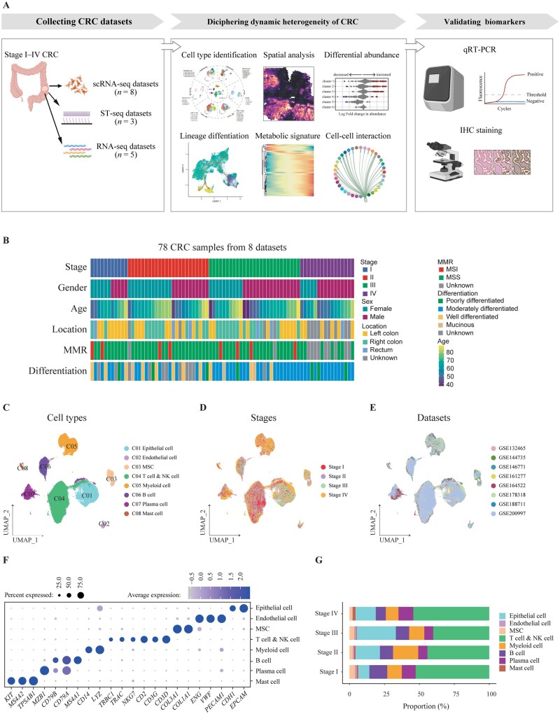
Eight single-cell RNA sequencing (scRNA-seq) data sets of CRC were included. Milo was utilized to reveal the differential abundance of cell clusters during progression. The differentiation trajectory was imputed by using the Palantir algorithm and metabolic states were assessed by using scMetabolism. Three spatial transcription sequencing (ST-seq) data sets of CRC were used to validate cell-type abundances and colocalization. Cancer-associated regulatory hubs were defined as communication networks affecting tumor biological behaviors. Finally, quantitative reverse transcription polymerase chain reaction and immunohistochemistry staining were performed for validation.
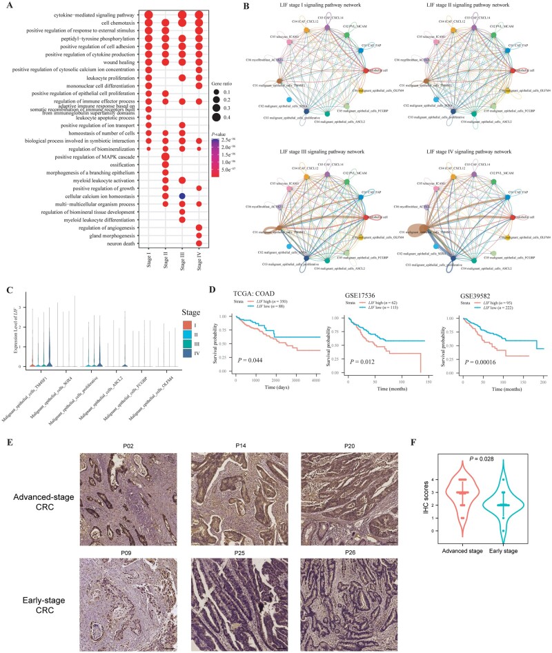
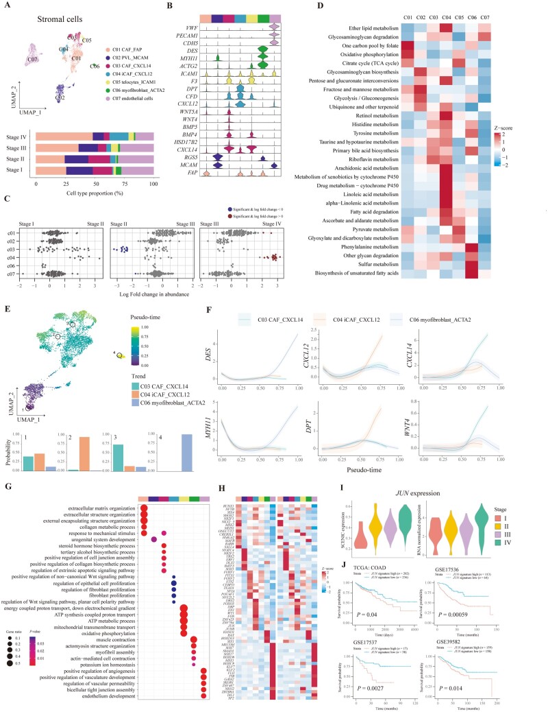
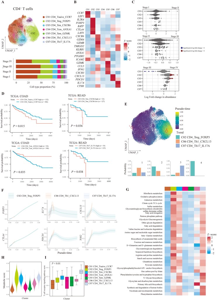
TM4SF1, SOX4, and MKI67 tumor cells; CXCL12 cancer-associated fibroblasts; CD4 resident memory T cells; Treg; IgA plasma cells; and several myeloid subsets were enriched in stage IV CRC, most of which were associated with overall survival of patients. Trajectory analysis indicated that tumor cells from patients with advanced-stage CRC were less differentiated, when metabolic heterogeneity showed a highest metabolic signature in terminal states of stromal cells, T cells, and myeloid cells. Moreover, ST-seq validated cell-type abundance in a spatial context and also revealed the correlation of immune infiltration between tertiary lymphoid structures and tumors followed by validation in our cohort. Importantly, analysis of cancer-associated regulatory hubs revealed a cascade of activated pathways including leukocyte apoptotic process, MAPK pathway, myeloid leukocyte differentiation, and angiogenesis during CRC progression.
Tumor heterogeneity was dynamic during progression, with the enrichment of immunosuppressive Treg, myeloid cells, and fibrotic cells. The differential state of tumor cells was associated with cancer staging. Assessment of cancer-associated regulatory hubs suggested impaired antitumor immunity and increased metastatic ability during CRC progression.
肿瘤异质性由肿瘤细胞和微环境共同导致。结直肠癌(CRC)进展过程中肿瘤异质性的动态变化尚未阐明。
纳入8个CRC的单细胞RNA测序(scRNA-seq)数据集。利用Milo揭示进展过程中细胞簇的差异丰度。使用Palantir算法推断分化轨迹,并使用scMetabolism评估代谢状态。使用3个CRC的空间转录组测序(ST-seq)数据集验证细胞类型丰度和共定位情况。将癌症相关调控枢纽定义为影响肿瘤生物学行为的通信网络。最后,进行定量逆转录聚合酶链反应和免疫组织化学染色以进行验证。
TM4SF1、SOX4和MKI67肿瘤细胞;CXCL12癌症相关成纤维细胞;CD4驻留记忆T细胞;调节性T细胞(Treg);IgA浆细胞;以及几个髓系亚群在IV期CRC中富集,其中大多数与患者的总生存期相关。轨迹分析表明,晚期CRC患者的肿瘤细胞分化程度较低,而代谢异质性在基质细胞、T细胞和髓系细胞的终末状态显示出最高的代谢特征。此外,ST-seq在空间背景下验证了细胞类型丰度,还揭示了三级淋巴结构与肿瘤之间免疫浸润的相关性,随后在我们的队列中进行了验证。重要的是,对癌症相关调控枢纽的分析揭示了CRC进展过程中一系列激活的通路,包括白细胞凋亡过程、丝裂原活化蛋白激酶(MAPK)通路、髓系白细胞分化和血管生成。
肿瘤异质性在进展过程中是动态变化的,免疫抑制性Treg、髓系细胞和纤维化细胞会富集。肿瘤细胞的差异状态与癌症分期相关。对癌症相关调控枢纽的评估表明,CRC进展过程中抗肿瘤免疫受损且转移能力增强。