• 文献检索
  • 文档翻译
  • 深度研究
  • 学术资讯
  • Suppr Zotero 插件Zotero 插件
  • 邀请有礼
  • 套餐&价格
  • 历史记录
应用&插件
Suppr Zotero 插件Zotero 插件浏览器插件Mac 客户端Windows 客户端微信小程序
定价
高级版会员购买积分包购买API积分包
服务
文献检索文档翻译深度研究API 文档MCP 服务
关于我们
关于 Suppr公司介绍联系我们用户协议隐私条款
关注我们

Suppr 超能文献

核心技术专利:CN118964589B侵权必究
粤ICP备2023148730 号-1Suppr @ 2026

文献检索

告别复杂PubMed语法,用中文像聊天一样搜索,搜遍4000万医学文献。AI智能推荐,让科研检索更轻松。

立即免费搜索

文件翻译

保留排版,准确专业,支持PDF/Word/PPT等文件格式,支持 12+语言互译。

免费翻译文档

深度研究

AI帮你快速写综述,25分钟生成高质量综述,智能提取关键信息,辅助科研写作。

立即免费体验

使用知识蒸馏框架在磁共振图像上自动检测脑微出血

Automated detection of cerebral microbleeds on MR images using knowledge distillation framework.

作者信息

Sundaresan Vaanathi, Arthofer Christoph, Zamboni Giovanna, Murchison Andrew G, Dineen Robert A, Rothwell Peter M, Auer Dorothee P, Wang Chaoyue, Miller Karla L, Tendler Benjamin C, Alfaro-Almagro Fidel, Sotiropoulos Stamatios N, Sprigg Nikola, Griffanti Ludovica, Jenkinson Mark

机构信息

Department of Computational and Data Sciences, Indian Institute of Science, Bengaluru, Karnataka, India.

Wellcome Centre for Integrative Neuroimaging, Oxford Centre for Functional MRI of the Brain, Nuffield Department of Clinical Neurosciences, University of Oxford, Oxford, United Kingdom.

出版信息

Front Neuroinform. 2023 Jul 10;17:1204186. doi: 10.3389/fninf.2023.1204186. eCollection 2023.

DOI:10.3389/fninf.2023.1204186
PMID:37492242
原文链接:
https://pmc.ncbi.nlm.nih.gov/articles/PMC10363739/
Abstract

INTRODUCTION

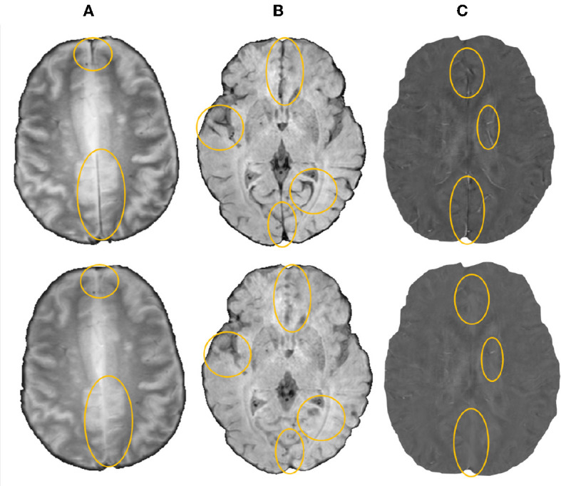
Cerebral microbleeds (CMBs) are associated with white matter damage, and various neurodegenerative and cerebrovascular diseases. CMBs occur as small, circular hypointense lesions on T2*-weighted gradient recalled echo (GRE) and susceptibility-weighted imaging (SWI) images, and hyperintense on quantitative susceptibility mapping (QSM) images due to their paramagnetic nature. Accurate automated detection of CMBs would help to determine quantitative imaging biomarkers (e.g., CMB count) on large datasets. In this work, we propose a fully automated, deep learning-based, 3-step algorithm, using structural and anatomical properties of CMBs from any single input image modality (e.g., GRE/SWI/QSM) for their accurate detections.

METHODS

In our method, the first step consists of an initial candidate detection step that detects CMBs with high sensitivity. In the second step, candidate discrimination step is performed using a knowledge distillation framework, with a multi-tasking teacher network that guides the student network to classify CMB and non-CMB instances in an offline manner. Finally, a morphological clean-up step further reduces false positives using anatomical constraints. We used four datasets consisting of different modalities specified above, acquired using various protocols and with a variety of pathological and demographic characteristics.

RESULTS

On cross-validation within datasets, our method achieved a cluster-wise true positive rate (TPR) of over 90% with an average of <2 false positives per subject. The knowledge distillation framework improves the cluster-wise TPR of the student model by 15%. Our method is flexible in terms of the input modality and provides comparable cluster-wise TPR and better cluster-wise precision compared to existing state-of-the-art methods. When evaluating across different datasets, our method showed good generalizability with a cluster-wise TPR >80 % with different modalities. The python implementation of the proposed method is openly available.

摘要

引言

脑微出血(CMB)与白质损伤以及各种神经退行性和脑血管疾病相关。在T2 *加权梯度回波(GRE)和磁敏感加权成像(SWI)图像上,CMB表现为小的圆形低信号病变,由于其顺磁性性质,在定量磁敏感图谱(QSM)图像上为高信号。准确自动检测CMB有助于在大型数据集中确定定量成像生物标志物(例如,CMB计数)。在这项工作中,我们提出了一种基于深度学习的全自动三步算法,利用来自任何单一输入图像模态(例如,GRE/SWI/QSM)的CMB的结构和解剖特性进行准确检测。

方法

在我们的方法中,第一步包括一个初始候选检测步骤,该步骤以高灵敏度检测CMB。第二步是候选判别步骤,使用知识蒸馏框架,通过一个多任务教师网络以离线方式指导学生网络对CMB和非CMB实例进行分类。最后,一个形态学清理步骤利用解剖学约束进一步减少假阳性。我们使用了四个数据集,这些数据集由上述不同模态组成,采用各种协议采集,具有各种病理和人口统计学特征。

结果

在数据集内的交叉验证中,我们的方法实现了超过90%的聚类真阳性率(TPR),平均每个受试者<2个假阳性。知识蒸馏框架将学生模型的聚类TPR提高了15%。我们的方法在输入模态方面具有灵活性,与现有的最先进方法相比,提供了相当的聚类TPR和更好的聚类精度。在跨不同数据集评估时,我们的方法表现出良好的通用性,不同模态下的聚类TPR>80%。所提出方法的Python实现是公开可用的。

https://cdn.ncbi.nlm.nih.gov/pmc/blobs/a978/10363739/408af6150356/fninf-17-1204186-g0011.jpg
https://cdn.ncbi.nlm.nih.gov/pmc/blobs/a978/10363739/eca2e114e88f/fninf-17-1204186-g0001.jpg
https://cdn.ncbi.nlm.nih.gov/pmc/blobs/a978/10363739/85d6f93ed031/fninf-17-1204186-g0002.jpg
https://cdn.ncbi.nlm.nih.gov/pmc/blobs/a978/10363739/80abc900764e/fninf-17-1204186-g0003.jpg
https://cdn.ncbi.nlm.nih.gov/pmc/blobs/a978/10363739/b51712fad26c/fninf-17-1204186-g0004.jpg
https://cdn.ncbi.nlm.nih.gov/pmc/blobs/a978/10363739/bc4769bd69ec/fninf-17-1204186-g0005.jpg
https://cdn.ncbi.nlm.nih.gov/pmc/blobs/a978/10363739/4173c432bc69/fninf-17-1204186-g0006.jpg
https://cdn.ncbi.nlm.nih.gov/pmc/blobs/a978/10363739/79ee8943df34/fninf-17-1204186-g0007.jpg
https://cdn.ncbi.nlm.nih.gov/pmc/blobs/a978/10363739/b5cfdc44f0bd/fninf-17-1204186-g0008.jpg
https://cdn.ncbi.nlm.nih.gov/pmc/blobs/a978/10363739/059bb8fcffdd/fninf-17-1204186-g0009.jpg
https://cdn.ncbi.nlm.nih.gov/pmc/blobs/a978/10363739/ed0ea27f10d5/fninf-17-1204186-g0010.jpg
https://cdn.ncbi.nlm.nih.gov/pmc/blobs/a978/10363739/408af6150356/fninf-17-1204186-g0011.jpg

相似文献

1
Automated detection of cerebral microbleeds on MR images using knowledge distillation framework.使用知识蒸馏框架在磁共振图像上自动检测脑微出血
Front Neuroinform. 2023 Jul 10;17:1204186. doi: 10.3389/fninf.2023.1204186. eCollection 2023.
2
Automated Detection of Candidate Subjects With Cerebral Microbleeds Using Machine Learning.使用机器学习自动检测患有脑微出血的候选受试者
Front Neuroinform. 2022 Jan 20;15:777828. doi: 10.3389/fninf.2021.777828. eCollection 2021.
3
Deep-Learning-Based MRI Microbleeds Detection for Cerebral Small Vessel Disease on Quantitative Susceptibility Mapping.基于深度学习的磁共振定量磁化率成像脑小血管病微出血检测。
J Magn Reson Imaging. 2024 Sep;60(3):1165-1175. doi: 10.1002/jmri.29198. Epub 2023 Dec 27.
4
Automated detection of cerebral microbleeds in MR images: A two-stage deep learning approach.基于两阶段深度学习的脑微出血磁共振图像自动检测方法
Neuroimage Clin. 2020;28:102464. doi: 10.1016/j.nicl.2020.102464. Epub 2020 Oct 13.
5
CMB-HUNT: Automatic detection of cerebral microbleeds using a deep neural network.CMB-HUNT:使用深度神经网络自动检测脑微出血。
Comput Biol Med. 2022 Dec;151(Pt A):106233. doi: 10.1016/j.compbiomed.2022.106233. Epub 2022 Oct 27.
6
Enhanced Reader Confidence and Differentiation of Calcification from Cerebral Microbleed Diagnosis Using QSM Relative to SWI.与磁敏感加权成像(SWI)相比,利用定量磁敏感图(QSM)增强读者信心并区分钙化与脑微出血诊断。
Clin Neuroradiol. 2024 Dec 17. doi: 10.1007/s00062-024-01478-0.
7
Susceptibility-weighted imaging is more reliable than T2*-weighted gradient-recalled echo MRI for detecting microbleeds.磁敏感加权成像比 T2*-加权梯度回波 MRI 更可靠,可用于检测微出血。
Stroke. 2013 Oct;44(10):2782-6. doi: 10.1161/STROKEAHA.113.002267. Epub 2013 Aug 6.
8
Toward automated detection of microbleeds with anatomical scale localization using deep learning.利用深度学习实现具有解剖尺度定位的微出血自动检测。
Med Image Anal. 2025 Apr;101:103415. doi: 10.1016/j.media.2024.103415. Epub 2024 Nov 30.
9
Clinical Performance of Quantitative Susceptibility Mapping in Cerebral Microbleed Detection Relative to 2D GRE.相对于二维梯度回波成像,定量磁化率图谱在脑微出血检测中的临床性能
Clin Neuroradiol. 2025 May 23. doi: 10.1007/s00062-025-01529-0.
10
Automated Detection of Cerebral Microbleeds on Two-dimensional Gradient-recalled Echo T2* Weighted Images Using a Morphology Filter Bank and Convolutional Neural Network.使用形态滤波器组和卷积神经网络在二维梯度回波T2*加权图像上自动检测脑微出血
Magn Reson Med Sci. 2025 Apr 1;24(2):220-228. doi: 10.2463/mrms.mp.2023-0146. Epub 2024 Mar 15.

引用本文的文献

1
A robust deep learning framework for cerebral microbleeds recognition in GRE and SWI MRI.一种用于在梯度回波(GRE)和磁敏感加权成像(SWI)磁共振成像中识别脑微出血的强大深度学习框架。
Neuroimage Clin. 2025 Aug 27;48:103873. doi: 10.1016/j.nicl.2025.103873.
2
Automated characterisation of cerebral microbleeds using their size and spatial distribution on brain MRI.利用脑微出血在脑部磁共振成像上的大小和空间分布进行自动特征描述。
Eur Radiol Exp. 2025 Jan 13;9(1):5. doi: 10.1186/s41747-024-00544-z.
3
SHIVA-CMB: a deep-learning-based robust cerebral microbleed segmentation tool trained on multi-source T2*GRE- and susceptibility-weighted MRI.

本文引用的文献

1
Phenotypic and genetic associations of quantitative magnetic susceptibility in UK Biobank brain imaging.英国生物库脑成像中定量磁化率的表型和遗传关联。
Nat Neurosci. 2022 Jun;25(6):818-831. doi: 10.1038/s41593-022-01074-w. Epub 2022 May 23.
2
Effect of Tranexamic Acid Administration on Remote Cerebral Ischemic Lesions in Acute Spontaneous Intracerebral Hemorrhage: A Substudy of a Randomized Clinical Trial.氨甲环酸给药对急性自发性脑出血后远隔脑缺血损伤的影响:一项随机临床试验的亚研究。
JAMA Neurol. 2022 May 1;79(5):468-477. doi: 10.1001/jamaneurol.2022.0217.
3
Automated Detection of Candidate Subjects With Cerebral Microbleeds Using Machine Learning.
SHIVA-CMB:一种基于深度学习的强大脑微出血分割工具,在多源T2*GRE和磁敏感加权MRI上进行训练。
Sci Rep. 2024 Dec 28;14(1):30901. doi: 10.1038/s41598-024-81870-5.
4
Enhancement and evaluation for deep learning-based classification of volumetric neuroimaging with 3D-to-2D knowledge distillation.基于深度学习的容积神经影像学分类的增强和评估,使用 3D 到 2D 知识蒸馏。
Sci Rep. 2024 Nov 28;14(1):29611. doi: 10.1038/s41598-024-80938-6.
5
Deep learning-assisted IoMT framework for cerebral microbleed detection.用于脑微出血检测的深度学习辅助物联网框架。
Heliyon. 2023 Nov 25;9(12):e22879. doi: 10.1016/j.heliyon.2023.e22879. eCollection 2023 Dec.
使用机器学习自动检测患有脑微出血的候选受试者
Front Neuroinform. 2022 Jan 20;15:777828. doi: 10.3389/fninf.2021.777828. eCollection 2021.
4
Prevalence and Risk Factors of Cerebral Microbleeds: Analysis From the UK Biobank.脑微出血的患病率及危险因素:来自英国生物银行的分析
Neurology. 2021 Oct 11;97(15):e1493-e1502. doi: 10.1212/WNL.0000000000012673.
5
DEEPMIR: a deep neural network for differential detection of cerebral microbleeds and iron deposits in MRI.DEEPMIR:一种用于 MRI 中脑微出血和铁沉积差异检测的深度神经网络。
Sci Rep. 2021 Jul 8;11(1):14124. doi: 10.1038/s41598-021-93427-x.
6
Automated detection of cerebral microbleeds on T2*-weighted MRI.基于 T2*-加权 MRI 的脑微出血自动检测。
Sci Rep. 2021 Feb 17;11(1):4004. doi: 10.1038/s41598-021-83607-0.
7
A Two Cascaded Network Integrating Regional-based YOLO and 3D-CNN for Cerebral Microbleeds Detection.一种用于脑微出血检测的集成基于区域的YOLO和3D-CNN的两级级联网络。
Annu Int Conf IEEE Eng Med Biol Soc. 2020 Jul;2020:1055-1058. doi: 10.1109/EMBC44109.2020.9176073.
8
Cerebral Micro-Bleeding Detection Based on Densely Connected Neural Network.基于密集连接神经网络的脑微出血检测
Front Neurosci. 2019 May 17;13:422. doi: 10.3389/fnins.2019.00422. eCollection 2019.
9
Cerebral microbleed detection using Susceptibility Weighted Imaging and deep learning.基于磁敏感加权成像和深度学习的脑微出血检测。
Neuroimage. 2019 Sep;198:271-282. doi: 10.1016/j.neuroimage.2019.05.046. Epub 2019 May 20.
10
Toward Automatic Detection of Radiation-Induced Cerebral Microbleeds Using a 3D Deep Residual Network.利用三维深度残差网络自动检测放射性脑微出血
J Digit Imaging. 2019 Oct;32(5):766-772. doi: 10.1007/s10278-018-0146-z.