Rane Levendovszky Swati
Department of Radiology, School of Medicine, University of Washington, Seattle, WA, United States.
Front Neuroimaging. 2022 Jun 2;1:828767. doi: 10.3389/fnimg.2022.828767. eCollection 2022.
Alzheimer's disease (AD) is a degenerative disease characterized by pathological accumulation of amyloid and phosphorylated tau. Typically, the early stage of AD, also called mild cognitive impairment (MCI), shows amyloid pathology. A small but significant number of individuals with MCI do not exhibit amyloid pathology but have elevated phosphorylated tau levels (A-T+ MCI). We used CSF amyloid and phosphorylated tau to identify the individuals with A+T+ and A-T+ MCI as well as cognitively normal (A-T-) controls. To increase the sample size, we leveraged the Global Alzheimer's Association Interactive Network and identified 137 MCI+ and 61 A-T+ MCI participants. We compared baseline and longitudinal, hippocampal, and cortical atrophy between groups.
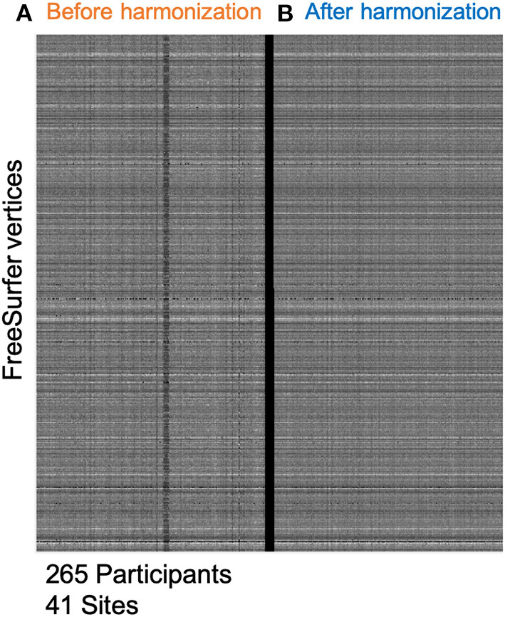
We applied ComBat harmonization to minimize site-related variability and used FreeSurfer for all measurements.
Harmonization reduced unwanted variability in cortical thickness by 3.4% and in hippocampal volume measurement by 10.3%. Cross-sectionally, widespread cortical thinning with age was seen in the A+T+ and A-T+ MCI groups ( < 0.0005). A decrease in the hippocampal volume with age was faster in both groups ( < 0.05) than in the controls. Longitudinally also, hippocampal atrophy rates were significant ( < 0.05) when compared with the controls. No longitudinal cortical thinning was observed in A-T+ MCI group.
A-T+ MCI participants showed similar baseline cortical thickness patterns with aging and longitudinal hippocampal atrophy rates as participants with A+T+ MCI, but did not show longitudinal cortical atrophy signature.
阿尔茨海默病(AD)是一种以淀粉样蛋白和磷酸化tau蛋白的病理性积聚为特征的退行性疾病。通常,AD的早期阶段,也称为轻度认知障碍(MCI),表现为淀粉样蛋白病理改变。一小部分但数量可观的MCI个体未表现出淀粉样蛋白病理改变,但磷酸化tau蛋白水平升高(A-T+ MCI)。我们使用脑脊液淀粉样蛋白和磷酸化tau蛋白来识别A+T+和A-T+ MCI个体以及认知正常(A-T-)的对照者。为了增加样本量,我们利用了全球阿尔茨海默病协会互动网络,确定了137名MCI+和61名A-T+ MCI参与者。我们比较了各组之间的基线以及纵向、海马和皮质萎缩情况。
我们应用ComBat归一化方法以最小化与研究中心相关的变异性,并使用FreeSurfer进行所有测量。
归一化使皮质厚度的不必要变异性降低了3.4%,海马体积测量的不必要变异性降低了10.3%。横断面分析显示,A+T+和A-T+ MCI组均出现随年龄增长的广泛皮质变薄(<0.0005)。两组海马体积随年龄的减小速度均快于对照组(<0.05)。纵向分析也显示,与对照组相比,海马萎缩率有显著差异(<0.05)。A-T+ MCI组未观察到纵向皮质变薄。
A-T+ MCI参与者在基线皮质厚度模式与衰老的关系以及纵向海马萎缩率方面与A+T+ MCI参与者相似,但未表现出纵向皮质萎缩特征。