Department of Anatomy, Physiology and Genetics, School of Medicine, Uniformed Services University of the Health Sciences, 4301 Jones Bridge Rd, Bethesda, MD, 20814, USA.
Henry M. Jackson Foundation for the Advancement of Military Medicine, Uniformed Services University of the Health Sciences, Bethesda, MD, USA.
Acta Neuropathol. 2023 Oct;146(4):585-610. doi: 10.1007/s00401-023-02622-9. Epub 2023 Aug 14.
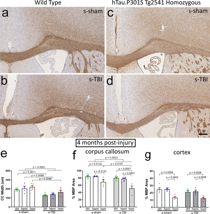
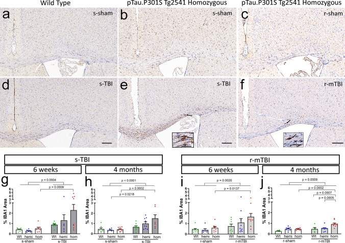
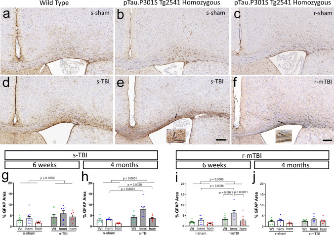
Traumatic brain injury (TBI) causes diffuse axonal injury which can produce chronic white matter pathology and subsequent post-traumatic neurodegeneration with poor patient outcomes. Tau modulates axon cytoskeletal functions and undergoes phosphorylation and mis-localization in neurodegenerative disorders. The effects of tau pathology on neurodegeneration after TBI are unclear. We used mice with neuronal expression of human mutant tau to examine effects of pathological tau on white matter pathology after TBI. Adult male and female hTau.P301S (Tg2541) transgenic and wild-type (Wt) mice received either moderate single TBI (s-TBI) or repetitive mild TBI (r-mTBI; once daily × 5), or sham procedures. Acutely, s-TBI produced more extensive axon damage in the corpus callosum (CC) as compared to r-mTBI. After s-TBI, significant CC thinning was present at 6 weeks and 4 months post-injury in Wt and transgenic mice, with homozygous tau expression producing additional pathology of late demyelination. In contrast, r-mTBI did not produce significant CC thinning except at the chronic time point of 4 months in homozygous mice, which exhibited significant CC atrophy (- 29.7%) with increased microgliosis. Serum neurofilament light quantification detected traumatic axonal injury at 1 day post-TBI in Wt and homozygous mice. At 4 months, high tau and neurofilament in homozygous mice implicated tau in chronic axon pathology. These findings did not have sex differences detected. Conclusions: Neuronal tau pathology differentially exacerbated CC pathology based on injury severity and chronicity. Ongoing CC atrophy from s-TBI became accompanied by late demyelination. Pathological tau significantly worsened CC atrophy during the chronic phase after r-mTBI.
创伤性脑损伤(TBI)导致弥漫性轴索损伤,可产生慢性白质病变和随后的创伤后神经退行性变,导致患者预后不良。tau 调节轴突细胞骨架功能,并在神经退行性疾病中发生磷酸化和定位错误。tau 病理学对 TBI 后神经退行性变的影响尚不清楚。我们使用表达人突变 tau 的神经元的小鼠来研究病理性 tau 对 TBI 后白质病变的影响。成年雄性和雌性 hTau.P301S(Tg2541)转基因和野生型(WT)小鼠接受中度单次 TBI(s-TBI)或重复轻度 TBI(r-mTBI;每天一次×5)或假手术。急性 s-TBI 在胼胝体(CC)中产生比 r-mTBI 更广泛的轴突损伤。在 s-TBI 后,WT 和转基因小鼠在 6 周和 4 个月伤后 CC 明显变薄,杂合子 tau 表达产生晚期脱髓鞘的额外病变。相比之下,r-mTBI 除了在 4 个月的慢性时间点外,不会导致 CC 明显变薄,杂合子小鼠的 CC 明显萎缩(-29.7%),伴有小胶质细胞增生增加。血清神经丝轻链定量检测在 WT 和杂合子小鼠的 TBI 后 1 天检测到创伤性轴索损伤。在 4 个月时,杂合子小鼠的 tau 和神经丝升高表明 tau 参与了慢性轴索病变。这些发现没有检测到性别差异。结论:神经元 tau 病理学根据损伤严重程度和慢性程度不同程度地加重 CC 病理学。s-TBI 持续的 CC 萎缩随后发生晚期脱髓鞘。r-mTBI 后的慢性阶段,病理性 tau 显著加重了 CC 萎缩。