Genes and Human Disease Program, Oklahoma Medical Research Foundation, 825 NE 13th Street, Oklahoma City, OK, 73104, USA.
Department of Physiology, University of Oklahoma Health Sciences Center, Oklahoma City, OK, USA.
J Neuroinflammation. 2023 Aug 16;20(1):188. doi: 10.1186/s12974-023-02870-2.
Microglia, the brain's principal immune cells, have been implicated in the pathogenesis of Alzheimer's disease (AD), a condition shown to affect more females than males. Although sex differences in microglial function and transcriptomic programming have been described across development and in disease models of AD, no studies have comprehensively identified the sex divergences that emerge in the aging mouse hippocampus. Further, existing models of AD generally develop pathology (amyloid plaques and tau tangles) early in life and fail to recapitulate the aged brain environment associated with late-onset AD. Here, we examined and compared transcriptomic and translatomic sex effects in young and old murine hippocampal microglia.
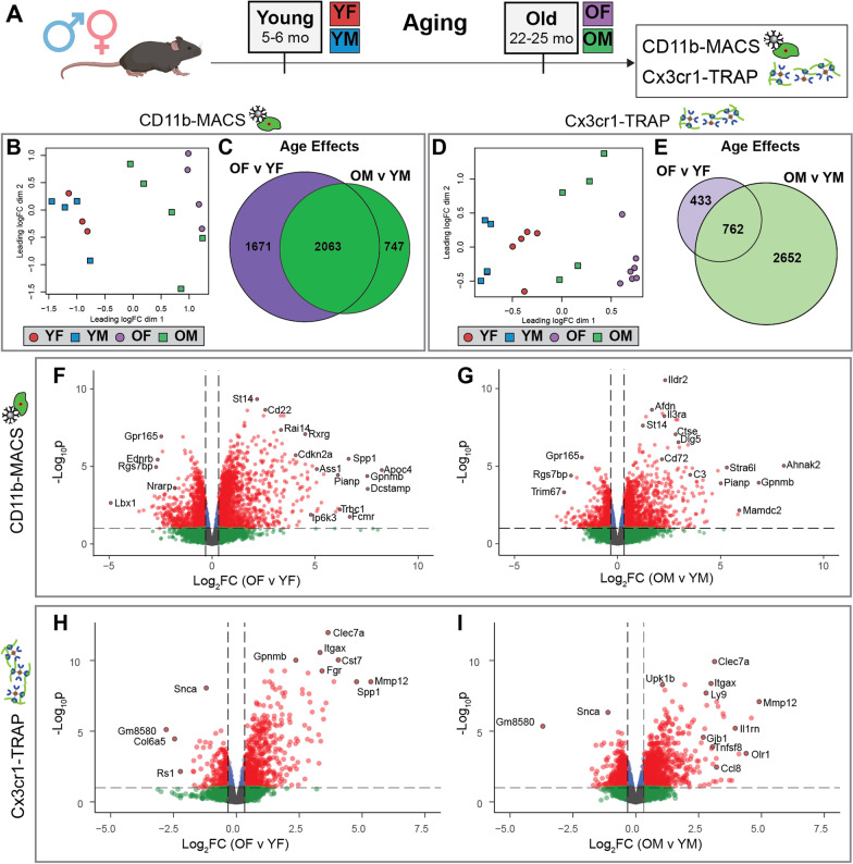
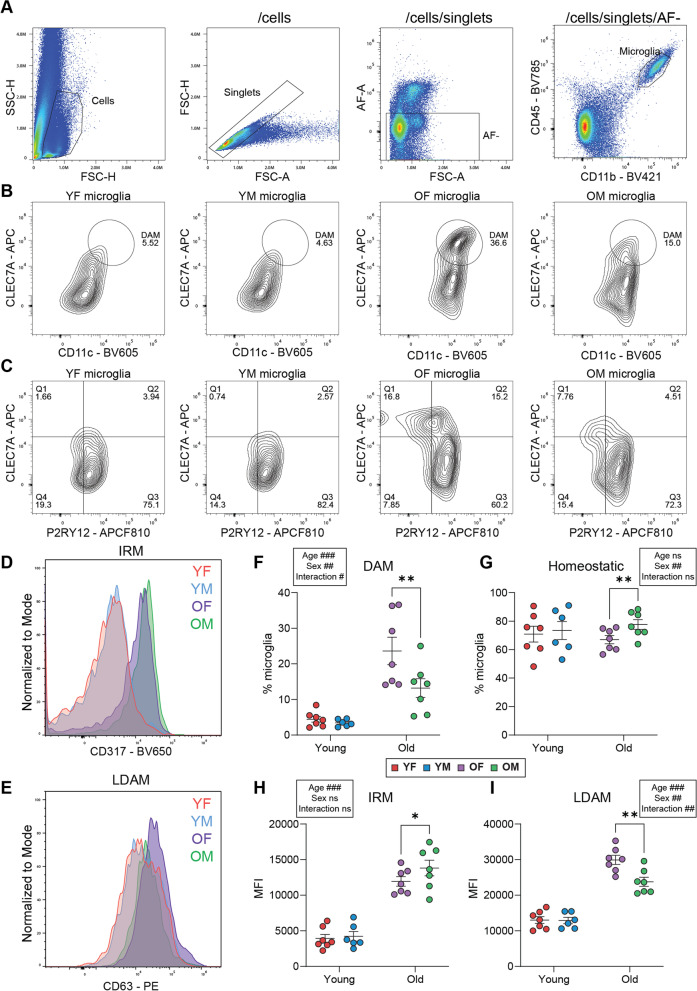
Hippocampal tissue from C57BL6/N and microglial NuTRAP mice of both sexes were collected at young (5-6 month-old [mo]) and old (22-25 mo) ages. Cell sorting and affinity purification techniques were used to isolate the microglial transcriptome and translatome for RNA-sequencing and differential expression analyses. Flow cytometry, qPCR, and imaging approaches were used to confirm the transcriptomic and translatomic findings.
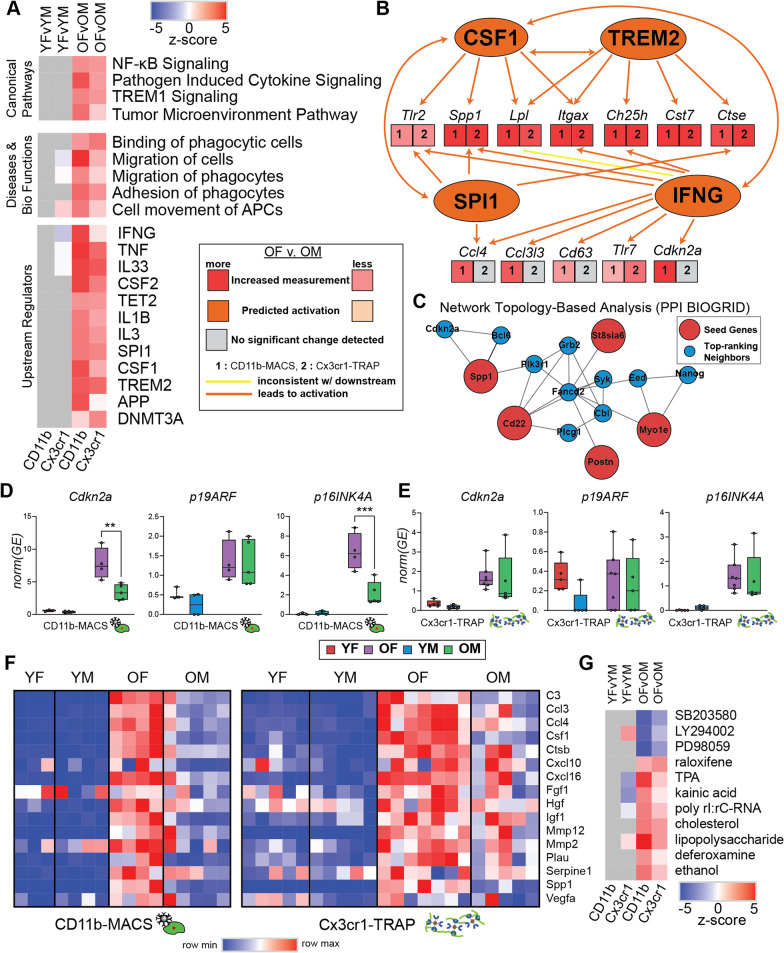
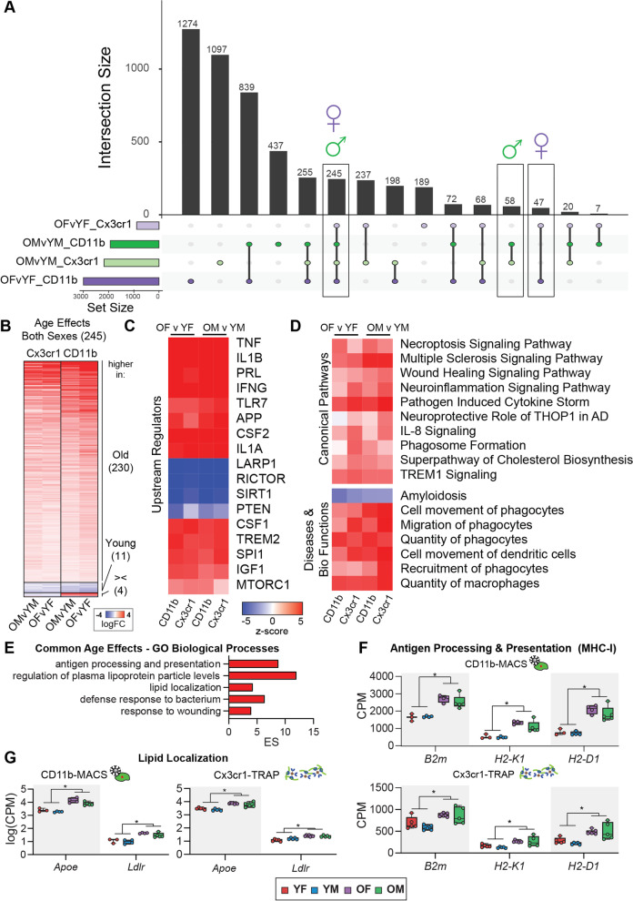
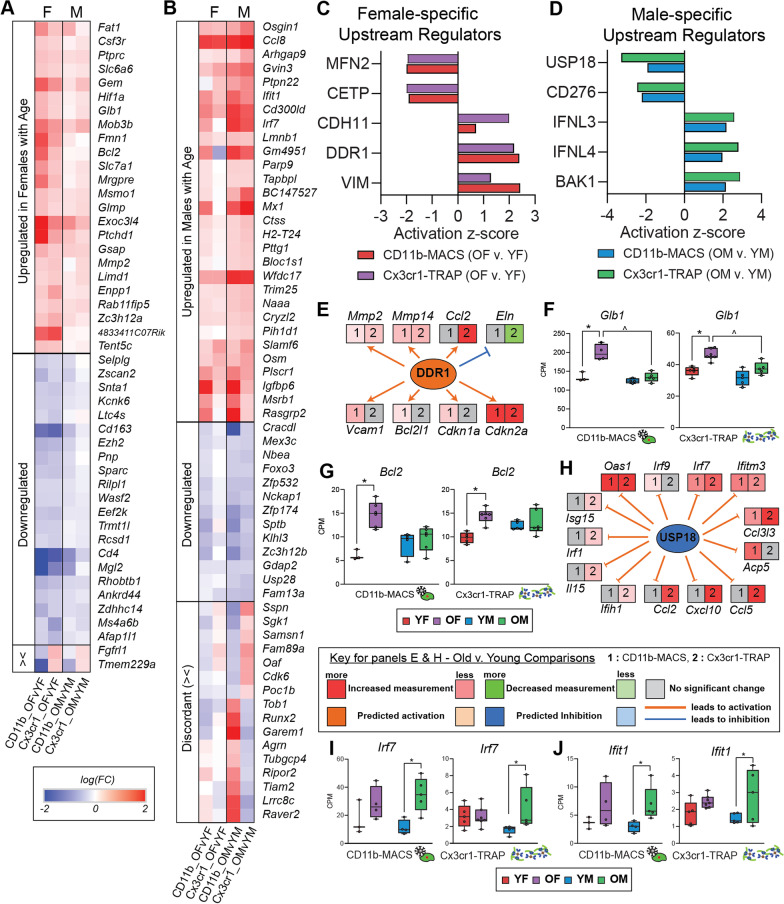
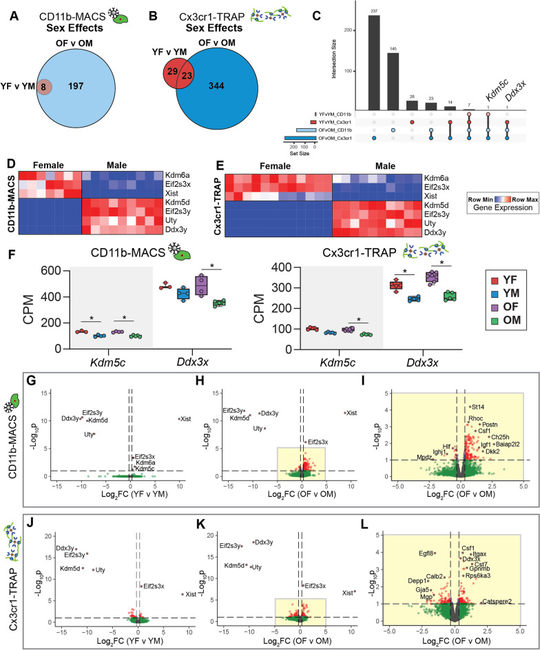
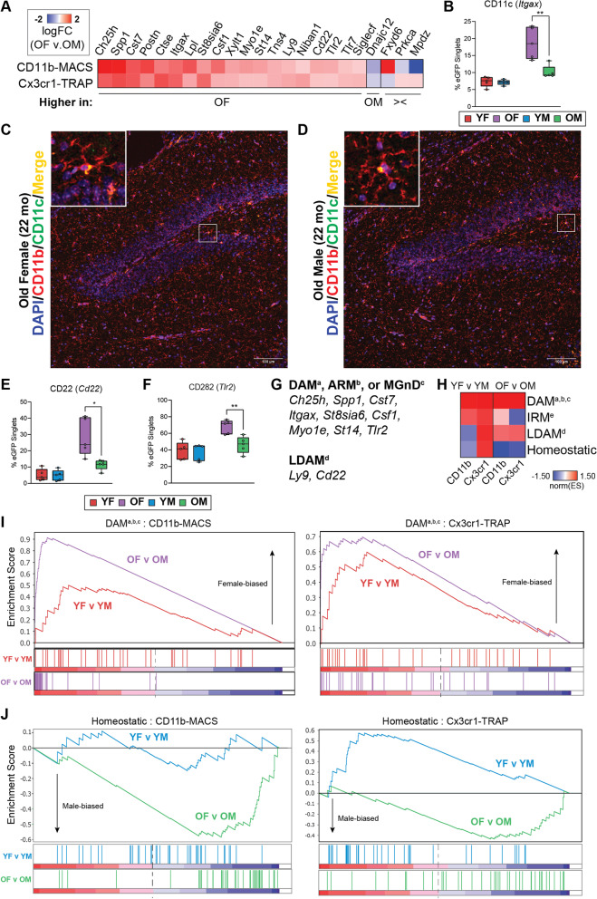
There were marginal sex differences identified in the young hippocampal microglia, with most differentially expressed genes (DEGs) restricted to the sex chromosomes. Both sex chromosomally and autosomally encoded sex differences emerged with aging. These sex DEGs identified at old age were primarily female-biased and enriched in senescent and disease-associated microglial signatures. Normalized gene expression values can be accessed through a searchable web interface ( https://neuroepigenomics.omrf.org/ ). Pathway analyses identified upstream regulators induced to a greater extent in females than in males, including inflammatory mediators IFNG, TNF, and IL1B, as well as AD-risk genes TREM2 and APP.
These data suggest that female microglia adopt disease-associated and senescent phenotypes in the aging mouse hippocampus, even in the absence of disease pathology, to a greater extent than males. This sexually divergent microglial phenotype may explain the difference in susceptibility and disease progression in the case of AD pathology. Future studies will need to explore sex differences in microglial heterogeneity in response to AD pathology and determine how sex-specific regulators (i.e., sex chromosomal or hormonal) elicit these sex effects.
小胶质细胞是大脑的主要免疫细胞,已被牵连到阿尔茨海默病(AD)的发病机制中,这种疾病表现为女性比男性更容易患病。尽管在 AD 疾病模型中,已经在发育过程中和在疾病模型中描述了小胶质细胞功能和转录组编程方面的性别差异,但没有研究全面确定在衰老的小鼠海马体中出现的性别差异。此外,现有的 AD 模型通常在生命早期发展病理学(淀粉样斑块和tau 缠结),并且未能重现与晚发性 AD 相关的老年大脑环境。在这里,我们检查并比较了年轻和年老的小鼠海马体小胶质细胞中的转录组和转译组的性别差异。
从 C57BL6/N 和 NuTRAP 小鼠的两性中收集年轻(5-6 月龄[mo])和年老(22-25 mo)的海马组织。使用细胞分选和亲和纯化技术分离小胶质细胞的转录组和转译组,进行 RNA-seq 和差异表达分析。使用流式细胞术,qPCR 和成像方法来确认转录组和转译组的发现。
在年轻的海马体小胶质细胞中发现了微小的性别差异,大多数差异表达的基因(DEGs)仅限于性染色体。随着年龄的增长,不仅出现了性染色体和常染色体编码的性别差异。在老年时发现的这些性别 DEGs 主要是雌性偏倚的,并富集了衰老和与疾病相关的小胶质细胞特征。通过可搜索的网络界面(https://neuroepigenomics.omrf.org/)可以访问标准化的基因表达值。途径分析确定了雌性中诱导程度更高的上游调节剂,包括炎症介质 IFNG、TNF 和 IL1B,以及 AD 风险基因 TREM2 和 APP。
这些数据表明,即使在没有疾病病理学的情况下,雌性小胶质细胞在衰老的小鼠海马体中也会表现出与疾病相关的和衰老的表型,比雄性更明显。这种性别分化的小胶质细胞表型可能解释了 AD 病理学中易感性和疾病进展的差异。未来的研究将需要探索 AD 病理学对小胶质细胞异质性的性别差异,并确定性别特异性调节剂(即性染色体或激素)如何引发这些性别效应。