State Key Laboratory of Modern Optical Instrumentations, Centre for Optical and Electromagnetic Research, College of Optical Science and Engineering, International Research Center for Advanced Photonics, Zhejiang University, Hangzhou, 310058, China.
College of Veterinary Medicine, Jilin University, Changchun, 130062, China.
Nat Commun. 2023 Aug 18;14(1):5017. doi: 10.1038/s41467-023-40728-6.
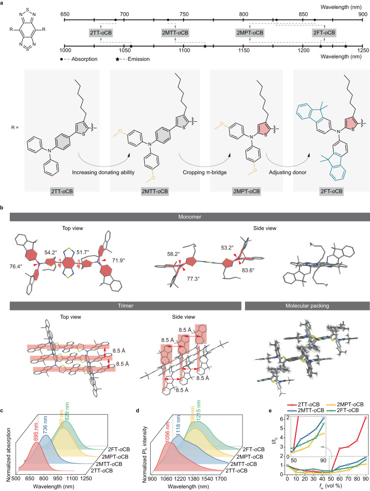
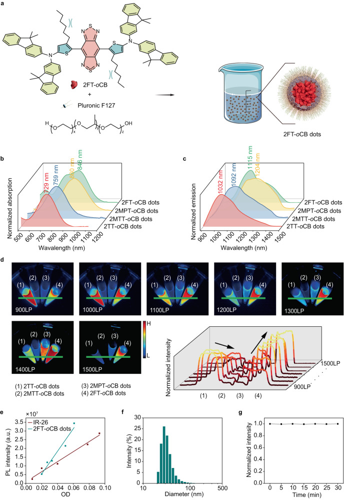
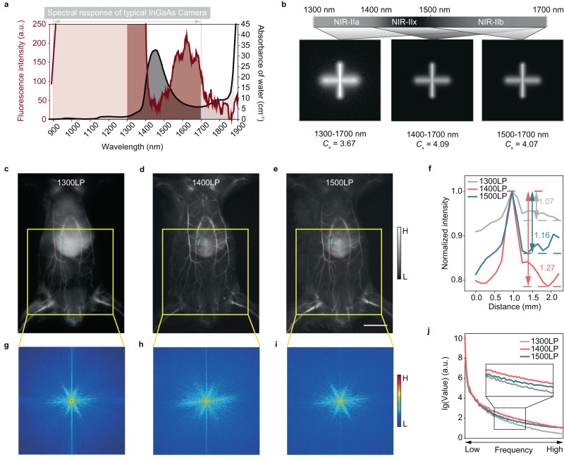
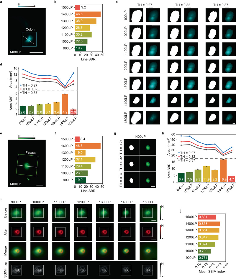
The limited signal of long-wavelength near-infrared-II (NIR-II, 900-1880 nm) fluorophores and the strong background caused by the diffused photons make high-contrast fluorescence imaging in vivo with deep tissue disturbed still challenging. Here, we develop NIR-II fluorescent small molecules with aggregation-induced emission properties, high brightness, and maximal emission beyond 1200 nm by enhancing electron-donating ability and reducing the donor-acceptor (D-A) distance, to complement the scarce bright long-wavelength emissive organic dyes. The convincing single-crystal evidence of D-A-D molecular structure reveals the strong inhibition of the π-π stacking with ultralong molecular packing distance exceeding 8 Å. The delicately-designed nanofluorophores with bright fluorescent signals extending to 1900 nm match the background-suppressed imaging window, enabling the signal-to-background ratio of the tissue image to reach over 100 with the tissue thickness of ~4-6 mm. In addition, the intraluminal lesions with strong negatively stained can be identified with almost zero background. This method can provide new avenues for future long-wavelength NIR-II molecular design and biomedical imaging of deep and highly scattering tissues.
长波长近红外二区(NIR-II,900-1880nm)荧光团的信号有限,漫射光子产生的强背景使得深部组织中的高对比度荧光成像是具有挑战性的。在这里,我们通过增强供电子能力和减小给体-受体(D-A)距离,开发出具有聚集诱导发射特性、高光亮度和最大发射超过 1200nm 的 NIR-II 荧光小分子,以补充稀少的明亮长波长发射有机染料。D-A-D 分子结构的令人信服的单晶证据表明,π-π 堆积受到强烈抑制,超长分子堆积距离超过 8Å。精心设计的纳米荧光团具有延伸至 1900nm 的明亮荧光信号,与背景抑制成像窗口匹配,使组织图像的信号与背景比达到 100 以上,组织厚度约为 4-6mm。此外,具有强负染的管腔内病变可以几乎无背景识别。该方法可为未来的长波长近红外二区分子设计和深部高散射组织的生物医学成像提供新途径。