Tamaddon Ali Mohammad, Bashiri Rahman, Najafi Haniyeh, Mousavi Khadijeh, Jafari Mahboobeh, Borandeh Sedigheh, Aghdaie Mahdokht H, Shafiee Mina, Abolmaali Samira Sadat, Azarpira Negar
Pharmaceutical Nanotechnology Department, Shiraz University of Medical Sciences, Shiraz, PO Box 71345-1583, Iran.
Center for Nanotechnology in Drug Delivery, Shiraz University of Medical Sciences, Shiraz, PO Box 71345-1583, Iran.
Heliyon. 2023 Aug 19;9(8):e19153. doi: 10.1016/j.heliyon.2023.e19153. eCollection 2023 Aug.
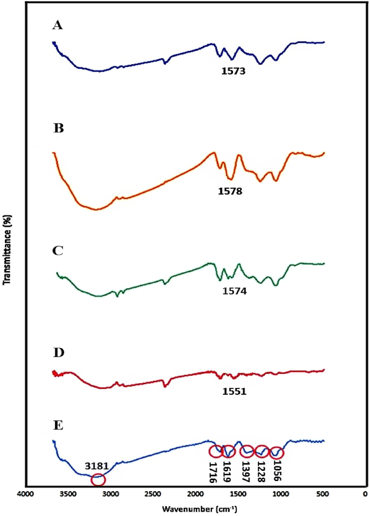
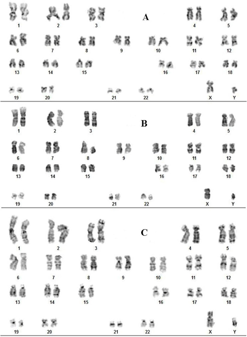
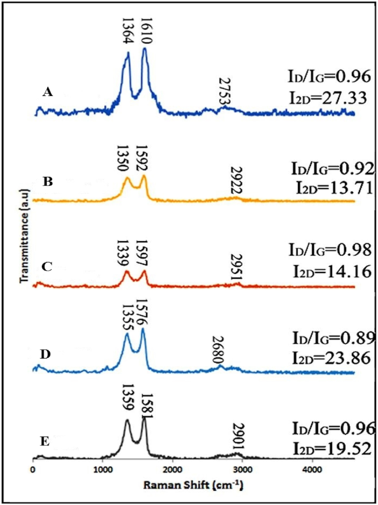
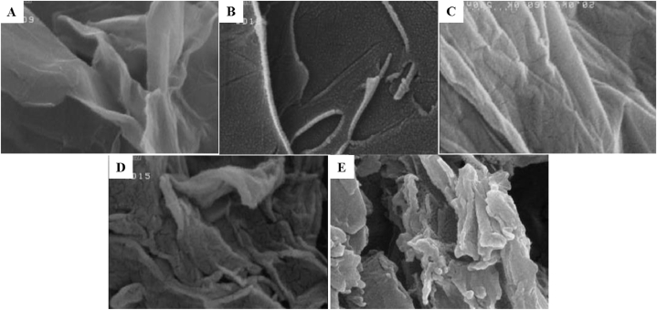
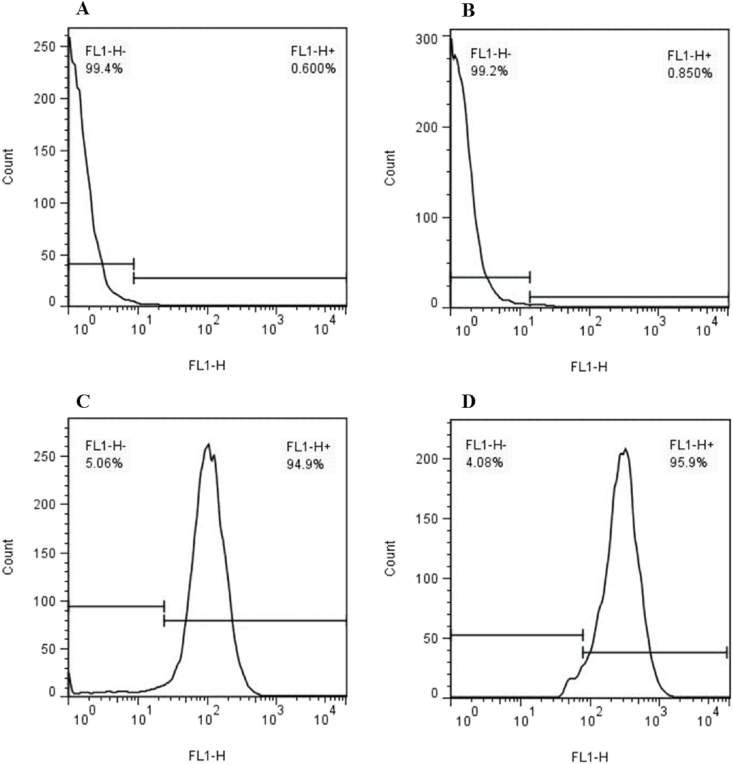
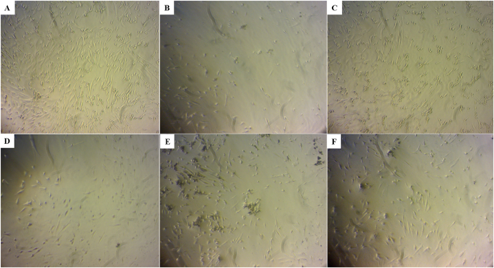
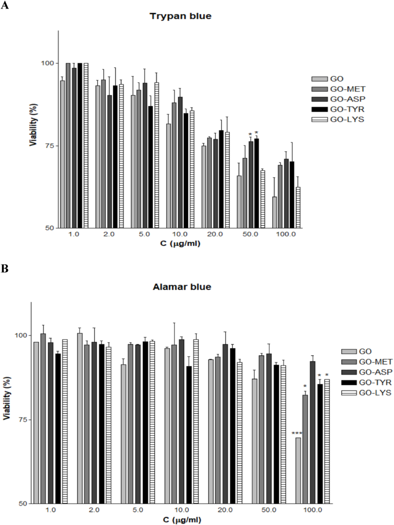
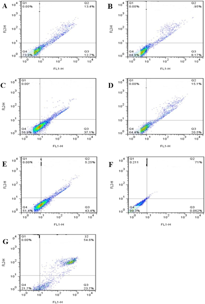
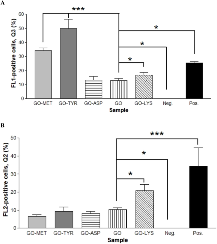
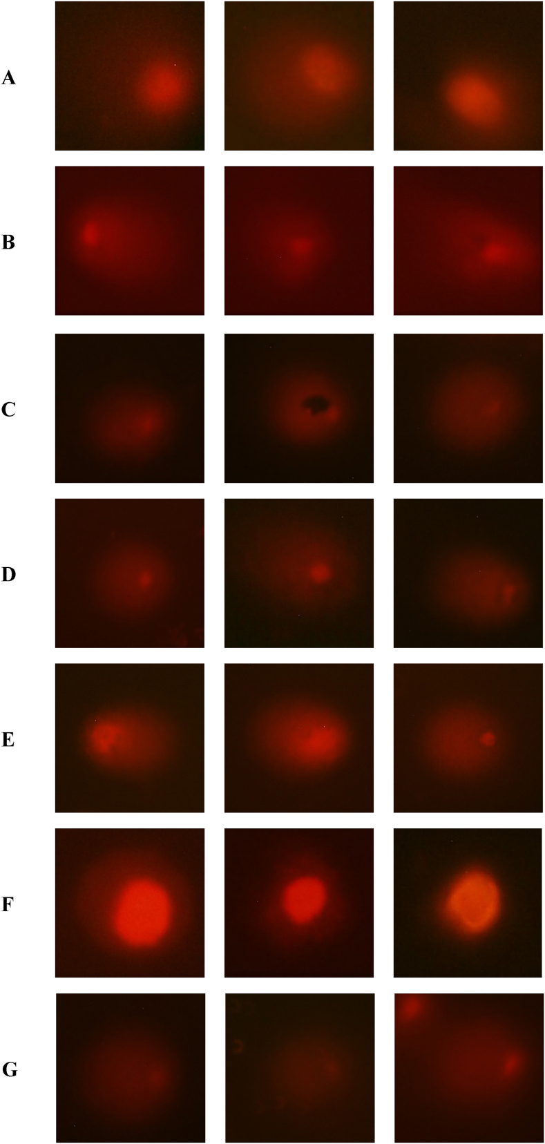
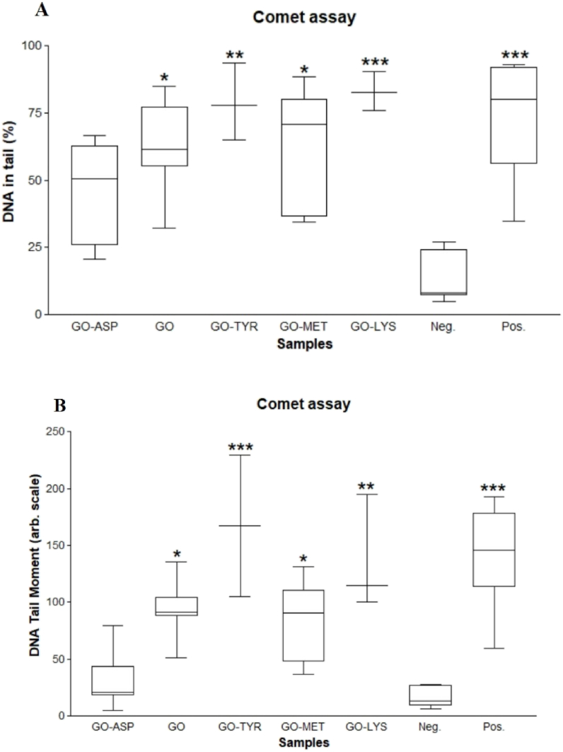
Graphene and its derivatives have gained popularity due to their numerous applications in various fields, such as biomedicine. Recent reports have revealed the severe toxic effects of these nanomaterials on cells and organs. In general, the chemical composition and surface chemistry of nanomaterials affect their biocompatibility. Therefore, the purpose of the present study was to evaluate the cytotoxicity and genotoxicity of graphene oxide (GO) synthesized by Hummer's method and functionalized by different amino acids such as lysine, methionine, aspartate, and tyrosine. The obtained nanosheets were identified by FT-IR, EDX, RAMAN, FE-SEM, and DLS techniques. In addition, trypan blue and Alamar blue methods were used to assess the cytotoxicity of mesenchymal stem cells extracted from human embryonic umbilical cord Wharton jelly (WJ-MSCs). The annexin V staining procedure was used to determine apoptotic and necrotic death. In addition, COMET and karyotyping techniques were used to assess the extent of DNA and chromosome damage. The results of the cytotoxicity assay showed that amino acid modifications significantly reduced the concentration-dependent cytotoxicity of GO to varying degrees. The GO modified with aspartic acid had the lowest cytotoxicity. There was no evidence of chromosomal damage in the karyotyping method, but in the comet assay, the samples modified with tyrosine and lysine showed the greatest DNA damage and rate of apoptosis. Overall, the aspartic acid-modified GO caused the least cellular and genetic damage to WJ-MSCs, implying its superior biomedical applications such as cell therapy and tissue engineering over GO.
石墨烯及其衍生物因其在生物医学等各个领域的众多应用而受到广泛关注。最近的报道揭示了这些纳米材料对细胞和器官的严重毒性作用。一般来说,纳米材料的化学成分和表面化学会影响其生物相容性。因此,本研究的目的是评估通过Hummer法合成并经赖氨酸、蛋氨酸、天冬氨酸和酪氨酸等不同氨基酸功能化的氧化石墨烯(GO)的细胞毒性和遗传毒性。通过傅里叶变换红外光谱(FT-IR)、能谱分析(EDX)、拉曼光谱(RAMAN)、场发射扫描电子显微镜(FE-SEM)和动态光散射(DLS)技术对所得纳米片进行了鉴定。此外,采用台盼蓝和alamar蓝法评估从人胚胎脐带华通氏胶中提取的间充质干细胞(WJ-MSCs)的细胞毒性。采用膜联蛋白V染色程序来确定凋亡和坏死死亡情况。此外,采用彗星试验和核型分析技术评估DNA和染色体损伤程度。细胞毒性试验结果表明,氨基酸修饰在不同程度上显著降低了GO的浓度依赖性细胞毒性。用天冬氨酸修饰的GO细胞毒性最低。核型分析方法中没有染色体损伤的证据,但在彗星试验中,用酪氨酸和赖氨酸修饰的样品显示出最大的DNA损伤和凋亡率。总体而言,天冬氨酸修饰的GO对WJ-MSCs造成的细胞和遗传损伤最小,这意味着其在细胞治疗和组织工程等生物医学应用方面优于GO。