Department of Radiology, University of Pennsylvania, Philadelphia, PA, USA.
Department of Bioengineering, University of Pennsylvania, Philadelphia, PA, USA.
Sci Rep. 2023 Sep 9;13(1):14895. doi: 10.1038/s41598-023-41969-7.
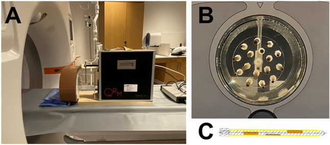
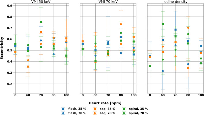
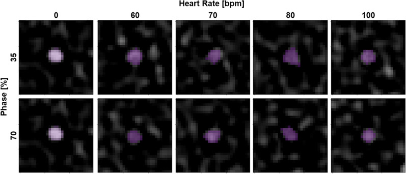
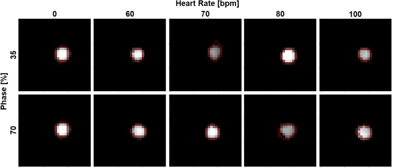
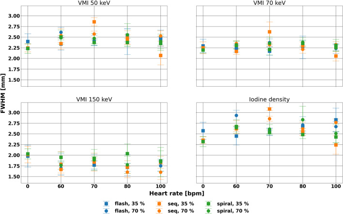
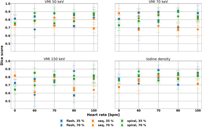
We evaluate stability of spectral results at different heart rates, acquisition modes, and cardiac phases in first-generation clinical dual-source photon-counting CT (PCCT). A cardiac motion simulator with a coronary stenosis mimicking a 50% eccentric calcium plaque was scanned at five different heart rates (0, 60-100 bpm) with the three available cardiac scan modes (high pitch prospectively ECG-triggered spiral, prospectively ECG-triggered axial, retrospectively ECG-gated spiral). Subsequently, full width half max (FWHM) of the stenosis, Dice score (DSC) for the stenosed region, and eccentricity of the non-stenosed region were calculated for virtual monoenergetic images (VMI) at 50, 70, and 150 keV and iodine density maps at both diastole and systole. FWHM averaged differences of - 0.20, - 0.28, and - 0.15 mm relative to static FWHM at VMI 150 keV across acquisition parameters for high pitch prospectively ECG-triggered spiral, prospectively ECG-triggered axial, and retrospectively ECG-gated spiral scans, respectively. Additionally, there was no effect of heart rate and acquisition mode on FWHM at diastole (p-values < 0.001). DSC demonstrated similarity among parameters with standard deviations of 0.08, 0.09, 0.11, and 0.08 for VMI 50, 70, and 150 keV, and iodine density maps, respectively, with insignificant differences at diastole (p-values < 0.01). Similarly, eccentricity illustrated small differences across heart rate and acquisition mode for each spectral result. Consistency of spectral results at different heart rates and acquisition modes for different cardiac phase demonstrates the added benefit of spectral results from PCCT to dual-source CT to further increase confidence in quantification and advance cardiovascular diagnostics.
我们评估第一代临床双源光子计数 CT (PCCT) 在不同心率、采集模式和心动周期下光谱结果的稳定性。使用具有模拟 50%偏心钙斑块的冠状动脉狭窄的心脏运动模拟器,以 5 种不同的心率(0、60-100 bpm)和 3 种可用的心脏扫描模式(高心率前瞻性心电图触发螺旋、前瞻性心电图触发轴向、回顾性心电图门控螺旋)进行扫描。随后,计算虚拟单能图像(VMI)50、70 和 150 keV 以及舒张期和收缩期碘密度图中狭窄处的全宽半高(FWHM)、狭窄区域的 Dice 评分(DSC)和非狭窄区域的偏心度。相对于 VMI 150 keV 的静态 FWHM,高心率前瞻性心电图触发螺旋、前瞻性心电图触发轴向和回顾性心电图门控螺旋扫描的 FWHM 平均差异分别为-0.20、-0.28 和-0.15mm。此外,心率和采集模式对舒张期 FWHM 没有影响(p 值<0.001)。DSC 表明各参数之间具有相似性,VMI 50、70 和 150 keV 以及碘密度图的标准偏差分别为 0.08、0.09、0.11 和 0.08,舒张期无显著差异(p 值<0.01)。同样,偏心度在每个光谱结果中在心率和采集模式之间显示出较小的差异。在不同的心率和采集模式下,在不同的心动周期下,光谱结果的一致性表明 PCCT 对双源 CT 的光谱结果具有额外的益处,可以进一步提高定量的可信度并推进心血管诊断。