Department of Ophthalmology, Wilmer Eye Institute, Johns Hopkins University, Baltimore, Maryland, United States of America.
McKusick-Nathans Institute of the Department of Genetic Medicine, Johns Hopkins School of Medicine, Baltimore, Maryland, United States of America.
PLoS Genet. 2023 Oct 11;19(10):e1010905. doi: 10.1371/journal.pgen.1010905. eCollection 2023 Oct.
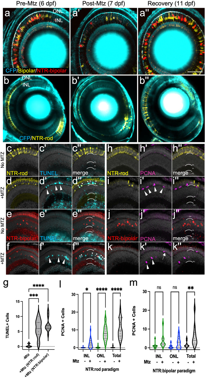
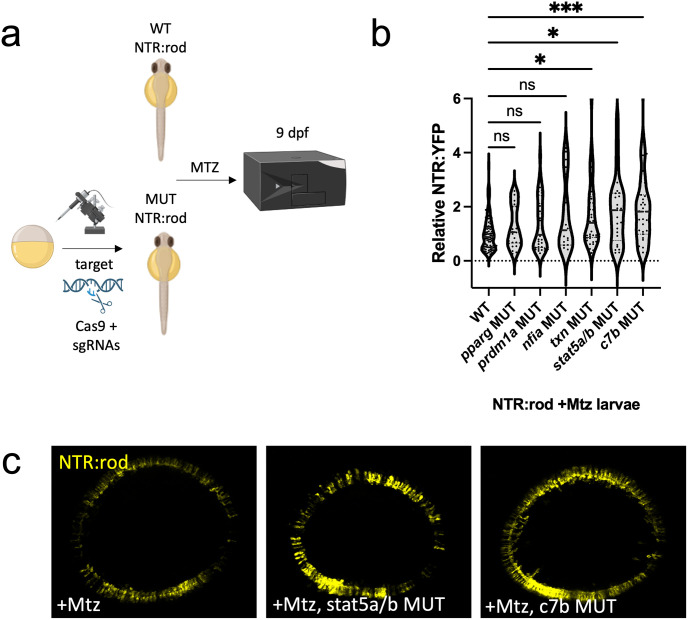
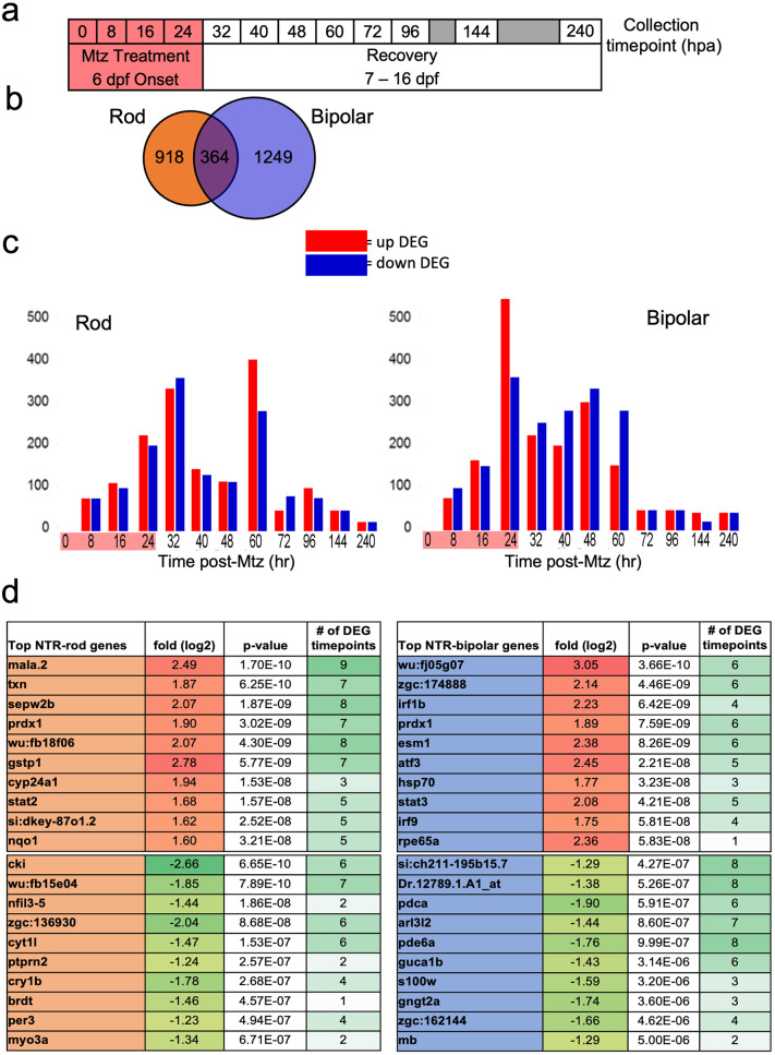
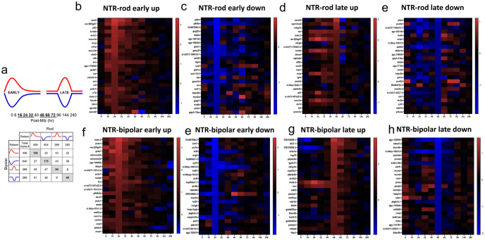
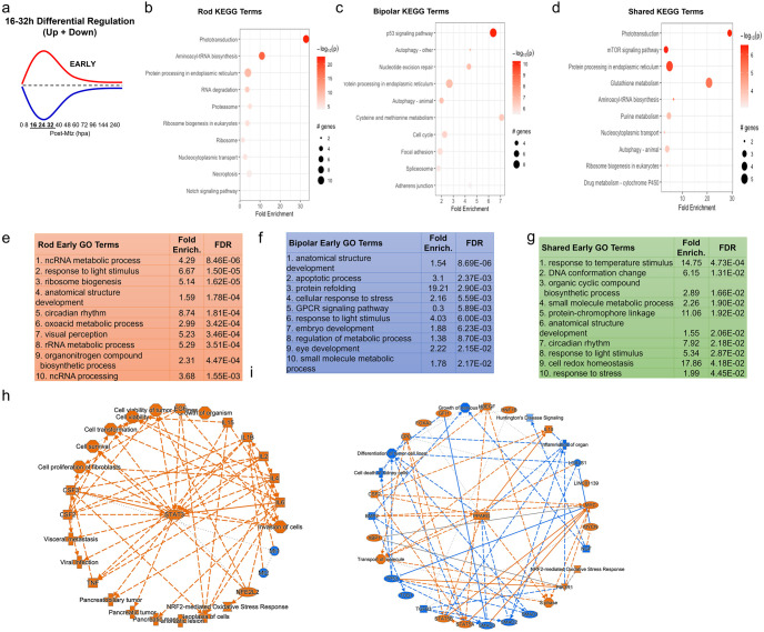
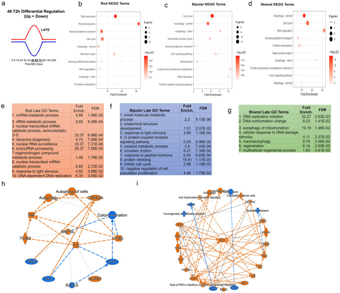
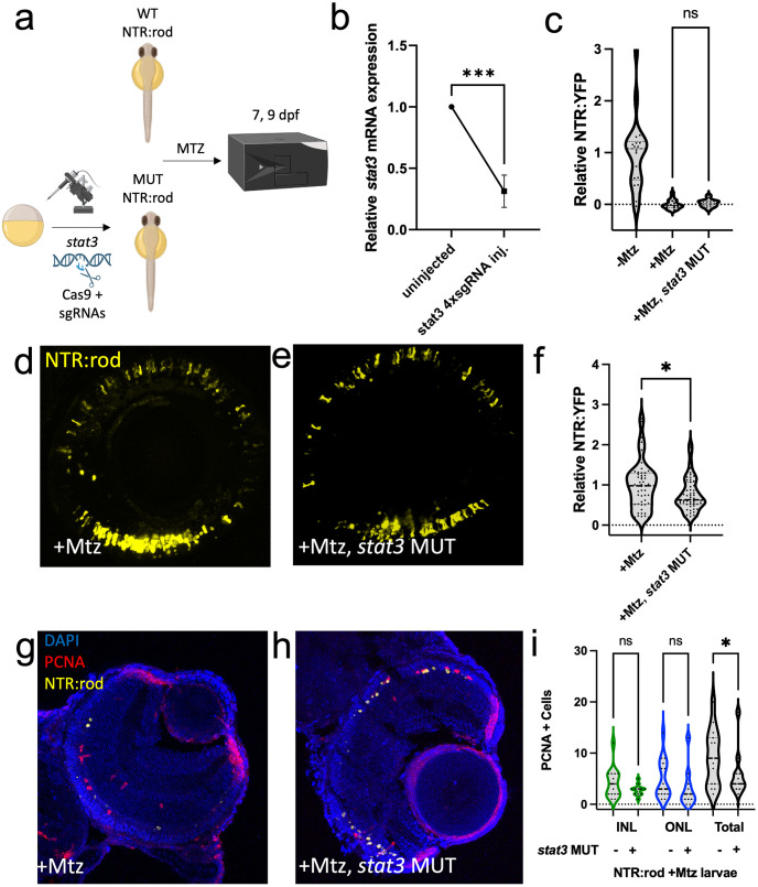
Retinal Müller glia (MG) can act as stem-like cells to generate new neurons in both zebrafish and mice. In zebrafish, retinal regeneration is innate and robust, resulting in the replacement of lost neurons and restoration of visual function. In mice, exogenous stimulation of MG is required to reveal a dormant and, to date, limited regenerative capacity. Zebrafish studies have been key in revealing factors that promote regenerative responses in the mammalian eye. Increased understanding of how the regenerative potential of MG is regulated in zebrafish may therefore aid efforts to promote retinal repair therapeutically. Developmental signaling pathways are known to coordinate regeneration following widespread retinal cell loss. In contrast, less is known about how regeneration is regulated in the context of retinal degenerative disease, i.e., following the loss of specific retinal cell types. To address this knowledge gap, we compared transcriptomic responses underlying regeneration following targeted loss of rod photoreceptors or bipolar cells. In total, 2,531 differentially expressed genes (DEGs) were identified, with the majority being paradigm specific, including during early MG activation phases, suggesting the nature of the injury/cell loss informs the regenerative process from initiation onward. For example, early modulation of Notch signaling was implicated in the rod but not bipolar cell ablation paradigm and components of JAK/STAT signaling were implicated in both paradigms. To examine candidate gene roles in rod cell regeneration, including several immune-related factors, CRISPR/Cas9 was used to create G0 mutant larvae (i.e., "crispants"). Rod cell regeneration was inhibited in stat3 crispants, while mutating stat5a/b, c7b and txn accelerated rod regeneration kinetics. These data support emerging evidence that discrete responses follow from selective retinal cell loss and that the immune system plays a key role in regulating "fate-biased" regenerative processes.
视网膜 Müller 胶质细胞 (MG) 可以作为干细胞,在斑马鱼和小鼠中产生新的神经元。在斑马鱼中,视网膜再生是先天的且强大的,导致丢失的神经元被替代,视觉功能得到恢复。在小鼠中,需要外源性刺激 MG 才能揭示其休眠的、迄今为止有限的再生能力。斑马鱼研究对于揭示促进哺乳动物眼睛再生反应的因素至关重要。因此,增加对 MG 再生潜力如何在斑马鱼中受到调节的理解,可能有助于促进视网膜修复的治疗。发育信号通路已知可协调广泛的视网膜细胞丢失后的再生。相比之下,对于再生如何在视网膜退行性疾病的背景下受到调节,即特定视网膜细胞类型丢失后,了解得较少。为了弥补这一知识空白,我们比较了靶向性丧失视杆细胞或双极细胞后再生的转录组反应。总共鉴定出 2531 个差异表达基因 (DEG),其中大多数是特定于范式的,包括在 MG 早期激活阶段,这表明损伤/细胞丢失的性质从启动开始就决定了再生过程。例如, Notch 信号的早期调节被认为与视杆细胞消融范式有关,但与双极细胞消融范式无关,JAK/STAT 信号的成分被认为与这两个范式都有关。为了研究候选基因在视杆细胞再生中的作用,包括几种免疫相关因子,我们使用 CRISPR/Cas9 技术创建了 G0 突变幼虫(即“crispants”)。在 stat3 crispants 中,视杆细胞再生受到抑制,而突变 stat5a/b、c7b 和 txn 则加速了视杆细胞的再生动力学。这些数据支持了一个新兴的证据,即选择性的视网膜细胞丢失会引发不同的反应,而免疫系统在调节“偏向命运”的再生过程中起着关键作用。