• 文献检索
  • 文档翻译
  • 深度研究
  • 学术资讯
  • Suppr Zotero 插件Zotero 插件
  • 邀请有礼
  • 套餐&价格
  • 历史记录
应用&插件
Suppr Zotero 插件Zotero 插件浏览器插件Mac 客户端Windows 客户端微信小程序
定价
高级版会员购买积分包购买API积分包
服务
文献检索文档翻译深度研究API 文档MCP 服务
关于我们
关于 Suppr公司介绍联系我们用户协议隐私条款
关注我们

Suppr 超能文献

核心技术专利:CN118964589B侵权必究
粤ICP备2023148730 号-1Suppr @ 2026

文献检索

告别复杂PubMed语法,用中文像聊天一样搜索,搜遍4000万医学文献。AI智能推荐,让科研检索更轻松。

立即免费搜索

文件翻译

保留排版,准确专业,支持PDF/Word/PPT等文件格式,支持 12+语言互译。

免费翻译文档

深度研究

AI帮你快速写综述,25分钟生成高质量综述,智能提取关键信息,辅助科研写作。

立即免费体验

用于阵列断层成像的计算性突触抗体表征工具

A Computational Synaptic Antibody Characterization Tool for Array Tomography.

作者信息

Simhal Anish K, Gong Belvin, Trimmer James S, Weinberg Richard J, Smith Stephen J, Sapiro Guillermo, Micheva Kristina D

机构信息

Electrical and Computer Engineering, Duke University, Durham, NC, United States.

Department of Neurobiology, Physiology and Behavior, University of California, Davis, Davis, CA, United States.

出版信息

Front Neuroanat. 2018 Jul 17;12:51. doi: 10.3389/fnana.2018.00051. eCollection 2018.

DOI:10.3389/fnana.2018.00051
PMID:30065633
原文链接:https://pmc.ncbi.nlm.nih.gov/articles/PMC6057115/
Abstract

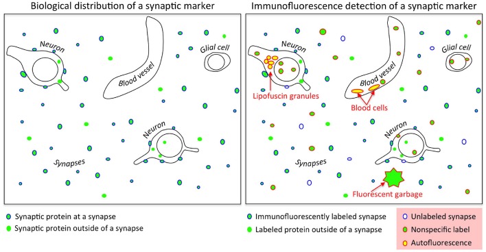
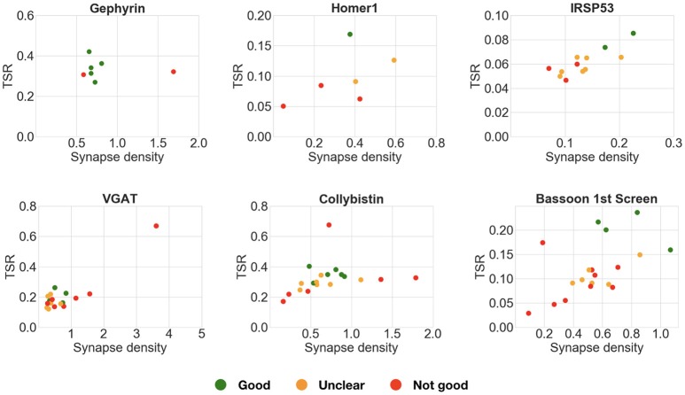
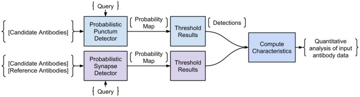
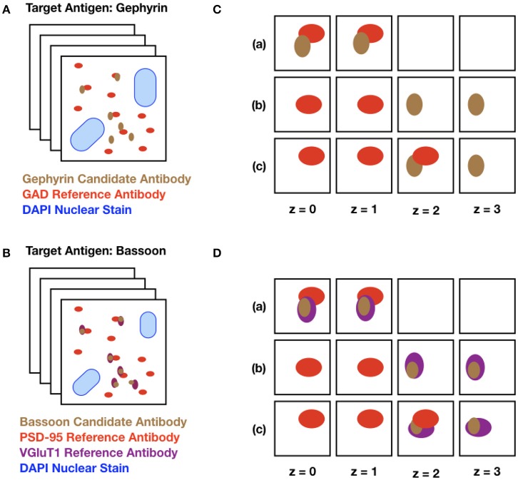
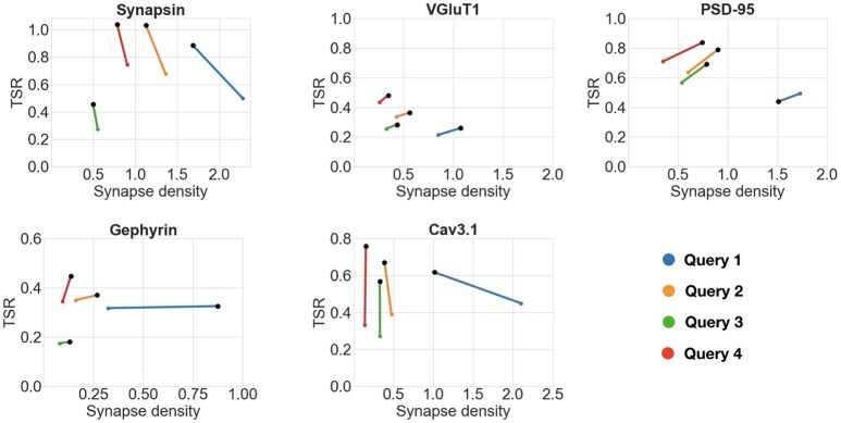
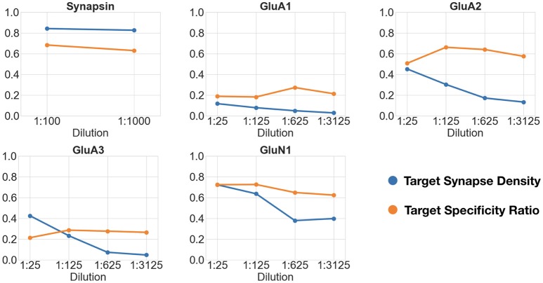
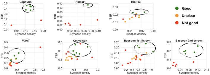
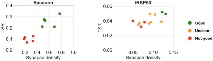
Application-specific validation of antibodies is a critical prerequisite for their successful use. Here we introduce an automated framework for characterization and screening of antibodies against synaptic molecules for high-resolution immunofluorescence array tomography (AT). The proposed Synaptic Antibody Characterization Tool (SACT) is designed to provide an automatic, robust, flexible, and efficient tool for antibody characterization at scale. SACT automatically detects puncta of immunofluorescence labeling from candidate antibodies and determines whether a punctum belongs to a synapse. The molecular composition and size of the target synapses expected to contain the antigen is determined by the user, based on biological knowledge. Operationally, the presence of a synapse is defined by the colocalization or adjacency of the candidate antibody punctum to one or more reference antibody puncta. The outputs of SACT are automatically computed measurements such as target synapse density and target specificity ratio that reflect the sensitivity and specificity of immunolabeling with a given candidate antibody. These measurements provide an objective way to characterize and compare the performance of different antibodies against the same target, and can be used to objectively select the antibodies best suited for AT and potentially for other immunolabeling applications.

摘要

抗体的应用特异性验证是其成功使用的关键前提。在此,我们介绍一种自动化框架,用于针对高分辨率免疫荧光阵列断层扫描(AT)对针对突触分子的抗体进行表征和筛选。所提出的突触抗体表征工具(SACT)旨在提供一种用于大规模抗体表征的自动、稳健、灵活且高效的工具。SACT自动检测候选抗体的免疫荧光标记斑点,并确定一个斑点是否属于突触。预期包含抗原的目标突触的分子组成和大小由用户根据生物学知识确定。在操作上,突触的存在由候选抗体斑点与一个或多个参考抗体斑点的共定位或相邻性来定义。SACT的输出是自动计算的测量值,如目标突触密度和目标特异性比率,这些值反映了用给定候选抗体进行免疫标记的敏感性和特异性。这些测量提供了一种客观的方法来表征和比较针对同一目标的不同抗体的性能,并且可用于客观地选择最适合AT以及潜在地适用于其他免疫标记应用的抗体。

https://cdn.ncbi.nlm.nih.gov/pmc/blobs/ccbb/6057115/094f4f164a8b/fnana-12-00051-g0011.jpg
https://cdn.ncbi.nlm.nih.gov/pmc/blobs/ccbb/6057115/9fd295a5b038/fnana-12-00051-g0001.jpg
https://cdn.ncbi.nlm.nih.gov/pmc/blobs/ccbb/6057115/4068bbe59f38/fnana-12-00051-g0002.jpg
https://cdn.ncbi.nlm.nih.gov/pmc/blobs/ccbb/6057115/2ac24eb8ede9/fnana-12-00051-g0003.jpg
https://cdn.ncbi.nlm.nih.gov/pmc/blobs/ccbb/6057115/214f0596a262/fnana-12-00051-g0004.jpg
https://cdn.ncbi.nlm.nih.gov/pmc/blobs/ccbb/6057115/ac980d6a4ce8/fnana-12-00051-g0005.jpg
https://cdn.ncbi.nlm.nih.gov/pmc/blobs/ccbb/6057115/d76ce3c7059a/fnana-12-00051-g0006.jpg
https://cdn.ncbi.nlm.nih.gov/pmc/blobs/ccbb/6057115/198258fd5a32/fnana-12-00051-g0007.jpg
https://cdn.ncbi.nlm.nih.gov/pmc/blobs/ccbb/6057115/ad8d7d5be80b/fnana-12-00051-g0008.jpg
https://cdn.ncbi.nlm.nih.gov/pmc/blobs/ccbb/6057115/7cf8658ba77a/fnana-12-00051-g0009.jpg
https://cdn.ncbi.nlm.nih.gov/pmc/blobs/ccbb/6057115/c9b253b563ed/fnana-12-00051-g0010.jpg
https://cdn.ncbi.nlm.nih.gov/pmc/blobs/ccbb/6057115/094f4f164a8b/fnana-12-00051-g0011.jpg

相似文献

1
A Computational Synaptic Antibody Characterization Tool for Array Tomography.用于阵列断层成像的计算性突触抗体表征工具
Front Neuroanat. 2018 Jul 17;12:51. doi: 10.3389/fnana.2018.00051. eCollection 2018.
2
SynQuant: an automatic tool to quantify synapses from microscopy images.SynQuant:一种自动工具,用于从显微镜图像中定量突触。
Bioinformatics. 2020 Mar 1;36(5):1599-1606. doi: 10.1093/bioinformatics/btz760.
3
Colocalization of synapse marker proteins evaluated by STED-microscopy reveals patterns of neuronal synapse distribution in vitro.通过受激发射损耗显微镜(STED)评估的突触标记蛋白共定位揭示了体外神经元突触分布模式。
J Neurosci Methods. 2016 Nov 1;273:149-159. doi: 10.1016/j.jneumeth.2016.09.001. Epub 2016 Sep 9.
4
Probabilistic fluorescence-based synapse detection.基于概率荧光的突触检测。
PLoS Comput Biol. 2017 Apr 17;13(4):e1005493. doi: 10.1371/journal.pcbi.1005493. eCollection 2017 Apr.
5
Learning-guided automatic three dimensional synapse quantification for drosophila neurons.果蝇神经元的学习引导自动三维突触定量分析
BMC Bioinformatics. 2015 May 28;16:177. doi: 10.1186/s12859-015-0616-y.
6
Automated criteria-based selection and analysis of fluorescent synaptic puncta.基于标准的荧光突触小体自动选择与分析。
J Neurosci Methods. 2006 Apr 15;152(1-2):32-9. doi: 10.1016/j.jneumeth.2005.08.008. Epub 2005 Sep 28.
7
Fully-Automatic Synapse Prediction and Validation on a Large Data Set.全自动突触预测及其在大数据集上的验证。
Front Neural Circuits. 2018 Oct 29;12:87. doi: 10.3389/fncir.2018.00087. eCollection 2018.
8
Mapping synapses by conjugate light-electron array tomography.通过共轭光电子阵列断层扫描绘制突触图谱。
J Neurosci. 2015 Apr 8;35(14):5792-807. doi: 10.1523/JNEUROSCI.4274-14.2015.
9
Differential distribution of synaptotagmin immunoreactivity among synapses in the goldfish, salamander, and mouse retina.金鱼、蝾螈和小鼠视网膜中突触间突触结合蛋白免疫反应性的差异分布。
Vis Neurosci. 2003 Jan-Feb;20(1):37-49. doi: 10.1017/s095252380320105x.
10
Molecular specificity of defined types of amacrine synapse in cat retina.猫视网膜中特定类型无长突细胞突触的分子特异性
J Neurosci. 1986 May;6(5):1314-24. doi: 10.1523/JNEUROSCI.06-05-01314.1986.

引用本文的文献

1
Data-driven synapse classification reveals a logic of glutamate receptor diversity.数据驱动的突触分类揭示了谷氨酸受体多样性的逻辑。
bioRxiv. 2025 Jan 14:2024.12.11.628056. doi: 10.1101/2024.12.11.628056.
2
Developing a Toolbox of Antibodies Validated for Array Tomography-Based Imaging of Brain Synapses.开发一套经阵列层析成像技术验证的用于检测脑突触的抗体工具。
eNeuro. 2023 Dec 22;10(12). doi: 10.1523/ENEURO.0290-23.2023. Print 2023 Dec.
3
Developing a Toolbox of Antibodies Validated for Array Tomography-Based Imaging of Brain Synapses.

本文引用的文献

1
: High-Throughput Quantification of Fluorescent Synaptic Protein Puncta by Machine Learning.: 基于机器学习的荧光突触蛋白斑的高通量定量分析。
eNeuro. 2017 Dec 6;4(6). doi: 10.1523/ENEURO.0219-17.2017. eCollection 2017 Nov-Dec.
2
Detection of axonal synapses in 3D two-photon images.三维双光子图像中轴突突触的检测。
PLoS One. 2017 Sep 5;12(9):e0183309. doi: 10.1371/journal.pone.0183309. eCollection 2017.
3
Probabilistic fluorescence-based synapse detection.基于概率荧光的突触检测。
开发用于基于阵列断层扫描的脑突触成像的经过验证的抗体工具箱。
bioRxiv. 2023 Nov 8:2023.06.28.546920. doi: 10.1101/2023.06.28.546920.
4
SynQuant: an automatic tool to quantify synapses from microscopy images.SynQuant:一种自动工具,用于从显微镜图像中定量突触。
Bioinformatics. 2020 Mar 1;36(5):1599-1606. doi: 10.1093/bioinformatics/btz760.
5
Multifaceted Changes in Synaptic Composition and Astrocytic Involvement in a Mouse Model of Fragile X Syndrome.脆性 X 综合征小鼠模型中突触组成的多方面变化和星形胶质细胞的参与。
Sci Rep. 2019 Sep 25;9(1):13855. doi: 10.1038/s41598-019-50240-x.
6
Effects of early life stress on cocaine conditioning and AMPA receptor composition are sex-specific and driven by TNF.早期生活应激对可卡因条件作用和 AMPA 受体组成的影响具有性别特异性,并且由 TNF 驱动。
Brain Behav Immun. 2019 May;78:41-51. doi: 10.1016/j.bbi.2019.01.006. Epub 2019 Jan 14.
7
Q&A: Array tomography.常见问题解答:面绘制技术
BMC Biol. 2018 Sep 6;16(1):98. doi: 10.1186/s12915-018-0560-1.
PLoS Comput Biol. 2017 Apr 17;13(4):e1005493. doi: 10.1371/journal.pcbi.1005493. eCollection 2017 Apr.
4
A proposal for validation of antibodies.一项抗体验证提案。
Nat Methods. 2016 Oct;13(10):823-7. doi: 10.1038/nmeth.3995. Epub 2016 Sep 5.
5
Developing high-quality mouse monoclonal antibodies for neuroscience research - approaches, perspectives and opportunities.开发用于神经科学研究的高质量小鼠单克隆抗体——方法、观点与机遇
N Biotechnol. 2016 Sep 25;33(5 Pt A):551-64. doi: 10.1016/j.nbt.2015.11.007. Epub 2015 Nov 28.
6
Reproducibility crisis: Blame it on the antibodies.可重复性危机:归咎于抗体。
Nature. 2015 May 21;521(7552):274-6. doi: 10.1038/521274a.
7
Synaptic molecular imaging in spared and deprived columns of mouse barrel cortex with array tomography.利用阵列断层扫描技术对小鼠桶状皮层中保留和剥夺柱的突触分子成像。
Sci Data. 2014 Dec 23;1:140046. doi: 10.1038/sdata.2014.46. eCollection 2014.
8
Mapping synapses by conjugate light-electron array tomography.通过共轭光电子阵列断层扫描绘制突触图谱。
J Neurosci. 2015 Apr 8;35(14):5792-807. doi: 10.1523/JNEUROSCI.4274-14.2015.
9
Automated detection of synapses in serial section transmission electron microscopy image stacks.在连续切片透射电子显微镜图像堆栈中自动检测突触。
PLoS One. 2014 Feb 6;9(2):e87351. doi: 10.1371/journal.pone.0087351. eCollection 2014.
10
Automated analysis of a diverse synapse population.多样化突触群体的自动化分析。
PLoS Comput Biol. 2013;9(3):e1002976. doi: 10.1371/journal.pcbi.1002976. Epub 2013 Mar 28.