• 文献检索
  • 文档翻译
  • 深度研究
  • 学术资讯
  • Suppr Zotero 插件Zotero 插件
  • 邀请有礼
  • 套餐&价格
  • 历史记录
应用&插件
Suppr Zotero 插件Zotero 插件浏览器插件Mac 客户端Windows 客户端微信小程序
定价
高级版会员购买积分包购买API积分包
服务
文献检索文档翻译深度研究API 文档MCP 服务
关于我们
关于 Suppr公司介绍联系我们用户协议隐私条款
关注我们

Suppr 超能文献

核心技术专利:CN118964589B侵权必究
粤ICP备2023148730 号-1Suppr @ 2026

文献检索

告别复杂PubMed语法,用中文像聊天一样搜索,搜遍4000万医学文献。AI智能推荐,让科研检索更轻松。

立即免费搜索

文件翻译

保留排版,准确专业,支持PDF/Word/PPT等文件格式,支持 12+语言互译。

免费翻译文档

深度研究

AI帮你快速写综述,25分钟生成高质量综述,智能提取关键信息,辅助科研写作。

立即免费体验

利用深度学习对稀疏标记的三维电子显微镜图像中的细胞超微结构进行分割。

Segmentation of cellular ultrastructures on sparsely labeled 3D electron microscopy images using deep learning.

作者信息

Machireddy Archana, Thibault Guillaume, Loftis Kevin G, Stoltz Kevin, Bueno Cecilia E, Smith Hannah R, Riesterer Jessica L, Gray Joe W, Song Xubo

机构信息

Program of Computer Science and Electrical Engineering, Oregon Health and Science University, Portland, OR, United States.

Department of Biomedical Engineering, Oregon Health and Science University, Portland, OR, United States.

出版信息

Front Bioinform. 2023 Dec 15;3:1308708. doi: 10.3389/fbinf.2023.1308708. eCollection 2023.

DOI:10.3389/fbinf.2023.1308708
PMID:38162124
原文链接:https://pmc.ncbi.nlm.nih.gov/articles/PMC10754953/
Abstract

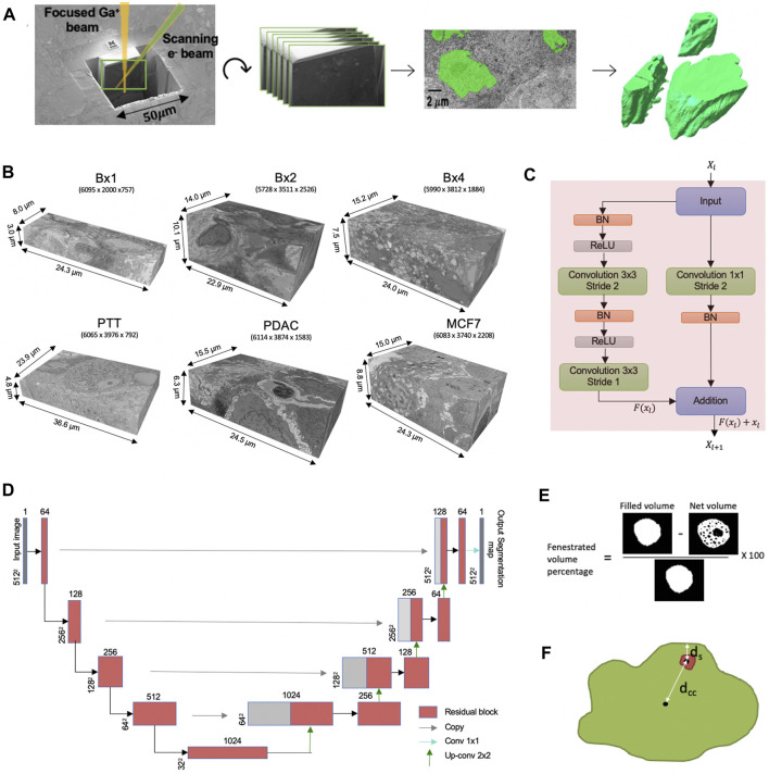
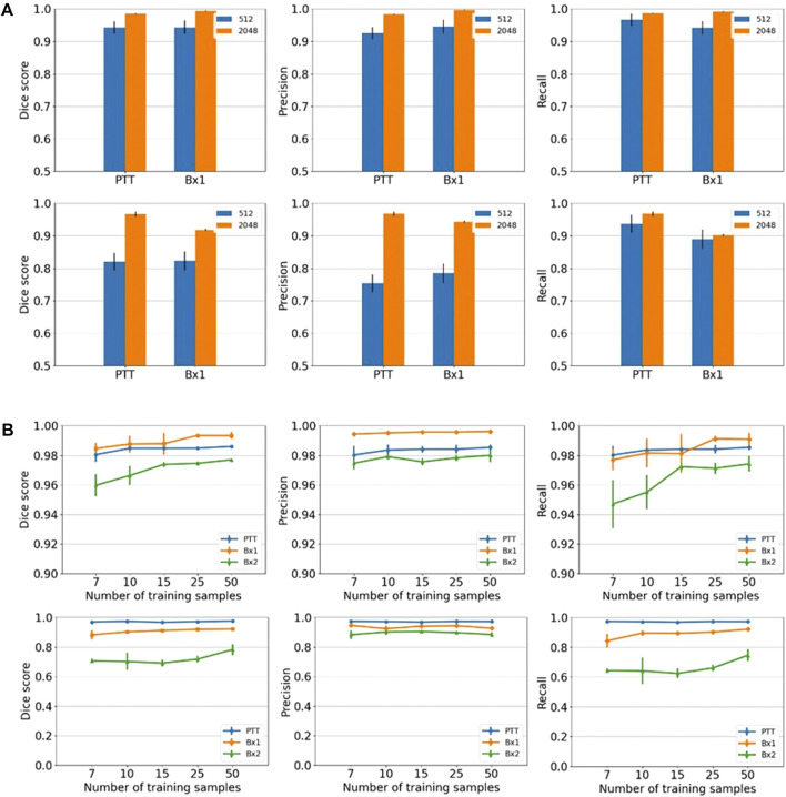
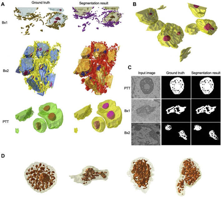
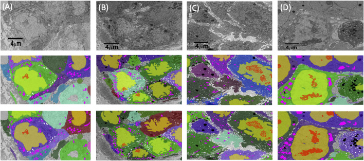
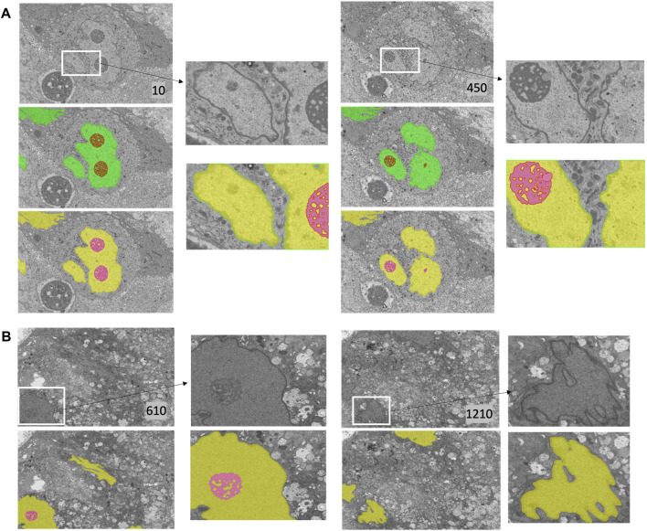
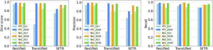
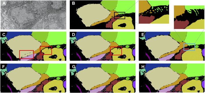
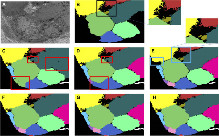
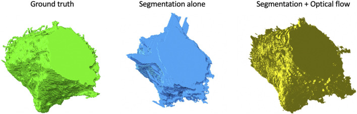
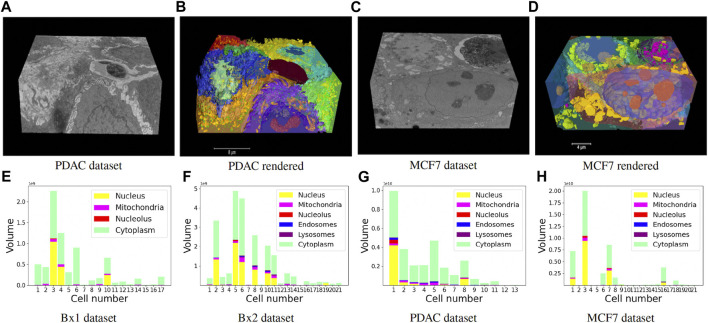
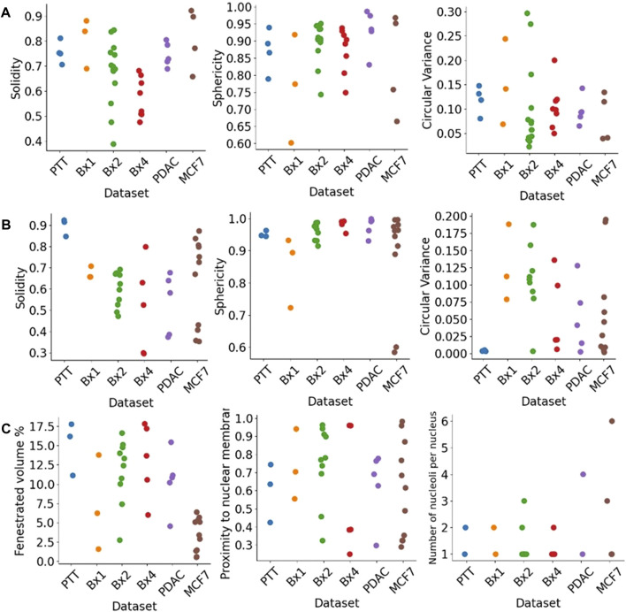
Focused ion beam-scanning electron microscopy (FIB-SEM) images can provide a detailed view of the cellular ultrastructure of tumor cells. A deeper understanding of their organization and interactions can shed light on cancer mechanisms and progression. However, the bottleneck in the analysis is the delineation of the cellular structures to enable quantitative measurements and analysis. We mitigated this limitation using deep learning to segment cells and subcellular ultrastructure in 3D FIB-SEM images of tumor biopsies obtained from patients with metastatic breast and pancreatic cancers. The ultrastructures, such as nuclei, nucleoli, mitochondria, endosomes, and lysosomes, are relatively better defined than their surroundings and can be segmented with high accuracy using a neural network trained with sparse manual labels. Cell segmentation, on the other hand, is much more challenging due to the lack of clear boundaries separating cells in the tissue. We adopted a multi-pronged approach combining detection, boundary propagation, and tracking for cell segmentation. Specifically, a neural network was employed to detect the intracellular space; optical flow was used to propagate cell boundaries across the z-stack from the nearest ground truth image in order to facilitate the separation of individual cells; finally, the filopodium-like protrusions were tracked to the main cells by calculating the intersection over union measure for all regions detected in consecutive images along z-stack and connecting regions with maximum overlap. The proposed cell segmentation methodology resulted in an average Dice score of 0.93. For nuclei, nucleoli, and mitochondria, the segmentation achieved Dice scores of 0.99, 0.98, and 0.86, respectively. The segmentation of FIB-SEM images will enable interpretative rendering and provide quantitative image features to be associated with relevant clinical variables.

摘要

聚焦离子束扫描电子显微镜(FIB-SEM)图像可以提供肿瘤细胞的细胞超微结构的详细视图。对其组织结构和相互作用的更深入理解可以揭示癌症机制和进展。然而,分析中的瓶颈在于细胞结构的描绘,以便进行定量测量和分析。我们利用深度学习对从转移性乳腺癌和胰腺癌患者获得的肿瘤活检组织的三维FIB-SEM图像中的细胞和亚细胞超微结构进行分割,从而缓解了这一限制。细胞核、核仁、线粒体、内体和溶酶体等超微结构相对于其周围环境定义得相对较好,可以使用经过稀疏手动标记训练的神经网络进行高精度分割。另一方面,由于组织中细胞之间缺乏清晰的边界,细胞分割更具挑战性。我们采用了一种多管齐下的方法,结合检测、边界传播和跟踪来进行细胞分割。具体而言,使用神经网络检测细胞内空间;利用光流从最近的真实图像跨z轴堆栈传播细胞边界,以促进单个细胞的分离;最后,通过计算沿z轴堆栈在连续图像中检测到的所有区域的交并比,并连接具有最大重叠的区域,将丝状伪足样突起追踪到主要细胞。所提出的细胞分割方法平均Dice评分为0.93。对于细胞核、核仁线粒体,分割的Dice评分分别为0.99、0.98和0.86。FIB-SEM图像的分割将实现解释性渲染,并提供与相关临床变量相关的定量图像特征。

https://cdn.ncbi.nlm.nih.gov/pmc/blobs/3339/10754953/48192ce17ede/fbinf-03-1308708-g012.jpg
https://cdn.ncbi.nlm.nih.gov/pmc/blobs/3339/10754953/3e9e49e8669a/fbinf-03-1308708-g001.jpg
https://cdn.ncbi.nlm.nih.gov/pmc/blobs/3339/10754953/aa8b5937fb47/fbinf-03-1308708-g002.jpg
https://cdn.ncbi.nlm.nih.gov/pmc/blobs/3339/10754953/3b8aad21041a/fbinf-03-1308708-g003.jpg
https://cdn.ncbi.nlm.nih.gov/pmc/blobs/3339/10754953/031a22352de1/fbinf-03-1308708-g004.jpg
https://cdn.ncbi.nlm.nih.gov/pmc/blobs/3339/10754953/bb14d6f7119d/fbinf-03-1308708-g005.jpg
https://cdn.ncbi.nlm.nih.gov/pmc/blobs/3339/10754953/31d837f82b4f/fbinf-03-1308708-g006.jpg
https://cdn.ncbi.nlm.nih.gov/pmc/blobs/3339/10754953/6070df24f307/fbinf-03-1308708-g007.jpg
https://cdn.ncbi.nlm.nih.gov/pmc/blobs/3339/10754953/3ed6c8103431/fbinf-03-1308708-g008.jpg
https://cdn.ncbi.nlm.nih.gov/pmc/blobs/3339/10754953/d9f71f27d758/fbinf-03-1308708-g009.jpg
https://cdn.ncbi.nlm.nih.gov/pmc/blobs/3339/10754953/5c1ce06beb39/fbinf-03-1308708-g010.jpg
https://cdn.ncbi.nlm.nih.gov/pmc/blobs/3339/10754953/a8eb4bda6049/fbinf-03-1308708-g011.jpg
https://cdn.ncbi.nlm.nih.gov/pmc/blobs/3339/10754953/48192ce17ede/fbinf-03-1308708-g012.jpg

相似文献

1
Segmentation of cellular ultrastructures on sparsely labeled 3D electron microscopy images using deep learning.利用深度学习对稀疏标记的三维电子显微镜图像中的细胞超微结构进行分割。
Front Bioinform. 2023 Dec 15;3:1308708. doi: 10.3389/fbinf.2023.1308708. eCollection 2023.
2
An Evaluation of Computational Learning-based Methods for the Segmentation of Nuclei in Cervical Cancer Cells from Microscopic Images.基于计算学习的方法在宫颈癌细胞显微图像细胞核分割中的评估。
Curr Comput Aided Drug Des. 2022;18(2):81-94. doi: 10.2174/1573409918666220208120756.
3
Automated segmentation of cardiomyocyte Z-disks from high-throughput scanning electron microscopy data.高通量扫描电子显微镜数据中心肌细胞 Z 盘的自动分割。
BMC Med Inform Decis Mak. 2019 Dec 19;19(Suppl 6):272. doi: 10.1186/s12911-019-0962-1.
4
Three-dimensional reconstruction of porous polymer films from FIB-SEM nanotomography data using random forests.利用随机森林从 FIB-SEM 纳米断层扫描数据对多孔聚合物薄膜进行三维重建。
J Microsc. 2021 Jan;281(1):76-86. doi: 10.1111/jmi.12950. Epub 2020 Aug 19.
5
Automatic three-dimensional segmentation of mouse embryonic stem cell nuclei by utilising multiple channels of confocal fluorescence images.利用共聚焦荧光图像的多个通道自动对小鼠胚胎干细胞核进行三维分割。
J Microsc. 2021 Jan;281(1):57-75. doi: 10.1111/jmi.12949. Epub 2020 Aug 8.
6
Znet: Deep Learning Approach for 2D MRI Brain Tumor Segmentation.Znet:二维 MRI 脑肿瘤分割的深度学习方法。
IEEE J Transl Eng Health Med. 2022 May 23;10:1800508. doi: 10.1109/JTEHM.2022.3176737. eCollection 2022.
7
Automated left ventricular myocardium segmentation using 3D deeply supervised attention U-net for coronary computed tomography angiography; CT myocardium segmentation.基于 3D 深度监督注意 U-Net 的冠状动脉 CT 血管造影自动左心室心肌分割;CT 心肌分割。
Med Phys. 2020 Apr;47(4):1775-1785. doi: 10.1002/mp.14066. Epub 2020 Feb 29.
8
Lung tumor segmentation in 4D CT images using motion convolutional neural networks.使用运动卷积神经网络进行 4D CT 图像中的肺部肿瘤分割。
Med Phys. 2021 Nov;48(11):7141-7153. doi: 10.1002/mp.15204. Epub 2021 Sep 13.
9
A novel adaptive cubic quasi-Newton optimizer for deep learning based medical image analysis tasks, validated on detection of COVID-19 and segmentation for COVID-19 lung infection, liver tumor, and optic disc/cup.一种用于深度学习的新型自适应三次拟牛顿优化器,在 COVID-19 检测和 COVID-19 肺部感染、肝脏肿瘤以及视盘/杯分割等医学图像分析任务中得到验证。
Med Phys. 2023 Mar;50(3):1528-1538. doi: 10.1002/mp.15969. Epub 2022 Oct 6.
10
Automated segmentation of the human supraclavicular fat depot via deep neural network in water-fat separated magnetic resonance images.通过深度神经网络在水脂分离磁共振图像中对人体锁骨上脂肪库进行自动分割。
Quant Imaging Med Surg. 2023 Jul 1;13(7):4699-4715. doi: 10.21037/qims-22-304. Epub 2023 Mar 14.

引用本文的文献

1
Efficient semi-supervised semantic segmentation of electron microscopy cancer images with sparse annotations.利用稀疏标注实现电子显微镜癌症图像的高效半监督语义分割
Front Bioinform. 2023 Dec 15;3:1308707. doi: 10.3389/fbinf.2023.1308707. eCollection 2023.
2
Efficient semi-supervised semantic segmentation of electron microscopy cancer images with sparse annotations.利用稀疏标注实现电子显微镜癌症图像的高效半监督语义分割。
bioRxiv. 2023 Nov 1:2023.10.30.563998. doi: 10.1101/2023.10.30.563998.
3
Online citizen science with the Zooniverse for analysis of biological volumetric data.

本文引用的文献

1
Automated large volume sample preparation for vEM.用于 vEM 的自动化大容量样品制备。
Methods Cell Biol. 2023;177:1-32. doi: 10.1016/bs.mcb.2023.01.009. Epub 2023 May 14.
2
Regulation of liver subcellular architecture controls metabolic homeostasis.调控肝亚细胞结构控制代谢稳态。
Nature. 2022 Mar;603(7902):736-742. doi: 10.1038/s41586-022-04488-5. Epub 2022 Mar 9.
3
An omic and multidimensional spatial atlas from serial biopsies of an evolving metastatic breast cancer.从不断演进的转移性乳腺癌的连续活检中获得的组学和多维空间图谱。
利用 Zooniverse 开展在线公民科学,分析生物体积数据。
Histochem Cell Biol. 2023 Sep;160(3):253-276. doi: 10.1007/s00418-023-02204-6. Epub 2023 Jun 7.
4
How innovations in methodology offer new prospects for volume electron microscopy.方法学创新如何为电子体视学带来新的前景。
J Microsc. 2022 Sep;287(3):114-137. doi: 10.1111/jmi.13134. Epub 2022 Jul 27.
Cell Rep Med. 2022 Feb 15;3(2):100525. doi: 10.1016/j.xcrm.2022.100525.
4
Deciphering tumour tissue organization by 3D electron microscopy and machine learning.通过 3D 电子显微镜和机器学习破译肿瘤组织的结构。
Commun Biol. 2021 Dec 13;4(1):1390. doi: 10.1038/s42003-021-02919-z.
5
Whole-cell organelle segmentation in volume electron microscopy.体积电子显微镜中的全细胞细胞器分割
Nature. 2021 Nov;599(7883):141-146. doi: 10.1038/s41586-021-03977-3. Epub 2021 Oct 6.
6
Whole-body integration of gene expression and single-cell morphology.整体基因表达与单细胞形态的整合。
Cell. 2021 Sep 2;184(18):4819-4837.e22. doi: 10.1016/j.cell.2021.07.017. Epub 2021 Aug 10.
7
3D FIB-SEM reconstruction of microtubule-organelle interaction in whole primary mouse β cells.全原生小鼠β细胞中微管-细胞器相互作用的 3D FIB-SEM 重建。
J Cell Biol. 2021 Feb 1;220(2). doi: 10.1083/jcb.202010039.
8
A workflow for visualizing human cancer biopsies using large-format electron microscopy.一种使用大格式电子显微镜可视化人类癌症活检样本的工作流程。
Methods Cell Biol. 2020;158:163-181. doi: 10.1016/bs.mcb.2020.01.005. Epub 2020 Mar 12.
9
Relevance of immune cell and tumor microenvironment imaging in the new era of immunotherapy.免疫细胞和肿瘤微环境成像在免疫治疗新时代的相关性。
J Exp Clin Cancer Res. 2020 May 18;39(1):89. doi: 10.1186/s13046-020-01586-y.
10
Tumor microenvironment complexity and therapeutic implications at a glance.肿瘤微环境的复杂性及其治疗意义简述。
Cell Commun Signal. 2020 Apr 7;18(1):59. doi: 10.1186/s12964-020-0530-4.