• 文献检索
  • 文档翻译
  • 深度研究
  • 学术资讯
  • Suppr Zotero 插件Zotero 插件
  • 邀请有礼
  • 套餐&价格
  • 历史记录
应用&插件
Suppr Zotero 插件Zotero 插件浏览器插件Mac 客户端Windows 客户端微信小程序
定价
高级版会员购买积分包购买API积分包
服务
文献检索文档翻译深度研究API 文档MCP 服务
关于我们
关于 Suppr公司介绍联系我们用户协议隐私条款
关注我们

Suppr 超能文献

核心技术专利:CN118964589B侵权必究
粤ICP备2023148730 号-1Suppr @ 2026

文献检索

告别复杂PubMed语法,用中文像聊天一样搜索,搜遍4000万医学文献。AI智能推荐,让科研检索更轻松。

立即免费搜索

文件翻译

保留排版,准确专业,支持PDF/Word/PPT等文件格式,支持 12+语言互译。

免费翻译文档

深度研究

AI帮你快速写综述,25分钟生成高质量综述,智能提取关键信息,辅助科研写作。

立即免费体验

基于深度学习的磁共振图像中盆腔危及器官的半监督自动分割方法

Semi-supervised auto-segmentation method for pelvic organ-at-risk in magnetic resonance images based on deep-learning.

作者信息

Li Xianan, Jia Lecheng, Lin Fengyu, Chai Fan, Liu Tao, Zhang Wei, Wei Ziquan, Xiong Weiqi, Li Hua, Zhang Min, Wang Yi

机构信息

Department of Radiation Oncology, Peking University People's Hospital, Beijing, China.

Radiotherapy laboratory, Shenzhen United Imaging Research Institute of Innovative Medical Equipment, Shenzhen, China.

出版信息

J Appl Clin Med Phys. 2024 Feb 22;25(3):e14296. doi: 10.1002/acm2.14296.

DOI:10.1002/acm2.14296
PMID:38386963
原文链接:https://pmc.ncbi.nlm.nih.gov/articles/PMC10929999/
Abstract

BACKGROUND AND PURPOSE

In radiotherapy, magnetic resonance (MR) imaging has higher contrast for soft tissues compared to computed tomography (CT) scanning and does not emit radiation. However, manual annotation of the deep learning-based automatic organ-at-risk (OAR) delineation algorithms is expensive, making the collection of large-high-quality annotated datasets a challenge. Therefore, we proposed the low-cost semi-supervised OAR segmentation method using small pelvic MR image annotations.

METHODS

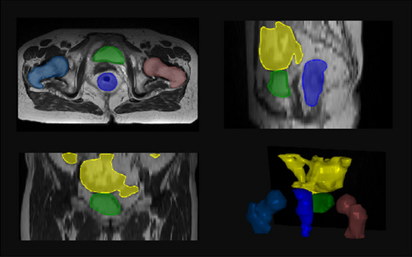
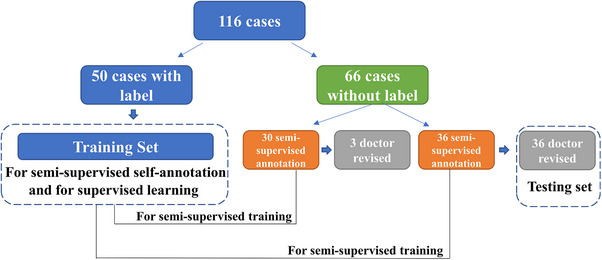
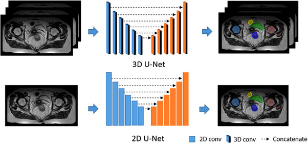
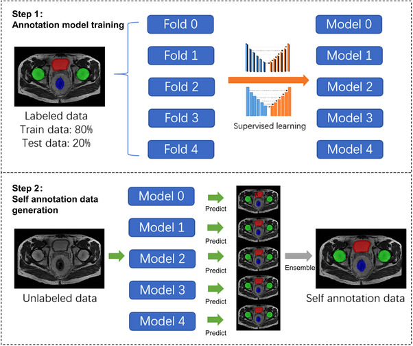
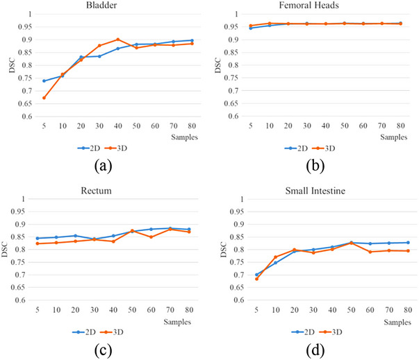
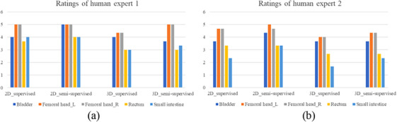
We trained a deep learning-based segmentation model using 116 sets of MR images from 116 patients. The bladder, femoral heads, rectum, and small intestine were selected as OAR regions. To generate the training set, we utilized a semi-supervised method and ensemble learning techniques. Additionally, we employed a post-processing algorithm to correct the self-annotation data. Both 2D and 3D auto-segmentation networks were evaluated for their performance. Furthermore, we evaluated the performance of semi-supervised method for 50 labeled data and only 10 labeled data.

RESULTS

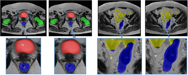
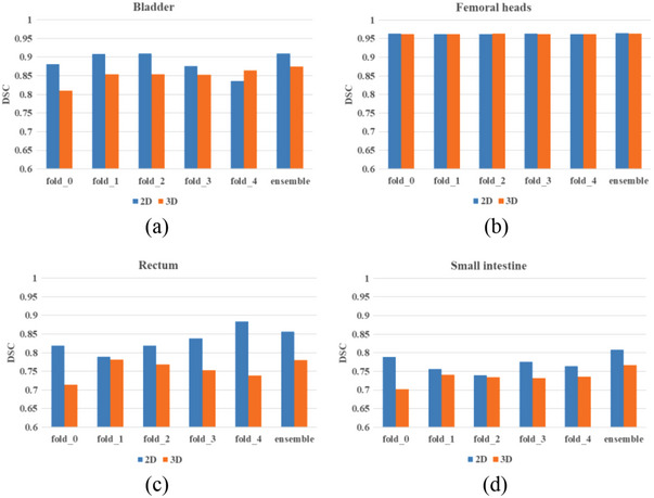
The Dice similarity coefficient (DSC) of the bladder, femoral heads, rectum and small intestine between segmentation results and reference masks is 0.954, 0.984, 0.908, 0.852 only using self-annotation and post-processing methods of 2D segmentation model. The DSC of corresponding OARs is 0.871, 0.975, 0.975, 0.783, 0.724 using 3D segmentation network, 0.896, 0.984, 0.890, 0.828 using 2D segmentation network and common supervised method.

CONCLUSION

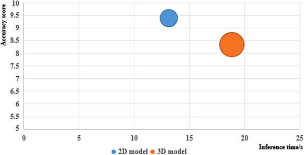
The outcomes of our study demonstrate that it is possible to train a multi-OAR segmentation model using small annotation samples and additional unlabeled data. To effectively annotate the dataset, ensemble learning and post-processing methods were employed. Additionally, when dealing with anisotropy and limited sample sizes, the 2D model outperformed the 3D model in terms of performance.

摘要

背景与目的

在放射治疗中,与计算机断层扫描(CT)相比,磁共振(MR)成像对软组织具有更高的对比度,且不发出辐射。然而,基于深度学习的自动危及器官(OAR)轮廓描绘算法的人工标注成本高昂,这使得收集大规模高质量标注数据集成为一项挑战。因此,我们提出了一种使用少量盆腔MR图像标注的低成本半监督OAR分割方法。

方法

我们使用来自116例患者的116组MR图像训练了一个基于深度学习的分割模型。选择膀胱、股骨头、直肠和小肠作为OAR区域。为了生成训练集,我们采用了半监督方法和集成学习技术。此外,我们使用了一种后处理算法来校正自标注数据。对二维和三维自动分割网络的性能进行了评估。此外,我们评估了半监督方法在50个标注数据和仅10个标注数据情况下的性能。

结果

仅使用二维分割模型的自标注和后处理方法时,分割结果与参考掩码之间膀胱、股骨头、直肠和小肠的骰子相似系数(DSC)分别为0.954、0.984、0.908、0.852。使用三维分割网络时,相应OAR的DSC分别为0.871、0.975、0.975、0.783、0.724;使用二维分割网络和普通监督方法时,相应OAR的DSC分别为0.896、0.984、0.890、0.828。

结论

我们的研究结果表明,使用少量标注样本和额外的未标注数据来训练多OAR分割模型是可行 的。为了有效地标注数据集,采用了集成学习和后处理方法。此外,在处理各向异性和样本量有限的情况时,二维模型在性能方面优于三维模型。

https://cdn.ncbi.nlm.nih.gov/pmc/blobs/7f14/10929999/b62b0f4e76f2/ACM2-25-e14296-g004.jpg
https://cdn.ncbi.nlm.nih.gov/pmc/blobs/7f14/10929999/04cb3d20f2d1/ACM2-25-e14296-g005.jpg
https://cdn.ncbi.nlm.nih.gov/pmc/blobs/7f14/10929999/f8f9fc2f9d4b/ACM2-25-e14296-g001.jpg
https://cdn.ncbi.nlm.nih.gov/pmc/blobs/7f14/10929999/9a81eccc820c/ACM2-25-e14296-g002.jpg
https://cdn.ncbi.nlm.nih.gov/pmc/blobs/7f14/10929999/3251bc276c98/ACM2-25-e14296-g006.jpg
https://cdn.ncbi.nlm.nih.gov/pmc/blobs/7f14/10929999/f3f03d5fff47/ACM2-25-e14296-g007.jpg
https://cdn.ncbi.nlm.nih.gov/pmc/blobs/7f14/10929999/0e3604705aeb/ACM2-25-e14296-g009.jpg
https://cdn.ncbi.nlm.nih.gov/pmc/blobs/7f14/10929999/8642ca1b5d0d/ACM2-25-e14296-g008.jpg
https://cdn.ncbi.nlm.nih.gov/pmc/blobs/7f14/10929999/0147d1fe7a82/ACM2-25-e14296-g003.jpg
https://cdn.ncbi.nlm.nih.gov/pmc/blobs/7f14/10929999/b62b0f4e76f2/ACM2-25-e14296-g004.jpg

相似文献

1
Semi-supervised auto-segmentation method for pelvic organ-at-risk in magnetic resonance images based on deep-learning.基于深度学习的磁共振图像中盆腔危及器官的半监督自动分割方法
J Appl Clin Med Phys. 2024 Feb 22;25(3):e14296. doi: 10.1002/acm2.14296.
2
Semi-supervised abdominal multi-organ segmentation by object-redrawing.通过对象重绘实现半监督腹部多器官分割
Med Phys. 2024 Nov;51(11):8334-8347. doi: 10.1002/mp.17364. Epub 2024 Aug 21.
3
Semi-supervised learning framework with shape encoding for neonatal ventricular segmentation from 3D ultrasound.基于形状编码的半监督学习框架,用于从 3D 超声中分割新生儿心室。
Med Phys. 2024 Sep;51(9):6134-6148. doi: 10.1002/mp.17242. Epub 2024 Jun 10.
4
Semi-supervised learning for automatic segmentation of the knee from MRI with convolutional neural networks.基于卷积神经网络的膝关节 MRI 半自动分割的半监督学习。
Comput Methods Programs Biomed. 2020 Jun;189:105328. doi: 10.1016/j.cmpb.2020.105328. Epub 2020 Jan 11.
5
Cross-modality deep learning: Contouring of MRI data from annotated CT data only.跨模态深度学习:仅从标注的CT数据对MRI数据进行轮廓提取。
Med Phys. 2021 Apr;48(4):1673-1684. doi: 10.1002/mp.14619. Epub 2020 Dec 13.
6
Automated segmentation of lesions and organs at risk on [Ga]Ga-PSMA-11 PET/CT images using self-supervised learning with Swin UNETR.使用基于 Swin UNETR 的自监督学习对 [Ga]Ga-PSMA-11 PET/CT 图像上的病变和危险器官进行自动分割。
Cancer Imaging. 2024 Feb 29;24(1):30. doi: 10.1186/s40644-024-00675-x.
7
A modality-collaborative convolution and transformer hybrid network for unpaired multi-modal medical image segmentation with limited annotations.一种用于具有有限标注的未配对多模态医学图像分割的模态协作卷积与Transformer混合网络。
Med Phys. 2023 Sep;50(9):5460-5478. doi: 10.1002/mp.16338. Epub 2023 Mar 15.
8
Patient-specific transfer learning for auto-segmentation in adaptive 0.35 T MRgRT of prostate cancer: a bi-centric evaluation.用于前列腺癌自适应 0.35 T MRgRT 自动分割的患者特异性迁移学习:双中心评估
Med Phys. 2023 Mar;50(3):1573-1585. doi: 10.1002/mp.16056. Epub 2022 Nov 7.
9
Reducing annotation burden in MR: A novel MR-contrast guided contrastive learning approach for image segmentation.减少磁共振成像中的标注负担:一种新的基于磁共振对比引导的对比学习方法用于图像分割。
Med Phys. 2024 Apr;51(4):2707-2720. doi: 10.1002/mp.16820. Epub 2023 Nov 13.
10
Semi Supervised Learning with Deep Embedded Clustering for Image Classification and Segmentation.用于图像分类和分割的深度嵌入聚类半监督学习
IEEE Access. 2019;7:11093-11104. doi: 10.1109/ACCESS.2019.2891970. Epub 2019 Jan 9.

引用本文的文献

1
How much data do you need? An analysis of pelvic multi-organ segmentation in a limited data context.你需要多少数据?有限数据背景下盆腔多器官分割的分析。
Phys Eng Sci Med. 2025 Mar;48(1):409-419. doi: 10.1007/s13246-024-01514-w. Epub 2025 Mar 11.

本文引用的文献

1
Patient-specific daily updated deep learning auto-segmentation for MRI-guided adaptive radiotherapy.基于患者的每日更新深度学习自动分割用于 MRI 引导自适应放疗。
Radiother Oncol. 2022 Dec;177:222-230. doi: 10.1016/j.radonc.2022.11.004. Epub 2022 Nov 12.
2
3D GAN image synthesis and dataset quality assessment for bacterial biofilm.用于细菌生物膜的 3D GAN 图像合成和数据集质量评估
Bioinformatics. 2022 Sep 30;38(19):4598-4604. doi: 10.1093/bioinformatics/btac529.
3
Clinical evaluation of deep learning-based clinical target volume three-channel auto-segmentation algorithm for adaptive radiotherapy in cervical cancer.
深度学习基础的临床靶区三通道自动分割算法在宫颈癌自适应放疗中的临床评估。
BMC Med Imaging. 2022 Jul 9;22(1):123. doi: 10.1186/s12880-022-00851-0.
4
Multiscale and Hierarchical Feature-Aggregation Network for Segmenting Medical Images.用于医学图像分割的多尺度和层次特征聚合网络。
Sensors (Basel). 2022 Apr 30;22(9):3440. doi: 10.3390/s22093440.
5
SSA-Net: Spatial self-attention network for COVID-19 pneumonia infection segmentation with semi-supervised few-shot learning.SSA-Net:基于半监督少样本学习的 COVID-19 肺炎感染分割的空间自注意力网络。
Med Image Anal. 2022 Jul;79:102459. doi: 10.1016/j.media.2022.102459. Epub 2022 Apr 22.
6
Transfer Learning-Based Autosegmentation of Primary Tumor Volumes of Glioblastomas Using Preoperative MRI for Radiotherapy Treatment.基于迁移学习的胶质母细胞瘤原发肿瘤体积自动分割:利用术前磁共振成像进行放射治疗
Front Oncol. 2022 Apr 14;12:856346. doi: 10.3389/fonc.2022.856346. eCollection 2022.
7
Utilizing pre-determined beam orientation information in dose prediction by 3D fully-connected network for intensity modulated radiotherapy.在用于调强放射治疗的三维全连接网络剂量预测中利用预先确定的射束方向信息。
Quant Imaging Med Surg. 2021 Dec;11(12):4742-4752. doi: 10.21037/qims-20-1076.
8
Focus U-Net: A novel dual attention-gated CNN for polyp segmentation during colonoscopy.聚焦 U-Net:一种新颖的双注意力门控 CNN,用于结肠镜检查中的息肉分割。
Comput Biol Med. 2021 Oct;137:104815. doi: 10.1016/j.compbiomed.2021.104815. Epub 2021 Sep 2.
9
Domain adversarial networks and intensity-based data augmentation for male pelvic organ segmentation in cone beam CT.基于域对抗网络和强度的锥形束 CT 中男性盆腔器官分割。
Comput Biol Med. 2021 Apr;131:104269. doi: 10.1016/j.compbiomed.2021.104269. Epub 2021 Feb 16.
10
nnU-Net: a self-configuring method for deep learning-based biomedical image segmentation.nnU-Net:一种基于深度学习的生物医学图像分割的自配置方法。
Nat Methods. 2021 Feb;18(2):203-211. doi: 10.1038/s41592-020-01008-z. Epub 2020 Dec 7.