• 文献检索
  • 文档翻译
  • 深度研究
  • 学术资讯
  • Suppr Zotero 插件Zotero 插件
  • 邀请有礼
  • 套餐&价格
  • 历史记录
应用&插件
Suppr Zotero 插件Zotero 插件浏览器插件Mac 客户端Windows 客户端微信小程序
定价
高级版会员购买积分包购买API积分包
服务
文献检索文档翻译深度研究API 文档MCP 服务
关于我们
关于 Suppr公司介绍联系我们用户协议隐私条款
关注我们

Suppr 超能文献

核心技术专利:CN118964589B侵权必究
粤ICP备2023148730 号-1Suppr @ 2026

文献检索

告别复杂PubMed语法,用中文像聊天一样搜索,搜遍4000万医学文献。AI智能推荐,让科研检索更轻松。

立即免费搜索

文件翻译

保留排版,准确专业,支持PDF/Word/PPT等文件格式,支持 12+语言互译。

免费翻译文档

深度研究

AI帮你快速写综述,25分钟生成高质量综述,智能提取关键信息,辅助科研写作。

立即免费体验

便携式超低场磁共振成像中基于稀疏采样的超分辨率技术

Super resolution using sparse sampling at portable ultra-low field MR.

作者信息

Donnay Corinne, Okar Serhat V, Tsagkas Charidimos, Gaitán María I, Poorman Megan, Reich Daniel S, Nair Govind

机构信息

Translational Neuroradiology Section, National Institutes of Health, National Institute of Neurological Disorders and Stroke, Bethesda, MD, United States.

Wellcome Centre for Integrative Neuroimaging, FMRIB, Nuffield Department of Clinical Neurosciences, University of Oxford, Oxford, United Kingdom.

出版信息

Front Neurol. 2024 May 24;15:1330203. doi: 10.3389/fneur.2024.1330203. eCollection 2024.

DOI:10.3389/fneur.2024.1330203
PMID:38854960
原文链接:https://pmc.ncbi.nlm.nih.gov/articles/PMC11157107/
Abstract

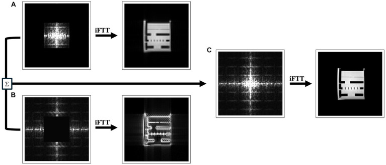
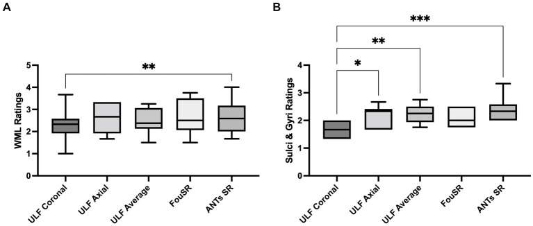
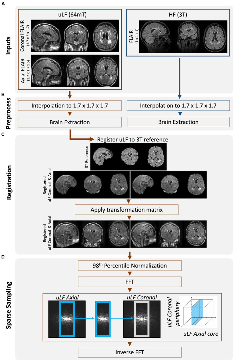
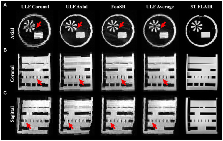
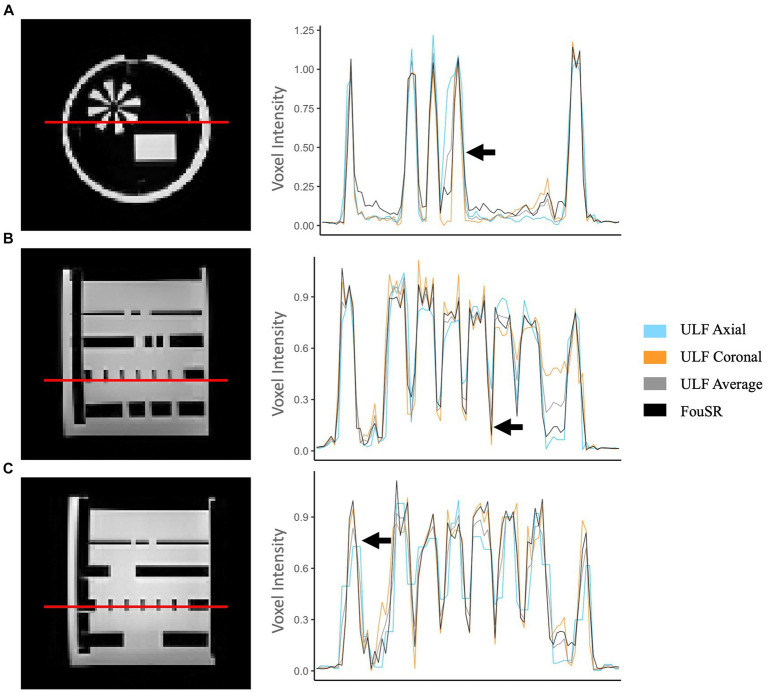
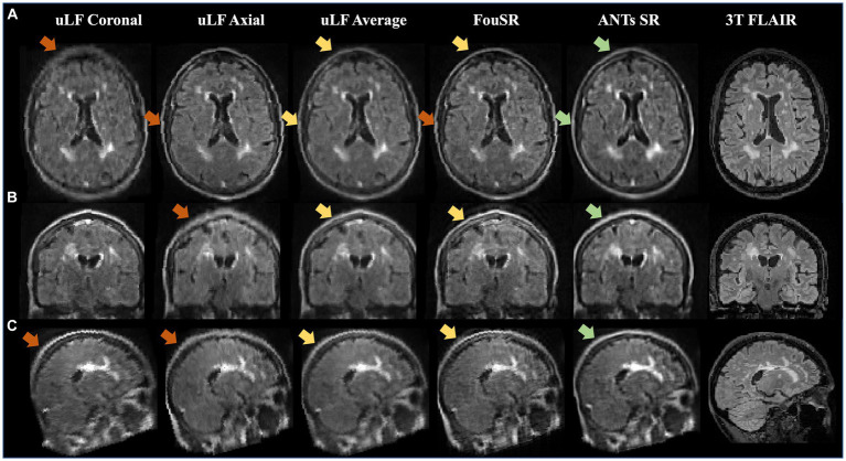
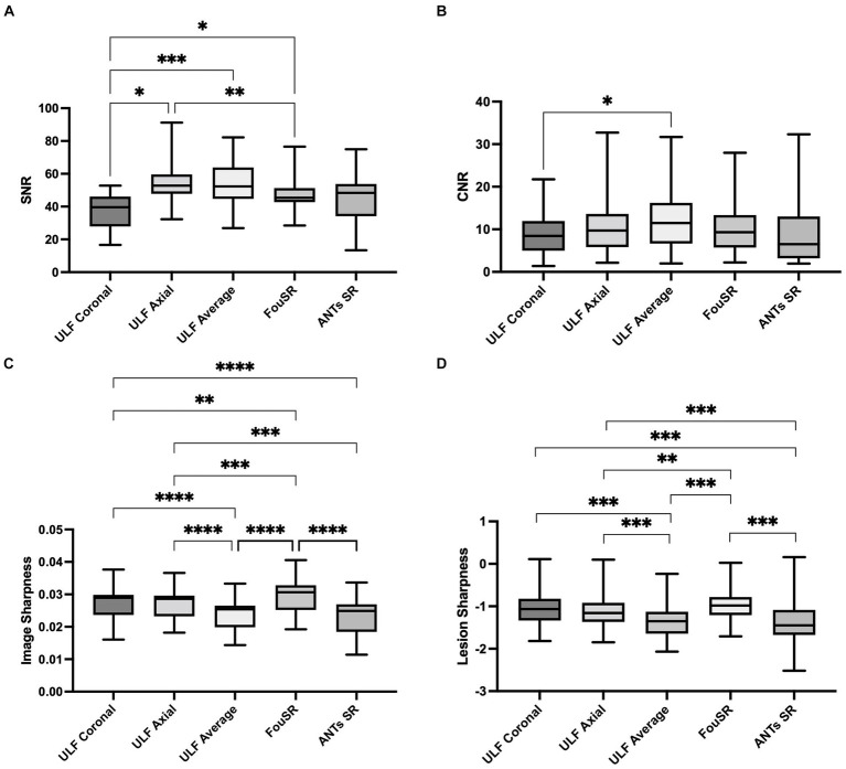
Ultra-low field (ULF) magnetic resonance imaging (MRI) holds the potential to make MRI more accessible, given its cost-effectiveness, reduced power requirements, and portability. However, signal-to-noise ratio (SNR) drops with field strength, necessitating imaging with lower resolution and longer scan times. This study introduces a novel Fourier-based Super Resolution (FouSR) approach, designed to enhance the resolution of ULF MRI images with minimal increase in total scan time. FouSR combines spatial frequencies from two orthogonal ULF images of anisotropic resolution to create an isotropic T2-weighted fluid-attenuated inversion recovery (FLAIR) image. We hypothesized that FouSR could effectively recover information from under-sampled slice directions, thereby improving the delineation of multiple sclerosis (MS) lesions and other significant anatomical features. Importantly, the FouSR algorithm can be implemented on the scanner with changes to the -space trajectory. Paired ULF (Hyperfine SWOOP, 0.064 tesla) and high field (Siemens, Skyra, 3 Tesla) FLAIR scans were collected on the same day from a phantom and a cohort of 10 participants with MS or suspected MS (6 female; mean ± SD age: 44.1 ± 4.1). ULF scans were acquired along both coronal and axial planes, featuring an in-plane resolution of 1.7 mm × 1.7 mm with a slice thickness of 5 mm. FouSR was evaluated against registered ULF coronal and axial scans, their average (ULF average) and a gold standard SR (ANTs SR). FouSR exhibited higher SNR (47.96 12.6) compared to ULF coronal (36.7 12.2) and higher lesion conspicuity (0.12 0.06) compared to ULF axial (0.13 0.07) but did not exhibit any significant differences contrast-to-noise-ratio (CNR) compared to other methods in patient scans. However, FouSR demonstrated superior image sharpness (0.025 0.0040) compared to all other techniques (ULF coronal 0.021 0.0037,  = 5.9, -adj. = 0.011; ULF axial 0.018 0.0026,  = 11.1, -adj. = 0.0001; ULF average 0.019 0.0034,  = 24.2, -adj. < 0.0001) and higher lesion sharpness (-0.97 0.31) when compared to the ULF average (-1.02 0.37, (543) = -10.174,  = <0.0001). Average blinded qualitative assessment by three experienced MS neurologists showed no significant difference in WML and sulci or gyri visualization between FouSR and other methods. FouSR can, in principle, be implemented on the scanner to produce clinically useful FLAIR images at higher resolution on the fly, providing a valuable tool for visualizing lesions and other anatomical structures in MS.

摘要

超低场(ULF)磁共振成像(MRI)具有使MRI更易于使用的潜力,因为其具有成本效益、降低的功率要求和便携性。然而,信噪比(SNR)会随着场强下降,这就需要以较低分辨率和更长扫描时间进行成像。本研究引入了一种基于傅里叶的新型超分辨率(FouSR)方法,旨在以总扫描时间的最小增加来提高ULF MRI图像的分辨率。FouSR将来自具有各向异性分辨率的两个正交ULF图像的空间频率组合起来,以创建一个各向同性的T2加权液体衰减反转恢复(FLAIR)图像。我们假设FouSR可以有效地从欠采样的切片方向恢复信息,从而改善多发性硬化症(MS)病变和其他重要解剖特征的描绘。重要的是,FouSR算法可以在扫描仪上通过改变k空间轨迹来实现。在同一天从一个体模和一组10名患有MS或疑似MS的参与者(6名女性;平均±标准差年龄:44.1±4.1)中收集配对的ULF(Hyperfine SWOOP,0.064特斯拉)和高场(西门子,Skyra,3特斯拉)FLAIR扫描。ULF扫描沿冠状面和轴面进行,面内分辨率为1.7毫米×1.7毫米,切片厚度为5毫米。将FouSR与配准后的ULF冠状面和轴面扫描、它们的平均值(ULF平均值)以及金标准超分辨率(ANTs SR)进行评估。与ULF冠状面(36.7±12.2)相比,FouSR表现出更高的SNR(47.96±12.6),与ULF轴面(0.13±0.07)相比,表现出更高的病变清晰度(0.12±0.06),但在患者扫描中与其他方法相比,在对比噪声比(CNR)方面没有表现出任何显著差异。然而,与所有其他技术相比,FouSR表现出更高的图像锐度(0.025±0.0040)(ULF冠状面0.021±0.0037,t = 5.9,p调整后 = = 0.011;ULF轴面0.018±0.0026,t = 11.1,p调整后 = = 0.0001;ULF平均值0.019±0.0034,t = 24.2,p调整后 < 0.0001),并且与ULF平均值(-1.02±0.37,t(543) = -10.174,p = <0.0001)相比,病变锐度更高(-0.97±0.3)。三位经验丰富的MS神经科医生进行的平均盲法定性评估显示,FouSR与其他方法在白质病变和脑沟或脑回可视化方面没有显著差异。原则上,FouSR可以在扫描仪上实现,以便即时生成具有更高分辨率的临床有用FLAIR图像,为可视化MS中的病变和其他解剖结构提供了一个有价值的工具。

https://cdn.ncbi.nlm.nih.gov/pmc/blobs/e936/11157107/9cff0240b7b0/fneur-15-1330203-g007.jpg
https://cdn.ncbi.nlm.nih.gov/pmc/blobs/e936/11157107/9808b547ee14/fneur-15-1330203-g001.jpg
https://cdn.ncbi.nlm.nih.gov/pmc/blobs/e936/11157107/fb2ec227ea46/fneur-15-1330203-g002.jpg
https://cdn.ncbi.nlm.nih.gov/pmc/blobs/e936/11157107/8436d3842d18/fneur-15-1330203-g003.jpg
https://cdn.ncbi.nlm.nih.gov/pmc/blobs/e936/11157107/bcbd62616d21/fneur-15-1330203-g004.jpg
https://cdn.ncbi.nlm.nih.gov/pmc/blobs/e936/11157107/7bd84e0eadc6/fneur-15-1330203-g005.jpg
https://cdn.ncbi.nlm.nih.gov/pmc/blobs/e936/11157107/cdc8319ceb10/fneur-15-1330203-g006.jpg
https://cdn.ncbi.nlm.nih.gov/pmc/blobs/e936/11157107/9cff0240b7b0/fneur-15-1330203-g007.jpg

相似文献

1
Super resolution using sparse sampling at portable ultra-low field MR.便携式超低场磁共振成像中基于稀疏采样的超分辨率技术
Front Neurol. 2024 May 24;15:1330203. doi: 10.3389/fneur.2024.1330203. eCollection 2024.
2
Sensitivity of portable low-field magnetic resonance imaging for multiple sclerosis lesions.便携式低场磁共振成像对多发性硬化病变的敏感性。
Neuroimage Clin. 2022;35:103101. doi: 10.1016/j.nicl.2022.103101. Epub 2022 Jun 27.
3
Combined Deep Learning-based Super-Resolution and Partial Fourier Reconstruction for Gradient Echo Sequences in Abdominal MRI at 3 Tesla: Shortening Breath-Hold Time and Improving Image Sharpness and Lesion Conspicuity.基于深度学习的超分辨率与部分傅里叶重建相结合用于3特斯拉腹部磁共振成像梯度回波序列:缩短屏气时间并提高图像清晰度和病变可见性
Acad Radiol. 2023 May;30(5):863-872. doi: 10.1016/j.acra.2022.06.003. Epub 2022 Jul 6.
4
FLAIR imaging for multiple sclerosis: a comparative MR study at 1.5 and 3.0 Tesla.用于多发性硬化症的液体衰减反转恢复序列成像:1.5 特斯拉和 3.0 特斯拉的对比磁共振研究
Eur Radiol. 2006 Apr;16(4):915-21. doi: 10.1007/s00330-005-0070-8. Epub 2005 Dec 20.
5
Ultra-High-Resolution Coronary CT Angiography With Photon-Counting Detector CT: Feasibility and Image Characterization.基于光子计数探测器 CT 的超高分辨率冠状动脉 CT 血管造影:可行性和图像特征。
Invest Radiol. 2022 Dec 1;57(12):780-788. doi: 10.1097/RLI.0000000000000897. Epub 2022 May 31.
6
Super-resolution reconstruction in frequency, image, and wavelet domains to reduce through-plane partial voluming in MRI.频率、图像和小波域中的超分辨率重建以减少MRI中的层间部分容积效应
Med Phys. 2015 Dec;42(12):6919-32. doi: 10.1118/1.4935149.
7
Development of neonatal-specific sequences for portable ultralow field magnetic resonance brain imaging: a prospective, single-centre, cohort study.用于便携式超低场磁共振脑成像的新生儿特异性序列的开发:一项前瞻性、单中心队列研究。
EClinicalMedicine. 2023 Oct 10;65:102253. doi: 10.1016/j.eclinm.2023.102253. eCollection 2023 Nov.
8
DeepFLAIR: A neural network approach to mitigate signal and contrast loss in temporal lobes at 7 Tesla FLAIR images.深 FLAIR:一种减轻 7T FLAIR 图像中颞叶信号和对比度损失的神经网络方法。
Magn Reson Imaging. 2024 Jul;110:57-68. doi: 10.1016/j.mri.2024.04.013. Epub 2024 Apr 15.
9
FLAIR*: a combined MR contrast technique for visualizing white matter lesions and parenchymal veins.FLAIR*:一种用于显示脑白质病变和脑实质静脉的联合磁共振对比技术。
Radiology. 2012 Dec;265(3):926-32. doi: 10.1148/radiol.12120208. Epub 2012 Oct 16.
10
Optimized 3D co-registration of ultra-low-field and high-field magnetic resonance images.优化超低磁场和高磁场磁共振图像的三维配准。
PLoS One. 2018 Mar 6;13(3):e0193890. doi: 10.1371/journal.pone.0193890. eCollection 2018.

引用本文的文献

1
Opportunities for Artificial Intelligence in Operational Medicine: Lessons from the United States Military.作战医学中人工智能的机遇:来自美国军方的经验教训。
Bioengineering (Basel). 2025 May 14;12(5):519. doi: 10.3390/bioengineering12050519.
2
High-Field-Blinded Assessment of Portable Ultra-Low-Field Brain MRI for Multiple Sclerosis.便携式超低场脑磁共振成像对多发性硬化症的高场盲法评估
J Neuroimaging. 2025 Jan-Feb;35(1):e70005. doi: 10.1111/jon.70005.
3
Proof of concept: Portable ultra-low-field magnetic resonance imaging for the diagnosis of epileptogenic brain pathologies.

本文引用的文献

1
Pushing the limits of low-cost ultra-low-field MRI by dual-acquisition deep learning 3D superresolution.双采集深度学习 3D 超分辨率技术推动低成本超低场 MRI 极限
Magn Reson Med. 2023 Aug;90(2):400-416. doi: 10.1002/mrm.29642. Epub 2023 Apr 3.
2
Quantitative Brain Morphometry of Portable Low-Field-Strength MRI Using Super-Resolution Machine Learning.基于超分辨率机器学习的便携式低场强 MRI 的定量脑形态测量
Radiology. 2023 Mar;306(3):e220522. doi: 10.1148/radiol.220522. Epub 2022 Nov 8.
3
Low-field MRI: Clinical promise and challenges.
概念验证:用于诊断致痫性脑部病变的便携式超低场磁共振成像
Epilepsia. 2024 Dec;65(12):3607-3618. doi: 10.1111/epi.18171. Epub 2024 Oct 29.
低场 MRI:临床的前景与挑战。
J Magn Reson Imaging. 2023 Jan;57(1):25-44. doi: 10.1002/jmri.28408. Epub 2022 Sep 19.
4
SynthStrip: skull-stripping for any brain image.SynthStrip:用于任何脑图像的头骨剥离。
Neuroimage. 2022 Oct 15;260:119474. doi: 10.1016/j.neuroimage.2022.119474. Epub 2022 Jul 13.
5
Deep learning for fast low-field MRI acquisitions.深度学习在快速低场 MRI 采集中的应用。
Sci Rep. 2022 Jul 6;12(1):11394. doi: 10.1038/s41598-022-14039-7.
6
Sensitivity of portable low-field magnetic resonance imaging for multiple sclerosis lesions.便携式低场磁共振成像对多发性硬化病变的敏感性。
Neuroimage Clin. 2022;35:103101. doi: 10.1016/j.nicl.2022.103101. Epub 2022 Jun 27.
7
Simultaneous high-resolution T -weighted imaging and quantitative T mapping at low magnetic field strengths using a multiple TE and multi-orientation acquisition approach.采用多回波和多方位采集方法在低磁场强度下进行同时高分辨率 T -加权成像和定量 T 映射。
Magn Reson Med. 2022 Sep;88(3):1273-1281. doi: 10.1002/mrm.29273. Epub 2022 May 12.
8
Deep learning-based single image super-resolution for low-field MR brain images.基于深度学习的低场磁共振脑图像单图像超分辨率。
Sci Rep. 2022 Apr 16;12(1):6362. doi: 10.1038/s41598-022-10298-6.
9
Development and evaluation of a high resolution 0.5mm isotropic T1-weighted template of the older adult brain.老年人大脑高分辨率 0.5mm 各向同性 T1 加权模板的建立与评估。
Neuroimage. 2022 Mar;248:118869. doi: 10.1016/j.neuroimage.2021.118869. Epub 2022 Jan 2.
10
A low-cost and shielding-free ultra-low-field brain MRI scanner.一种低成本、无屏蔽的超低场脑 MRI 扫描仪。
Nat Commun. 2021 Dec 14;12(1):7238. doi: 10.1038/s41467-021-27317-1.