• 文献检索
  • 文档翻译
  • 深度研究
  • 学术资讯
  • Suppr Zotero 插件Zotero 插件
  • 邀请有礼
  • 套餐&价格
  • 历史记录
应用&插件
Suppr Zotero 插件Zotero 插件浏览器插件Mac 客户端Windows 客户端微信小程序
定价
高级版会员购买积分包购买API积分包
服务
文献检索文档翻译深度研究API 文档MCP 服务
关于我们
关于 Suppr公司介绍联系我们用户协议隐私条款
关注我们

Suppr 超能文献

核心技术专利:CN118964589B侵权必究
粤ICP备2023148730 号-1Suppr @ 2026

文献检索

告别复杂PubMed语法,用中文像聊天一样搜索,搜遍4000万医学文献。AI智能推荐,让科研检索更轻松。

立即免费搜索

文件翻译

保留排版,准确专业,支持PDF/Word/PPT等文件格式,支持 12+语言互译。

免费翻译文档

深度研究

AI帮你快速写综述,25分钟生成高质量综述,智能提取关键信息,辅助科研写作。

立即免费体验

优化急性腹主动脉夹层的管理——基于人工智能的CT成像工作流程

Streamlining Acute Abdominal Aortic Dissection Management-An AI-based CT Imaging Workflow.

作者信息

Raj Anish, Allababidi Ahmad, Kayed Hany, Gerken Andreas L H, Müller Julia, Schoenberg Stefan O, Zöllner Frank G, Rink Johann S

机构信息

Computer Assisted Clinical Medicine, Medical Faculty Mannheim, Heidelberg University, Theodor-Kutzer-Ufer 1-3, D-68167, Mannheim, Germany.

Mannheim Institute for Intelligent Systems in Medicine, Medical Faculty Mannheim, Heidelberg University, Theodor-Kutzer-Ufer 1-3, D-68167, Mannheim, Germany.

出版信息

J Imaging Inform Med. 2024 Dec;37(6):2729-2739. doi: 10.1007/s10278-024-01164-0. Epub 2024 Jun 12.

DOI:10.1007/s10278-024-01164-0
PMID:38864947
原文链接:https://pmc.ncbi.nlm.nih.gov/articles/PMC11612133/
Abstract

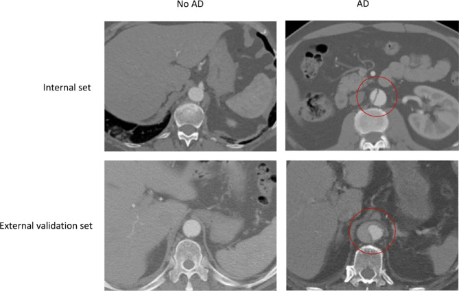
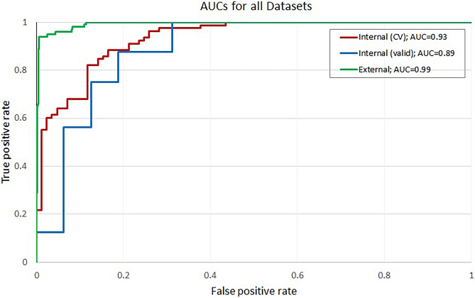
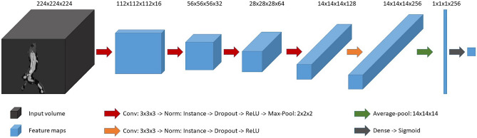
Life-threatening acute aortic dissection (AD) demands timely diagnosis for effective intervention. To streamline intrahospital workflows, automated detection of AD in abdominal computed tomography (CT) scans seems useful to assist humans. We aimed at creating a robust convolutional neural network (CNN)-based pipeline capable of real-time screening for signs of abdominal AD in CT. In this retrospective study, abdominal CT data from AD patients presenting with AD and from non-AD patients were collected (n 195, AD cases 94, mean age 65.9 years, female ratio 35.8%). A CNN-based algorithm was developed with the goal of enabling a robust, automated, and highly sensitive detection of abdominal AD. Two sets from internal (n = 32, AD cases 16) and external sources (n = 1189, AD cases 100) were procured for validation. The abdominal region was extracted, followed by the automatic isolation of the aorta region of interest (ROI) and highlighting of the membrane via edge extraction, followed by classification of the aortic ROI as dissected/healthy. A fivefold cross-validation was employed on the internal set, and an ensemble of the 5 trained models was used to predict the internal and external validation set. Evaluation metrics included receiver operating characteristic curve (AUC) and balanced accuracy. The AUC, balanced accuracy, and sensitivity scores of the internal dataset were 0.932 (CI 0.891-0.963), 0.860, and 0.885, respectively. For the internal validation dataset, the AUC, balanced accuracy, and sensitivity scores were 0.887 (CI 0.732-0.988), 0.781, and 0.875, respectively. Furthermore, for the external validation dataset, AUC, balanced accuracy, and sensitivity scores were 0.993 (CI 0.918-0.994), 0.933, and 1.000, respectively. The proposed automated pipeline could assist humans in expediting acute aortic dissection management when integrated into clinical workflows.

摘要

危及生命的急性主动脉夹层(AD)需要及时诊断以便进行有效干预。为了简化医院内部工作流程,利用腹部计算机断层扫描(CT)自动检测AD似乎有助于辅助人工诊断。我们旨在创建一个基于卷积神经网络(CNN)的强大流程,能够实时筛查CT图像中腹部AD的迹象。在这项回顾性研究中,收集了AD患者和非AD患者的腹部CT数据(n = 195,AD病例94例,平均年龄65.9岁,女性比例35.8%)。开发了一种基于CNN的算法,目标是实现对腹部AD进行强大、自动且高度敏感的检测。从内部(n = 32,AD病例16例)和外部来源(n = 1189,AD病例100例)获取了两组数据用于验证。提取腹部区域,然后自动分离主动脉感兴趣区域(ROI),通过边缘提取突出显示内膜,接着将主动脉ROI分类为夹层/健康。对内部数据集采用五折交叉验证,并使用5个训练模型的集成来预测内部和外部验证集。评估指标包括受试者工作特征曲线(AUC)和平衡准确率。内部数据集的AUC、平衡准确率和灵敏度得分分别为0.932(CI 0.891 - 0.963)、0.860和0.885。对于内部验证数据集,AUC、平衡准确率和灵敏度得分分别为0.887(CI 0.732 - 0.988)、0.781和0.875。此外,对于外部验证数据集,AUC、平衡准确率和灵敏度得分分别为0.993(CI 0.918 - 0.994)、0.933和1.000。当集成到临床工作流程中时,所提出的自动化流程可以帮助人工加速急性主动脉夹层的管理。

https://cdn.ncbi.nlm.nih.gov/pmc/blobs/9dcd/11612133/11e55a06a2a1/10278_2024_1164_Fig5_HTML.jpg
https://cdn.ncbi.nlm.nih.gov/pmc/blobs/9dcd/11612133/b2902b60876c/10278_2024_1164_Fig1_HTML.jpg
https://cdn.ncbi.nlm.nih.gov/pmc/blobs/9dcd/11612133/ed58a9f03967/10278_2024_1164_Fig2_HTML.jpg
https://cdn.ncbi.nlm.nih.gov/pmc/blobs/9dcd/11612133/26d15fa109b4/10278_2024_1164_Fig3_HTML.jpg
https://cdn.ncbi.nlm.nih.gov/pmc/blobs/9dcd/11612133/819617de0034/10278_2024_1164_Fig4_HTML.jpg
https://cdn.ncbi.nlm.nih.gov/pmc/blobs/9dcd/11612133/11e55a06a2a1/10278_2024_1164_Fig5_HTML.jpg

相似文献

1
Streamlining Acute Abdominal Aortic Dissection Management-An AI-based CT Imaging Workflow.优化急性腹主动脉夹层的管理——基于人工智能的CT成像工作流程
J Imaging Inform Med. 2024 Dec;37(6):2729-2739. doi: 10.1007/s10278-024-01164-0. Epub 2024 Jun 12.
2
Deep learning for automatic bowel-obstruction identification on abdominal CT.深度学习在腹部 CT 自动肠梗阻识别中的应用。
Eur Radiol. 2024 Sep;34(9):5842-5853. doi: 10.1007/s00330-024-10657-z. Epub 2024 Feb 22.
3
Deep learning algorithm for detection of aortic dissection on non-contrast-enhanced CT.深度学习算法在非增强 CT 上检测主动脉夹层。
Eur Radiol. 2021 Feb;31(2):1151-1159. doi: 10.1007/s00330-020-07213-w. Epub 2020 Aug 28.
4
Automatic Segmentation, Detection, and Diagnosis of Abdominal Aortic Aneurysm (AAA) Using Convolutional Neural Networks and Hough Circles Algorithm.使用卷积神经网络和霍夫圆算法对腹主动脉瘤(AAA)进行自动分割、检测和诊断
Cardiovasc Eng Technol. 2019 Sep;10(3):490-499. doi: 10.1007/s13239-019-00421-6. Epub 2019 Jun 19.
5
Development and Validation of a Convolutional Neural Network Model to Predict a Pathologic Fracture in the Proximal Femur Using Abdomen and Pelvis CT Images of Patients With Advanced Cancer.利用晚期癌症患者腹部和骨盆 CT 图像建立卷积神经网络模型预测股骨近端病理性骨折的研究
Clin Orthop Relat Res. 2023 Nov 1;481(11):2247-2256. doi: 10.1097/CORR.0000000000002771. Epub 2023 Aug 23.
6
Deep learning-aided diagnosis of acute abdominal aortic dissection by ultrasound images.基于超声图像的深度学习辅助诊断急性腹主动脉夹层。
Emerg Radiol. 2025 Apr;32(2):233-239. doi: 10.1007/s10140-025-02311-y. Epub 2025 Jan 17.
7
Deep Learning Prediction for Distal Aortic Remodeling After Thoracic Endovascular Aortic Repair in Stanford Type B Aortic Dissection.深度学习预测 Stanford B 型主动脉夹层胸主动脉腔内修复术后远端主动脉重塑。
J Endovasc Ther. 2024 Oct;31(5):910-918. doi: 10.1177/15266028231160101. Epub 2023 Mar 16.
8
Automated abdominal CT contrast phase detection using an interpretable and open-source artificial intelligence algorithm.使用可解释和开源人工智能算法进行自动腹部 CT 对比期检测。
Eur Radiol. 2024 Oct;34(10):6680-6687. doi: 10.1007/s00330-024-10769-6. Epub 2024 Apr 29.
9
Development and multicenter validation of deep convolutional neural network-based detection of colorectal cancer on abdominal CT.基于深度卷积神经网络的腹部 CT 结直肠癌检测的开发和多中心验证。
Eur Radiol. 2024 Sep;34(9):6182-6192. doi: 10.1007/s00330-023-10452-2. Epub 2024 Feb 1.
10
The diagnostic value of a non-contrast computed tomography scan-based radiomics model for acute aortic dissection.基于非对比 CT 扫描的放射组学模型对急性主动脉夹层的诊断价值。
Medicine (Baltimore). 2021 Jun 4;100(22):e26212. doi: 10.1097/MD.0000000000026212.

引用本文的文献

1
From promise to practice: a scoping review of AI applications in abdominal radiology.从承诺到实践:腹部放射学中人工智能应用的范围综述
Abdom Radiol (NY). 2025 Jul 28. doi: 10.1007/s00261-025-05144-y.
2
Multi-Stage Cascaded Deep Learning-Based Model for Acute Aortic Syndrome Detection: A Multisite Validation Study.基于多阶段级联深度学习的急性主动脉综合征检测模型:多中心验证研究
J Clin Med. 2025 Jul 7;14(13):4797. doi: 10.3390/jcm14134797.
3
Automated Detection and Differentiation of Stanford Type A and Type B Aortic Dissections in CTA Scans Using Deep Learning.

本文引用的文献

1
TotalSegmentator: Robust Segmentation of 104 Anatomic Structures in CT Images.全段分割器:CT图像中104种解剖结构的稳健分割
Radiol Artif Intell. 2023 Jul 5;5(5):e230024. doi: 10.1148/ryai.230024. eCollection 2023 Sep.
2
Analyzing Barriers and Enablers for the Acceptance of Artificial Intelligence Innovations into Radiology Practice: A Scoping Review.分析阻碍和推动放射科接受人工智能创新的因素:范围综述。
Tomography. 2023 Jul 28;9(4):1443-1455. doi: 10.3390/tomography9040115.
3
Artificial Intelligence Tool for Detection and Worklist Prioritization Reduces Time to Diagnosis of Incidental Pulmonary Embolism at CT.
使用深度学习在CTA扫描中自动检测和区分斯坦福A型和B型主动脉夹层
Diagnostics (Basel). 2024 Dec 25;15(1):12. doi: 10.3390/diagnostics15010012.
用于检测和工作列表优先级排序的人工智能工具可减少CT诊断偶然发现的肺栓塞的时间。
Radiol Cardiothorac Imaging. 2023 Apr 20;5(2):e220163. doi: 10.1148/ryct.220163. eCollection 2023 Apr.
4
Detection of acute thoracic aortic dissection based on plain chest radiography and a residual neural network (Resnet).基于普通胸部 X 射线摄影和残余神经网络(Resnet)检测急性胸主动脉夹层。
Sci Rep. 2022 Dec 19;12(1):21884. doi: 10.1038/s41598-022-26486-3.
5
Artificial Intelligence Applications in Aortic Dissection Imaging.人工智能在主动脉夹层成像中的应用。
Semin Roentgenol. 2022 Oct;57(4):357-363. doi: 10.1053/j.ro.2022.07.001. Epub 2022 Jul 30.
6
An Extra Set of Intelligent Eyes: Application of Artificial Intelligence in Imaging of Abdominopelvic Pathologies in Emergency Radiology.另一双智能之眼:人工智能在急诊放射学腹部盆腔病变成像中的应用
Diagnostics (Basel). 2022 May 30;12(6):1351. doi: 10.3390/diagnostics12061351.
7
Acceptance, Barriers, and Facilitators to Implementing Artificial Intelligence-Based Decision Support Systems in Emergency Departments: Quantitative and Qualitative Evaluation.急诊科实施基于人工智能的决策支持系统的接受度、障碍与促进因素:定量与定性评估
JMIR Form Res. 2022 Jun 13;6(6):e36501. doi: 10.2196/36501.
8
A Systematic Review and Bibliometric Analysis of Applications of Artificial Intelligence and Machine Learning in Vascular Surgery.人工智能和机器学习在血管外科应用的系统评价与文献计量分析
Ann Vasc Surg. 2022 Sep;85:395-405. doi: 10.1016/j.avsg.2022.03.019. Epub 2022 Mar 24.
9
Advanced Warning of Aortic Dissection on Non-Contrast CT: The Combination of Deep Learning and Morphological Characteristics.非增强CT上主动脉夹层的早期预警:深度学习与形态学特征的结合
Front Cardiovasc Med. 2022 Jan 5;8:762958. doi: 10.3389/fcvm.2021.762958. eCollection 2021.
10
: Multicenter aortic vessel tree CTA dataset collection with ground truth segmentation masks.多中心主动脉血管树CTA数据集收集及真实分割掩码
Data Brief. 2022 Jan 6;40:107801. doi: 10.1016/j.dib.2022.107801. eCollection 2022 Feb.