Gao Chuan, Wu Linyu, Wu Wei, Huang Yichao, Wang Xinyue, Sun Zhichao, Xu Maosheng, Gao Chen
The First Affiliated Hospital of Zhejiang Chinese Medical University (Zhejiang Provincial Hospital of Chinese Medicine), Hangzhou, China.
The First School of Clinical Medicine, Zhejiang Chinese Medical University, Hangzhou, China.
Eur Radiol. 2025 Jan;35(1):255-266. doi: 10.1007/s00330-024-10907-0. Epub 2024 Jul 10.
The accurate detection and precise segmentation of lung nodules on computed tomography are key prerequisites for early diagnosis and appropriate treatment of lung cancer. This study was designed to compare detection and segmentation methods for pulmonary nodules using deep-learning techniques to fill methodological gaps and biases in the existing literature.
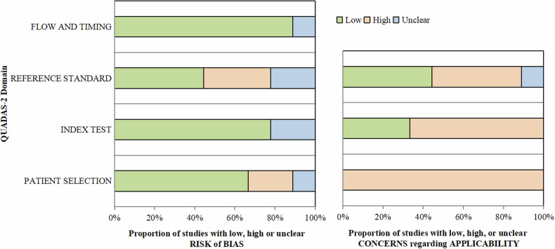
This study utilized a systematic review with the Preferred Reporting Items for Systematic Reviews and Meta-Analyses guidelines, searching PubMed, Embase, Web of Science Core Collection, and the Cochrane Library databases up to May 10, 2023. The Quality Assessment of Diagnostic Accuracy Studies 2 criteria was used to assess the risk of bias and was adjusted with the Checklist for Artificial Intelligence in Medical Imaging. The study analyzed and extracted model performance, data sources, and task-focus information.
After screening, we included nine studies meeting our inclusion criteria. These studies were published between 2019 and 2023 and predominantly used public datasets, with the Lung Image Database Consortium Image Collection and Image Database Resource Initiative and Lung Nodule Analysis 2016 being the most common. The studies focused on detection, segmentation, and other tasks, primarily utilizing Convolutional Neural Networks for model development. Performance evaluation covered multiple metrics, including sensitivity and the Dice coefficient.
This study highlights the potential power of deep learning in lung nodule detection and segmentation. It underscores the importance of standardized data processing, code and data sharing, the value of external test datasets, and the need to balance model complexity and efficiency in future research.
Deep learning demonstrates significant promise in autonomously detecting and segmenting pulmonary nodules. Future research should address methodological shortcomings and variability to enhance its clinical utility.
Deep learning shows potential in the detection and segmentation of pulmonary nodules. There are methodological gaps and biases present in the existing literature. Factors such as external validation and transparency affect the clinical application.
在计算机断层扫描上准确检测和精确分割肺结节是肺癌早期诊断和恰当治疗的关键前提。本研究旨在比较使用深度学习技术的肺结节检测和分割方法,以填补现有文献中的方法学空白和偏差。
本研究采用系统评价,并遵循系统评价和Meta分析的首选报告项目指南,检索截至2023年5月10日的PubMed、Embase、Web of Science核心合集和Cochrane图书馆数据库。使用诊断准确性研究的质量评估2标准来评估偏倚风险,并根据医学影像人工智能检查表进行调整。该研究分析并提取了模型性能、数据源和任务重点信息。
经过筛选,我们纳入了9项符合纳入标准的研究。这些研究发表于2019年至2023年之间,主要使用公共数据集,其中肺影像数据库联盟图像集和图像数据库资源倡议以及2016年肺结节分析是最常用的。这些研究专注于检测、分割和其他任务,主要利用卷积神经网络进行模型开发。性能评估涵盖多个指标,包括灵敏度和Dice系数。
本研究突出了深度学习在肺结节检测和分割中的潜在力量。它强调了标准化数据处理、代码和数据共享的重要性、外部测试数据集的价值以及在未来研究中平衡模型复杂性和效率的必要性。
深度学习在自主检测和分割肺结节方面显示出巨大前景。未来的研究应解决方法学上的不足和变异性,以提高其临床效用。
深度学习在肺结节的检测和分割中显示出潜力。现有文献中存在方法学空白和偏差。外部验证和透明度等因素影响临床应用。