• 文献检索
  • 文档翻译
  • 深度研究
  • 学术资讯
  • Suppr Zotero 插件Zotero 插件
  • 邀请有礼
  • 套餐&价格
  • 历史记录
应用&插件
Suppr Zotero 插件Zotero 插件浏览器插件Mac 客户端Windows 客户端微信小程序
定价
高级版会员购买积分包购买API积分包
服务
文献检索文档翻译深度研究API 文档MCP 服务
关于我们
关于 Suppr公司介绍联系我们用户协议隐私条款
关注我们

Suppr 超能文献

核心技术专利:CN118964589B侵权必究
粤ICP备2023148730 号-1Suppr @ 2026

文献检索

告别复杂PubMed语法,用中文像聊天一样搜索,搜遍4000万医学文献。AI智能推荐,让科研检索更轻松。

立即免费搜索

文件翻译

保留排版,准确专业,支持PDF/Word/PPT等文件格式,支持 12+语言互译。

免费翻译文档

深度研究

AI帮你快速写综述,25分钟生成高质量综述,智能提取关键信息,辅助科研写作。

立即免费体验

基于深度学习的 OCT 图像硬性渗出物检测及视网膜内层紊乱识别方法。

A deep learning approach to hard exudates detection and disorganization of retinal inner layers identification on OCT images.

机构信息

Ophthalmology Clinic, Department of Medicine and Ageing Science, University "G. D'Annunzio" of Chieti-Pescara, Via Dei Vestini Snc, 66100, Chieti, Italy.

Datamantix S.R.L. Artificial Intelligence Company, Via Paolo Sarpi, 14/15, 33100, Udine, Italy.

出版信息

Sci Rep. 2024 Jul 19;14(1):16652. doi: 10.1038/s41598-024-63844-9.

DOI:10.1038/s41598-024-63844-9
PMID:39030181
原文链接:https://pmc.ncbi.nlm.nih.gov/articles/PMC11271624/
Abstract

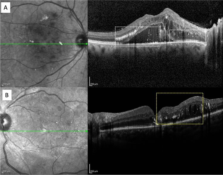
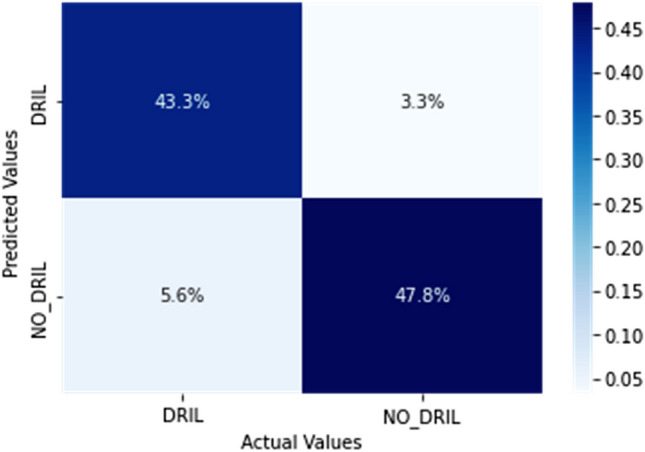
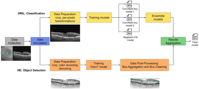
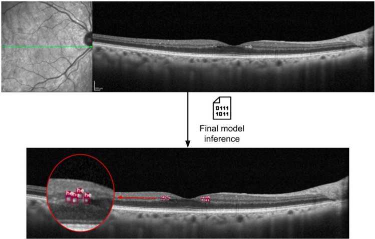
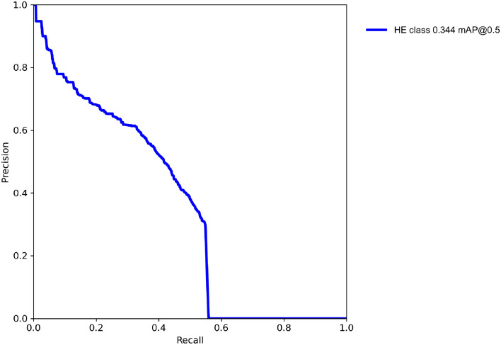
The purpose of the study was to detect Hard Exudates (HE) and classify Disorganization of Retinal Inner Layers (DRIL) implementing a Deep Learning (DL) system on optical coherence tomography (OCT) images of eyes with diabetic macular edema (DME). We collected a dataset composed of 442 OCT images on which we annotated 6847 HE and the presence of DRIL. A complex operational pipeline was defined to implement data cleaning and image transformations, and train two DL models. The state-of-the-art neural network architectures (Yolov7, ConvNeXt, RegNetX) and advanced techniques were exploited to aggregate the results (Ensemble learning, Edge detection) and obtain a final model. The DL approach reached good performance in detecting HE and classifying DRIL. Regarding HE detection the model got an AP@0.5 score equal to 34.4% with Precision of 48.7% and Recall of 43.1%; while for DRIL classification an Accuracy of 91.1% with Sensitivity and Specificity both of 91.1% and AUC and AUPR values equal to 91% were obtained. The P-value was lower than 0.05 and the Kappa coefficient was 0.82. The DL models proved to be able to identify HE and DRIL in eyes with DME with a very good accuracy and all the metrics calculated confirmed the system performance. Our DL approach demonstrated to be a good candidate as a supporting tool for ophthalmologists in OCT images analysis.

摘要

本研究旨在利用深度学习(DL)系统在糖尿病性黄斑水肿(DME)患者的光学相干断层扫描(OCT)图像上检测硬性渗出物(HE)并对视网膜内层紊乱(DRIL)进行分类。我们收集了一个由 442 张 OCT 图像组成的数据集,在这些图像上我们标注了 6847 个 HE 和 DRIL 的存在。定义了一个复杂的操作流程来实现数据清理和图像转换,并训练两个 DL 模型。利用最先进的神经网络架构(Yolov7、ConvNeXt、RegNetX)和先进技术来聚合结果(集成学习、边缘检测)并获得最终模型。DL 方法在检测 HE 和分类 DRIL 方面表现出良好的性能。在 HE 检测方面,该模型在 AP@0.5 上的得分达到 34.4%,精度为 48.7%,召回率为 43.1%;而在 DRIL 分类方面,准确率为 91.1%,灵敏度和特异性均为 91.1%,AUC 和 AUPR 值均为 91%。P 值小于 0.05,Kappa 系数为 0.82。DL 模型证明能够非常准确地识别 DME 患者的 HE 和 DRIL,所有计算的指标都证实了系统的性能。我们的 DL 方法被证明是 OCT 图像分析中眼科医生的一个很好的辅助工具。

https://cdn.ncbi.nlm.nih.gov/pmc/blobs/6ec1/11271624/5ab6610f82b0/41598_2024_63844_Fig5_HTML.jpg
https://cdn.ncbi.nlm.nih.gov/pmc/blobs/6ec1/11271624/7d72642e89cc/41598_2024_63844_Fig1_HTML.jpg
https://cdn.ncbi.nlm.nih.gov/pmc/blobs/6ec1/11271624/749e31fde16b/41598_2024_63844_Fig2_HTML.jpg
https://cdn.ncbi.nlm.nih.gov/pmc/blobs/6ec1/11271624/7cd7af78c326/41598_2024_63844_Fig3_HTML.jpg
https://cdn.ncbi.nlm.nih.gov/pmc/blobs/6ec1/11271624/1a2f5a3c6074/41598_2024_63844_Fig4_HTML.jpg
https://cdn.ncbi.nlm.nih.gov/pmc/blobs/6ec1/11271624/5ab6610f82b0/41598_2024_63844_Fig5_HTML.jpg

相似文献

1
A deep learning approach to hard exudates detection and disorganization of retinal inner layers identification on OCT images.基于深度学习的 OCT 图像硬性渗出物检测及视网膜内层紊乱识别方法。
Sci Rep. 2024 Jul 19;14(1):16652. doi: 10.1038/s41598-024-63844-9.
2
Deep Learning Algorithm Detects Presence of Disorganization of Retinal Inner Layers (DRIL)-An Early Imaging Biomarker in Diabetic Retinopathy.深度学习算法检测视网膜内层紊乱(DRIL)的存在 - 糖尿病视网膜病变的早期成像生物标志物。
Transl Vis Sci Technol. 2023 Jul 3;12(7):6. doi: 10.1167/tvst.12.7.6.
3
A new intelligent system based deep learning to detect DME and AMD in OCT images.基于深度学习的新型智能系统,用于检测 OCT 图像中的 DME 和 AMD。
Int Ophthalmol. 2024 Apr 23;44(1):191. doi: 10.1007/s10792-024-03115-8.
4
Pathological Neurovascular Unit Mapping onto Multimodal Imaging in Diabetic Macular Edema.病理性神经血管单元在糖尿病性黄斑水肿的多模态成像中的定位。
Medicina (Kaunas). 2023 May 7;59(5):896. doi: 10.3390/medicina59050896.
5
Fully automated detection of retinal disorders by image-based deep learning.基于图像的深度学习技术对视网膜疾病进行全自动检测。
Graefes Arch Clin Exp Ophthalmol. 2019 Mar;257(3):495-505. doi: 10.1007/s00417-018-04224-8. Epub 2019 Jan 4.
6
Disorganization of the retinal inner layers as a predictor of visual acuity in eyes with center-involved diabetic macular edema.视网膜内层结构紊乱作为累及黄斑中心的糖尿病性黄斑水肿患眼视力的预测指标。
JAMA Ophthalmol. 2014 Nov;132(11):1309-16. doi: 10.1001/jamaophthalmol.2014.2350.
7
Epiretinal Membrane Detection at the Ophthalmologist Level using Deep Learning of Optical Coherence Tomography.使用光学相干断层扫描的深度学习技术在眼科医生层面检测视网膜内界膜。
Sci Rep. 2020 May 21;10(1):8424. doi: 10.1038/s41598-020-65405-2.
8
Deep Ensemble Learning Based Objective Grading of Macular Edema by Extracting Clinically Significant Findings from Fused Retinal Imaging Modalities.基于深度集成学习的融合视网膜成像模态中临床显著发现提取的黄斑水肿客观分级。
Sensors (Basel). 2019 Jul 5;19(13):2970. doi: 10.3390/s19132970.
9
The Disorganization of Retinal Inner Layers Is Correlated to Müller Cells Impairment in Diabetic Macular Edema: An Imaging and Omics Study.视网膜内层次的紊乱与糖尿病性黄斑水肿中 Müller 细胞损伤相关:一项影像学和组学研究。
Int J Mol Sci. 2023 Jun 1;24(11):9607. doi: 10.3390/ijms24119607.
10
Machine learning techniques for diabetic macular edema (DME) classification on SD-OCT images.用于基于频域光学相干断层扫描(SD-OCT)图像的糖尿病性黄斑水肿(DME)分类的机器学习技术。
Biomed Eng Online. 2017 Jun 7;16(1):68. doi: 10.1186/s12938-017-0352-9.

引用本文的文献

1
Artificial intelligence derived grading of mustard gas induced corneal injury and opacity.人工智能辅助芥子气致角膜损伤和混浊的分级
Sci Rep. 2025 Jul 1;15(1):20359. doi: 10.1038/s41598-025-08042-x.
2
AI Quantification of Vascular Lesions in Mouse Fundus Fluorescein Angiography.小鼠眼底荧光血管造影中血管病变的人工智能量化
Transl Vis Sci Technol. 2025 Jun 2;14(6):4. doi: 10.1167/tvst.14.6.4.
3
Hyperreflective Retinal Foci (HRF): Definition and Role of an Invaluable OCT Sign.高反射性视网膜病灶(HRF):一种重要的光学相干断层扫描(OCT)征象的定义及作用

本文引用的文献

1
Diabetic Macular Edema Optical Coherence Tomography Biomarkers Detected with EfficientNetV2B1 and ConvNeXt.使用EfficientNetV2B1和ConvNeXt检测糖尿病性黄斑水肿光学相干断层扫描生物标志物
Diagnostics (Basel). 2023 Dec 28;14(1):76. doi: 10.3390/diagnostics14010076.
2
Clinical Utility of Spectral-Domain Optical Coherence Tomography Marker Disorganization of Retinal Inner Layers in Diabetic Retinopathy.光谱域光学相干断层扫描视网膜内层标记紊乱在糖尿病视网膜病变中的临床应用。
Ophthalmic Surg Lasers Imaging Retina. 2023 Dec;54(12):692-700. doi: 10.3928/23258160-20231031-02. Epub 2023 Dec 1.
3
Predicting OCT biological marker localization from weak annotations.
J Clin Med. 2025 Apr 27;14(9):3021. doi: 10.3390/jcm14093021.
从弱标注中预测 OCT 生物标志物定位。
Sci Rep. 2023 Nov 11;13(1):19667. doi: 10.1038/s41598-023-47019-6.
4
Automated evaluation of retinal hyperreflective foci changes in diabetic macular edema patients before and after intravitreal injection.糖尿病性黄斑水肿患者玻璃体内注射前后视网膜高反射灶变化的自动评估
Front Med (Lausanne). 2023 Oct 6;10:1280714. doi: 10.3389/fmed.2023.1280714. eCollection 2023.
5
Deep Learning Algorithm Detects Presence of Disorganization of Retinal Inner Layers (DRIL)-An Early Imaging Biomarker in Diabetic Retinopathy.深度学习算法检测视网膜内层紊乱(DRIL)的存在 - 糖尿病视网膜病变的早期成像生物标志物。
Transl Vis Sci Technol. 2023 Jul 3;12(7):6. doi: 10.1167/tvst.12.7.6.
6
Validation of an Automated Artificial Intelligence Algorithm for the Quantification of Major OCT Parameters in Diabetic Macular Edema.一种用于量化糖尿病性黄斑水肿主要光学相干断层扫描参数的自动化人工智能算法的验证
J Clin Med. 2023 Mar 9;12(6):2134. doi: 10.3390/jcm12062134.
7
Persistent diabetic macular edema: Definition, incidence, biomarkers, and treatment methods.持续性糖尿病性黄斑水肿:定义、发生率、生物标志物和治疗方法。
Surv Ophthalmol. 2023 Mar-Apr;68(2):147-174. doi: 10.1016/j.survophthal.2022.11.008. Epub 2022 Nov 24.
8
Diabetic Macular Edema: Current Understanding, Molecular Mechanisms and Therapeutic Implications.糖尿病性黄斑水肿:当前认识、分子机制及治疗意义。
Cells. 2022 Oct 25;11(21):3362. doi: 10.3390/cells11213362.
9
A New Approach for Detecting Fundus Lesions Using Image Processing and Deep Neural Network Architecture Based on YOLO Model.基于 YOLO 模型的图像处理和深度神经网络架构的眼底病变检测新方法。
Sensors (Basel). 2022 Aug 26;22(17):6441. doi: 10.3390/s22176441.
10
Hyperreflective Dots on OCT as a Predictor of Treatment Outcome in Diabetic Macular Edema: A Systematic Review.OCT 上的高反射点预测糖尿病性黄斑水肿的治疗效果:系统评价。
Ophthalmol Retina. 2022 Sep;6(9):814-827. doi: 10.1016/j.oret.2022.03.020. Epub 2022 Mar 30.