Department of Rehabilitation Medicine, Sichuan Provincial People's Hospital, School of Medicine, University of Electronic Science and Technology of China, Chengdu, 610072, People's Republic of China.
School of Medicine, University of Electronic Science and Technology of China, Chengdu, 610072, People's Republic of China.
Sci Rep. 2024 Aug 16;14(1):19027. doi: 10.1038/s41598-024-69862-x.
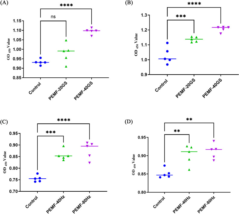
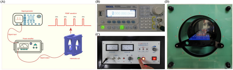
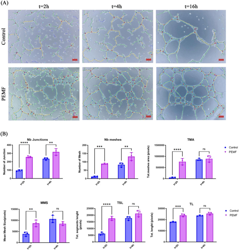
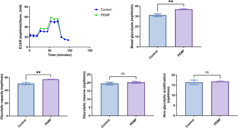
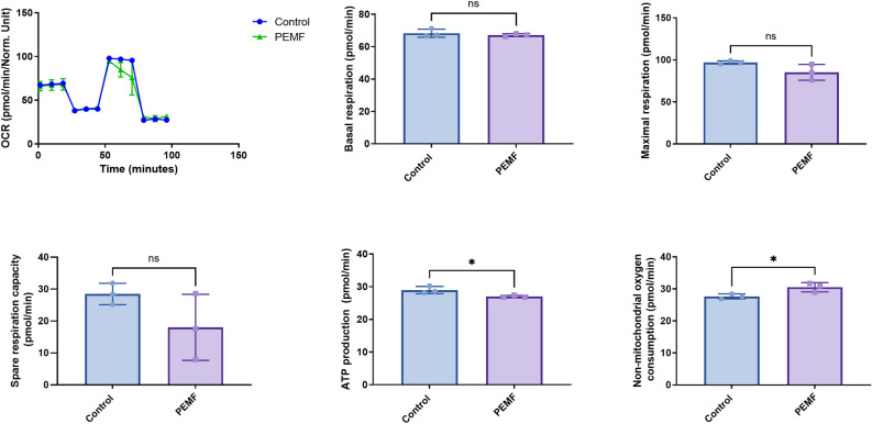
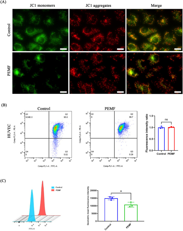
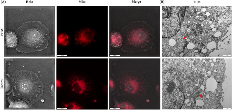
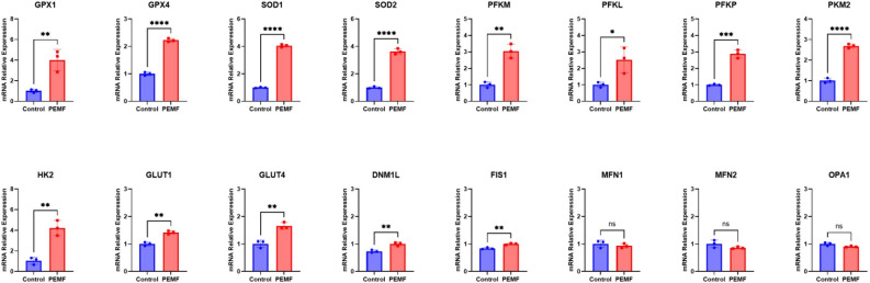
Pulsed electromagnetic field (PEMF) therapy has been extensively investigated in clinical studies for the treatment of angiogenesis-related diseases. However, there is a lack of research on the impact of PEMFs on energy metabolism and mitochondrial dynamics during angiogenesis. The present study included tube formation and CCK-8 assays. A Seahorse assay was conducted to analyze energy metabolism, and mitochondrial membrane potential assays, mitochondrial imaging, and reactive oxygen species assays were used to measure changes in mitochondrial structure and function in human umbilical vein endothelial cells (HUVECs) exposed to PEMFs. Real-time polymerase chain reaction was used to analyze the mRNA expression levels of antioxidants, glycolytic pathway-related genes, and genes associated with mitochondrial fission and fusion. The tube formation assay demonstrated a significantly greater tube network in the PEMF group compared to the control group. The glycolysis and mitochondrial stress tests revealed that PEMFs promoted a shift in the energy metabolism pattern of HUVECs from oxidative phosphorylation to aerobic glycolysis. Mitochondrial imaging revealed a wire-like mitochondrial morphology in the control group, and treatment with PEMFs led to shorter and more granular mitochondria. Our major findings indicate that exposure to PEMFs accelerates angiogenesis in HUVECs, likely by inducing energy metabolism reprogramming and mitochondrial fission.
脉冲电磁场(PEMF)治疗在临床研究中已被广泛研究用于治疗与血管生成相关的疾病。然而,目前缺乏关于 PEMF 对血管生成过程中能量代谢和线粒体动力学影响的研究。本研究包括管形成和 CCK-8 测定。使用 Seahorse 测定法分析能量代谢,并用线粒体膜电位测定法、线粒体成像和活性氧测定法来测量人脐静脉内皮细胞(HUVEC)中暴露于 PEMF 后线粒体结构和功能的变化。实时聚合酶链反应用于分析抗氧化剂、糖酵解途径相关基因以及与线粒体分裂和融合相关基因的 mRNA 表达水平。管形成测定表明,与对照组相比,PEMF 组的管网络明显更大。糖酵解和线粒体应激测试表明,PEMF 促进了 HUVEC 能量代谢模式从氧化磷酸化向有氧糖酵解的转变。线粒体成像显示对照组中存在线状线粒体形态,而 PEMF 处理导致线粒体变短且颗粒状增多。我们的主要发现表明,暴露于 PEMF 可加速 HUVEC 中的血管生成,可能通过诱导能量代谢重编程和线粒体分裂来实现。