• 文献检索
  • 文档翻译
  • 深度研究
  • 学术资讯
  • Suppr Zotero 插件Zotero 插件
  • 邀请有礼
  • 套餐&价格
  • 历史记录
应用&插件
Suppr Zotero 插件Zotero 插件浏览器插件Mac 客户端Windows 客户端微信小程序
定价
高级版会员购买积分包购买API积分包
服务
文献检索文档翻译深度研究API 文档MCP 服务
关于我们
关于 Suppr公司介绍联系我们用户协议隐私条款
关注我们

Suppr 超能文献

核心技术专利:CN118964589B侵权必究
粤ICP备2023148730 号-1Suppr @ 2026

文献检索

告别复杂PubMed语法,用中文像聊天一样搜索,搜遍4000万医学文献。AI智能推荐,让科研检索更轻松。

立即免费搜索

文件翻译

保留排版,准确专业,支持PDF/Word/PPT等文件格式,支持 12+语言互译。

免费翻译文档

深度研究

AI帮你快速写综述,25分钟生成高质量综述,智能提取关键信息,辅助科研写作。

立即免费体验

毁损对自动脑萎缩估计的影响。

Impact of defacing on automated brain atrophy estimation.

作者信息

Rubbert Christian, Wolf Luisa, Turowski Bernd, Hedderich Dennis M, Gaser Christian, Dahnke Robert, Caspers Julian

机构信息

University Dusseldorf, Medical Faculty, Department of Diagnostic and Interventional Radiology, D-40225, Dusseldorf, Germany.

Department of Diagnostic and Interventional Neuroradiology, School of Medicine, Technical University of Munich, 81675, Munich, Germany.

出版信息

Insights Imaging. 2022 Mar 26;13(1):54. doi: 10.1186/s13244-022-01195-7.

DOI:10.1186/s13244-022-01195-7
PMID:35348936
原文链接:https://pmc.ncbi.nlm.nih.gov/articles/PMC8964867/
Abstract

BACKGROUND

Defacing has become mandatory for anonymization of brain MRI scans; however, concerns regarding data integrity were raised. Thus, we systematically evaluated the effect of different defacing procedures on automated brain atrophy estimation.

METHODS

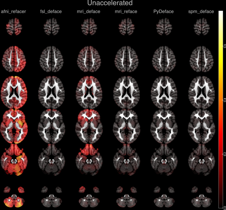
In total, 268 Alzheimer's disease patients were included from ADNI, which included unaccelerated (n = 154), within-session unaccelerated repeat (n = 67) and accelerated 3D T1 imaging (n = 114). Atrophy maps were computed using the open-source software veganbagel for every original, unmodified scan and after defacing using afni_refacer, fsl_deface, mri_deface, mri_reface, PyDeface or spm_deface, and the root-mean-square error (RMSE) between z-scores was calculated. RMSE values derived from unaccelerated and unaccelerated repeat imaging served as a benchmark. Outliers were defined as RMSE > 75th percentile and by using Grubbs's test.

RESULTS

Benchmark RMSE was 0.28 ± 0.1 (range 0.12-0.58, 75th percentile 0.33). Outliers were found for unaccelerated and accelerated T1 imaging using the 75th percentile cutoff: afni_refacer (unaccelerated: 18, accelerated: 16), fsl_deface (unaccelerated: 4, accelerated: 18), mri_deface (unaccelerated: 0, accelerated: 15), mri_reface (unaccelerated: 0, accelerated: 2) and spm_deface (unaccelerated: 0, accelerated: 7). PyDeface performed best with no outliers (unaccelerated mean RMSE 0.08 ± 0.05, accelerated mean RMSE 0.07 ± 0.05). The following outliers were found according to Grubbs's test: afni_refacer (unaccelerated: 16, accelerated: 13), fsl_deface (unaccelerated: 10, accelerated: 21), mri_deface (unaccelerated: 7, accelerated: 20), mri_reface (unaccelerated: 7, accelerated: 6), PyDeface (unaccelerated: 5, accelerated: 8) and spm_deface (unaccelerated: 10, accelerated: 12).

CONCLUSION

Most defacing approaches have an impact on atrophy estimation, especially in accelerated 3D T1 imaging. Only PyDeface showed good results with negligible impact on atrophy estimation.

摘要

背景

对脑部磁共振成像(MRI)扫描进行匿名化处理时,毁损已成为一项强制性要求;然而,人们对数据完整性提出了担忧。因此,我们系统地评估了不同毁损程序对脑萎缩自动估计的影响。

方法

总共从阿尔茨海默病神经影像倡议(ADNI)中纳入了268例阿尔茨海默病患者,其中包括未加速成像(n = 154)、同次扫描内未加速重复成像(n = 67)和加速三维T1成像(n = 114)。使用开源软件veganbagel对每幅原始未修改扫描图像以及使用afni_refacer、fsl_deface、mri_deface、mri_reface、PyDeface或spm_deface进行毁损后的图像计算萎缩图谱,并计算z分数之间的均方根误差(RMSE)。来自未加速成像和未加速重复成像的RMSE值作为基准。异常值定义为RMSE > 第75百分位数,并通过格拉布斯检验确定。

结果

基准RMSE为0.28 ± 0.1(范围0.12 - 0.58,第75百分位数为0.33)。使用第75百分位数临界值时,在未加速和加速T1成像中发现了异常值:afni_refacer(未加速:18例,加速:16例)、fsl_deface(未加速:4例,加速:18例)、mri_deface(未加速:0例,加速:15例)、mri_reface(未加速:0例,加速:2例)和spm_deface(未加速:0例,加速:7例)。PyDeface表现最佳,未发现异常值(未加速平均RMSE 0.08 ± 0.05,加速平均RMSE 0.07 ± 0.05)。根据格拉布斯检验发现了以下异常值:afni_refacer(未加速:16例,加速:13例)、fsl_deface(未加速:10例,加速:21例)、mri_deface(未加速:7例,加速:20例)、mri_reface(未加速:7例,加速:6例)、PyDeface(未加速:5例,加速:8例)和spm_deface(未加速:10例,加速:12例)。

结论

大多数毁损方法都会对萎缩估计产生影响,尤其是在加速三维T1成像中。只有PyDeface显示出良好的结果,对萎缩估计的影响可忽略不计。

https://cdn.ncbi.nlm.nih.gov/pmc/blobs/d4af/8964867/7d2c358ea0b9/13244_2022_1195_Fig4_HTML.jpg
https://cdn.ncbi.nlm.nih.gov/pmc/blobs/d4af/8964867/959b63aa77d7/13244_2022_1195_Fig1_HTML.jpg
https://cdn.ncbi.nlm.nih.gov/pmc/blobs/d4af/8964867/33b87725374f/13244_2022_1195_Fig2_HTML.jpg
https://cdn.ncbi.nlm.nih.gov/pmc/blobs/d4af/8964867/d1d1acda094b/13244_2022_1195_Fig3_HTML.jpg
https://cdn.ncbi.nlm.nih.gov/pmc/blobs/d4af/8964867/7d2c358ea0b9/13244_2022_1195_Fig4_HTML.jpg

相似文献

1
Impact of defacing on automated brain atrophy estimation.毁损对自动脑萎缩估计的影响。
Insights Imaging. 2022 Mar 26;13(1):54. doi: 10.1186/s13244-022-01195-7.
2
Segmentation stability of human head and neck cancer medical images for radiotherapy applications under de-identification conditions: Benchmarking data sharing and artificial intelligence use-cases.去识别化条件下用于放射治疗的人类头颈癌医学图像的分割稳定性:基准数据共享和人工智能用例
Front Oncol. 2023 Feb 28;13:1120392. doi: 10.3389/fonc.2023.1120392. eCollection 2023.
3
Multisite Comparison of MRI Defacing Software Across Multiple Cohorts.多队列中MRI去标识软件的多中心比较
Front Psychiatry. 2021 Feb 24;12:617997. doi: 10.3389/fpsyt.2021.617997. eCollection 2021.
4
PyFaceWipe: a new defacing tool for almost any MRI contrast.PyFaceWipe:一种新的去面工具,几乎适用于任何 MRI 对比。
MAGMA. 2024 Dec;37(6):993-1003. doi: 10.1007/s10334-024-01170-x. Epub 2024 Jun 21.
5
Deep learning-based defacing tool for CT angiography: CTA-DEFACE.基于深度学习的 CT 血管造影图像篡改工具:CTA-DEFACE。
Eur Radiol Exp. 2024 Oct 9;8(1):111. doi: 10.1186/s41747-024-00510-9.
6
Assessing the Impact of Defacing Algorithms on Brain Volumetry Accuracy in MRI Analyses.评估毁损算法对MRI分析中脑容量测量准确性的影响。
Dement Neurocogn Disord. 2024 Jul;23(3):127-135. doi: 10.12779/dnd.2024.23.3.127. Epub 2024 May 8.
7
Accelerated vs. unaccelerated serial MRI based TBM-SyN measurements for clinical trials in Alzheimer's disease.基于加速与非加速序列磁共振成像的丘脑底核同步测量在阿尔茨海默病临床试验中的应用
Neuroimage. 2015 Jun;113:61-9. doi: 10.1016/j.neuroimage.2015.03.026. Epub 2015 Mar 20.
8
Reproducibility evaluation of the effects of MRI defacing on brain segmentation.MRI去标识对脑部分割影响的可重复性评估
J Med Imaging (Bellingham). 2023 Nov;10(6):064001. doi: 10.1117/1.JMI.10.6.064001. Epub 2023 Nov 8.
9
A reproducibility evaluation of the effects of MRI defacing on brain segmentation.MRI去标识化对脑部分割影响的可重复性评估
medRxiv. 2023 May 21:2023.05.15.23289995. doi: 10.1101/2023.05.15.23289995.
10
Systematic evaluation of the impact of defacing on quality and volumetric assessments on T1-weighted MR-images.对T1加权磁共振图像上毁损对质量和容积评估的影响进行系统评价。
J Neuroradiol. 2022 May;49(3):250-257. doi: 10.1016/j.neurad.2021.03.001. Epub 2021 Mar 13.

引用本文的文献

1
Automated quality control of T1-weighted brain MRI scans for clinical research datasets: methods comparison and design of a quality prediction classifier.用于临床研究数据集的T1加权脑磁共振成像扫描的自动质量控制:方法比较与质量预测分类器设计
Imaging Neurosci (Camb). 2025 May 28;3. doi: 10.1162/IMAG.a.4. eCollection 2025.
2
Bridging Huntington's disease research with big data science: Harmonized neuroimaging datasets from multiple studies.将亨廷顿舞蹈症研究与大数据科学相衔接:来自多项研究的统一神经影像数据集。
Imaging Neurosci (Camb). 2024 Dec 16;2. doi: 10.1162/imag_a_00395. eCollection 2024.
3
Analysis of Factors Affecting Quality in Structural Magnetic Resonance Images.

本文引用的文献

1
Reproducibility of Brain Volume Changes in Longitudinal Voxel-Based Morphometry Between Non-Accelerated and Accelerated Magnetic Resonance Imaging.基于体素的形态测量学在非加速和加速磁共振成像中脑容量变化的重复性。
J Alzheimers Dis. 2021;83(1):281-290. doi: 10.3233/JAD-210596.
2
Systematic evaluation of the impact of defacing on quality and volumetric assessments on T1-weighted MR-images.对T1加权磁共振图像上毁损对质量和容积评估的影响进行系统评价。
J Neuroradiol. 2022 May;49(3):250-257. doi: 10.1016/j.neurad.2021.03.001. Epub 2021 Mar 13.
3
Multisite Comparison of MRI Defacing Software Across Multiple Cohorts.
结构磁共振图像质量的影响因素分析
Hum Brain Mapp. 2025 Aug 1;46(11):e70271. doi: 10.1002/hbm.70271.
4
Empowering Data Sharing in Neuroscience: A Deep Learning Deidentification Method for Pediatric Brain MRIs.助力神经科学中的数据共享:一种用于儿科脑磁共振成像的深度学习去识别方法。
AJNR Am J Neuroradiol. 2025 May 2;46(5):964-972. doi: 10.3174/ajnr.A8581.
5
OpenMAP-T1: A Rapid Deep-Learning Approach to Parcellate 280 Anatomical Regions to Cover the Whole Brain.OpenMAP-T1:一种快速深度学习方法,可将 280 个解剖区域分割为覆盖整个大脑的区域。
Hum Brain Mapp. 2024 Nov;45(16):e70063. doi: 10.1002/hbm.70063.
6
Implementation and validation of face de-identification (de-facing) in ADNI4.在 ADNI4 中实现和验证人脸去识别(去面)。
Alzheimers Dement. 2024 Nov;20(11):8048-8061. doi: 10.1002/alz.14303. Epub 2024 Oct 11.
7
Assessing the Impact of Defacing Algorithms on Brain Volumetry Accuracy in MRI Analyses.评估毁损算法对MRI分析中脑容量测量准确性的影响。
Dement Neurocogn Disord. 2024 Jul;23(3):127-135. doi: 10.12779/dnd.2024.23.3.127. Epub 2024 May 8.
8
Summary of the National Cancer Institute 2023 Virtual Workshop on Medical Image De-identification-Part 2: Pathology Whole Slide Image De-identification, De-facing, the Role of AI in Image De-identification, and the NCI MIDI Datasets and Pipeline.美国国立癌症研究所2023年医学图像去识别化虚拟研讨会总结 - 第二部分:病理学全切片图像去识别化、面部去除、人工智能在图像去识别化中的作用以及美国国立癌症研究所MIDI数据集和流程
J Imaging Inform Med. 2025 Feb;38(1):16-30. doi: 10.1007/s10278-024-01183-x. Epub 2024 Jul 9.
9
PyFaceWipe: a new defacing tool for almost any MRI contrast.PyFaceWipe:一种新的去面工具,几乎适用于任何 MRI 对比。
MAGMA. 2024 Dec;37(6):993-1003. doi: 10.1007/s10334-024-01170-x. Epub 2024 Jun 21.
10
Prediction of therapy response of breast cancer patients with machine learning based on clinical data and imaging data derived from breast [F]FDG-PET/MRI.基于来自乳腺 [F]FDG-PET/MRI 的临床数据和影像学数据,应用机器学习预测乳腺癌患者的治疗反应。
Eur J Nucl Med Mol Imaging. 2024 Apr;51(5):1451-1461. doi: 10.1007/s00259-023-06513-9. Epub 2023 Dec 22.
多队列中MRI去标识软件的多中心比较
Front Psychiatry. 2021 Feb 24;12:617997. doi: 10.3389/fpsyt.2021.617997. eCollection 2021.
4
Changing the face of neuroimaging research: Comparing a new MRI de-facing technique with popular alternatives.改变神经影像学研究的面貌:比较一种新的 MRI 去面技术与流行的替代方法。
Neuroimage. 2021 May 1;231:117845. doi: 10.1016/j.neuroimage.2021.117845. Epub 2021 Feb 11.
5
Automated age- and sex-specific volumetric estimation of regional brain atrophy: workflow and feasibility.基于年龄和性别的区域脑萎缩自动容积估算:工作流程和可行性。
Eur Radiol. 2021 Feb;31(2):1043-1048. doi: 10.1007/s00330-020-07196-8. Epub 2020 Aug 27.
6
MAGNIMS consensus recommendations on the use of brain and spinal cord atrophy measures in clinical practice.MAGNIMS 关于在临床实践中使用脑和脊髓萎缩测量的共识建议。
Nat Rev Neurol. 2020 Mar;16(3):171-182. doi: 10.1038/s41582-020-0314-x. Epub 2020 Feb 24.
7
Normative brain volume reports may improve differential diagnosis of dementing neurodegenerative diseases in clinical practice.正常脑容量报告可能有助于改善临床实践中痴呆性神经退行性疾病的鉴别诊断。
Eur Radiol. 2020 May;30(5):2821-2829. doi: 10.1007/s00330-019-06602-0. Epub 2020 Jan 30.
8
Facing privacy in neuroimaging: removing facial features degrades performance of image analysis methods.神经影像学中的隐私问题:去除面部特征会降低图像分析方法的性能。
Eur Radiol. 2020 Feb;30(2):1062-1074. doi: 10.1007/s00330-019-06459-3. Epub 2019 Nov 5.
9
Identification of Anonymous MRI Research Participants with Face-Recognition Software.使用面部识别软件识别匿名MRI研究参与者
N Engl J Med. 2019 Oct 24;381(17):1684-1686. doi: 10.1056/NEJMc1908881.
10
Huntington's disease: Brain imaging in Huntington's disease.亨廷顿病:亨廷顿病的脑影像学。
Prog Mol Biol Transl Sci. 2019;165:321-369. doi: 10.1016/bs.pmbts.2019.04.004. Epub 2019 May 15.